Open Access
Research
Studies on HDL associated enzymes under experimental
hypercholesterolemia: possible modulation on selenium
supplementation
Harman D Kaur and Mohinder P Bansal*
Address: Department of Biophysics, Panjab University, Chandigarh 160014, India
Email: Harman D Kaur - [email protected]; Mohinder P Bansal* - [email protected] * Corresponding author
Abstract
Background: Atherosclerosis is a chronic disorder of the arterial wall that starts by formation of fatty streaks and gradually evolves into atherosclerotic plaques. High-density lipoproteins (HDL) blood levels are inversely correlated with atherosclerosis. This beneficial effect of HDL has been partly attributed to its antioxidant properties mediated by paraoxonase1 (PON1) or platelet-activating factor acetylhydrolase (PAF-AH). The present study was aimed to study HDL associated enzymes i.e. PON1 and PAF-AH under experimental hypercholesterolemia and their possible modulation on selenium (Se; an antioxidant) supplementation. Male Sprague Dawley rats were divided into three groups and fed on the control diet, high fat diet (HFD) and HFD + Se respectively for the period of 4 months.
Results: Cholesterol, triglycerides, HDL and LDL levels were significantly increased by HFD feeding. Selenium supplementation lowered the triglyceride level, whereas the other lipid values remained unchanged. Serum selenium levels were reduced by 31% and ROS levels in the liver were 2-fold increased by HFD. Se supplementation, however, diminished the HFD-induced ROS levels by 29%. Furthermore, Se also improved the HFD-mediated reduction of serum PON1 enzyme activity by 34% and PON1 protein levels by 21%. However, no significant effect of Se was detected on the reduced PAF-AH proteins levels in HFD fed rats. mRNA expression of PON1 and PAF-AH in the liver was not affected in the Se treated groups.
Conclusion: Se supplementation appears to be protective in hypercholesterolemia by restoring the antioxidant properties of the HDL associated enzyme i.e. PON1 whereas biological system aims towards maintaining the same PAF-AH levels even on selenium supplementation indicating its probable role in both anti and pro-atherogenic activities. Therefore, Se supplementation might be a valuable approach to limit the adverse effects of hypercholesterolemia and may need further investigations.
Background
Hypercholesterolemia represents one of the important and recognized risk factor for atherosclerosis [1]. There are
compelling evidences indicating the importance of type of fats than the total amount of the fats with respect to the risk of the cardiovascular diseases [2]. Cholesterol is
trans-Published: 16 December 2009
Lipids in Health and Disease 2009, 8:55 doi:10.1186/1476-511X-8-55
Received: 13 October 2009 Accepted: 16 December 2009
This article is available from: http://www.lipidworld.com/content/8/1/55 © 2009 Kaur and Bansal; licensee BioMed Central Ltd.
related to the risk for atherosclerotic events [3] and are found to possess anti-atherogenic activity [4]. Among the risk factors, total cholesterol/HDL cholesterol ratio is con-sidered to be the most predictive for atherosclerosis [5]. The protective effect of the HDL is related partly to enzymes associated with HDL [6-8] and due to its partici-pation in reverse cholesterol transport [9].
Paraoxonase1 (PON1) is one of the enzymes associated with HDL [10]. PON1 was shown to protect against oxi-dative stress [11,12], a phenomenon that can be attrib-uted to its ability to modulate oxidized lipids in LDL and HDL [13,14], in macrophages [15,16] and also in athero-sclerotic plaques [17]. PON is capable of hydrolyzing lipid peroxides in LDL [18]. Serum HDL-associated PON1 reduces oxidative stress in lipoproteins, in macrophages and in atherosclerotic lesion, whereas PON2 acts as an antioxidant at the cellular and not humoral level. The attenuation to atherosclerosis is related to the nutritional anti-oxidative induced increase in HDL-PON activity [19].
PAF-AH is the major enzyme responsible for the catabo-lism of PAF and PAF like lipids that are also the potent mediators of inflammation [20,21]. Genetic deficiency of PAF-AH in defined human populations increases the severity of atherosclerosis and other syndromes [22]. PAF-AH has marked preference for phospholipids with short chain moieties at syn-2 position and, with the exception of PAF, it can equally hydrolyze oxidized phospholipids containing at syn-2 position polyunsaturated fatty acyl residues [23]. However, during hydrolysis the oxidized phospholipids, PAF-AH liberate the bioactive oxidized free fatty acids [24] and generates lysophopsphatidylcho-line, both of which are implicated in the biological actions of ox-LDL [25]. Thus, PAF-AH could play both pro-atherogenic and anti-atherogenic role.
Selenium, an essential trace element, is associated with cardiovascular diseases since years. Selenium deficiency is related to increase in plasma cholesterol levels [26,27], cardiac myopathy [28], other cardiovascular disease and ischemic heart diseases [29,30]. Selenium supplementa-tion leads to decrease in total cholesterol and triglyceride levels [31,32].
Keeping these in view, in the present study influence of selenium was explored on HDL associated enzymes, PON1 and PAF-AH.
Results
Selenium levelsSelenium levels were estimated in the serum of rats from all the groups after 4 months of diet feeding schedule
(Fig-group. However, an apparent increase (P < 0.001) in Se was observed on HFD + Se (1 ppm) supplementation in comparison to the rats fed on only HFD diet.
Lipid profile
Lipid profile analysis was done in serum (Table 1). Total cholesterol (P < 0.001), triglycerides (P < 0.05) and HDL cholesterol (P < 0.05) levels were found to be significantly increased in HFD group in comparison to control group. A highly significant increase (P < 0.001) was observed in LDL-cholesterol and total cholesterol/HDL-cholesterol ratio in HFD group in comparison to the control group. However, no significant change in various lipid profile parameters except decrease in triglycerides levels was observed in HFD + Se group in comparison to the only HFD fed group.
ROS (Reactive oxygen species) levels
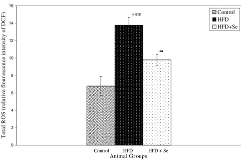
ROS levels in liver homogenates were estimated using a fluorescence probe DCFH-DA. Oxidation of DCFH-DA to DCF was measured as an index of total ROS. A highly sig-nificant (P < 0.001) increase in ROS levels was observed in HFD group in comparison to the control group. How-ever, a significant (P < 0.01) decrease in ROS levels were observed in HFD+Se fed group in comparison to HFD fed group (Figure 2).
PON1 activity
PON1 activity was estimated in serum using substrate paraoxon. A highly significant (P < 0.001) decrease in the level of PON1 activity was observed in HFD fed group in comparison to control group. However, a significant (P < 0.01) increase in the level of PON1 was observed in HFD+Se fed group in comparison to HFD fed group (Fig-ure 3).
mRNA expression of PON1 and PAF-AH
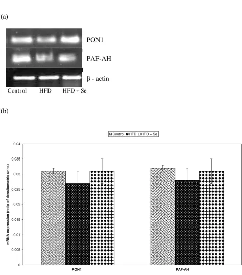
Statistically no significant changes in the mRNA expres-sion of PON1 and PAF-AH were observed in HFD or HFD + Se fed groups in comparison to the control group or HFD fed group respectively Figure 4a, Figure 4b.
Protein expression of PON1 and PAF-AH by ELISA
Discussion
Hypercholesterolemia represents one of the very impor-tant and recognized risk factor for atherosclerosis [33]. Abnormally high cholesterol levels (high concentration of LDL and low concentration of HDL) are strongly associ-ated with the cardiovascular diseases. High cholesterol diet leads to the cholesterol deposition in the arterial walls [34]. Compelling evidences indicate the importance of the type of fats than the total amount of the fats with respect to the risk of the cardiovascular diseases [35]. Con-trolled clinical trials have shown that replacing saturated fats with unsaturated is more effective in lowering serum cholesterol and reducing the risk of cardiovascular dis-eases than simply reducing total fat consumption [36].
Selenium, an essential trace element is proved to be pro-tective against cardiovascular diseases [37]. In the earlier experiments in the authors laboratory, it was found that selenium supplementation at 1 ppm level along with high fat diet (HFD) feeding in rats inhibited the incidence of atherosclerosis as studied by Scanning electron micros-copy (SEM) of aorta [38]. In the present study, interest-ingly the serum selenium levels decreased in HFD group in comparison to the control group. On the other hand, selenium level, as expected, increased when we gave exter-nal supplementation of 1 ppm as sodium selenite in HFD + Se fed group.
Serum selenium levels
Figure 1
Serum selenium levels. *P < 0.05 represents comparison between control and HFD, aaaP < 0.001 represents comparison between HFD and HFD + Se group.
0 0.5 1 1.5 2 2.5 3
Animal Gr oups
μg
S
e
/ m
l
se
ru
m
Control
HFD
HFD+Se
ªªª
*
Excess of lipids in the serum derived from endogenous synthesis/dietary sources initiate atherosclerosis by accu-mulation in the cells of the arterial wall and provoking atheroma growth [39]. In the present study, total choles-terol and triglycerides levels were found to be significantly increased in HFD group in comparison to control group.
Triglyceride levels were found to be greatly reduced in HFD + Se fed group in comparison to HFD fed group. These findings suggest the hypercholesterolemic state as reported earlier [40-42]. The selenium potential against hypercholesterolemia is supported by some other research groups as well [41,43].
Lipid profile (mg/dL) Control HFD HFD + Se
Total cholesterol 71.79 ± 13.66 451.66 ± 26.73*** 438.20 ± 53.91
Triglycerides 82.50 ± 6.96 91.98 ± 0.02* 81.26 ± 11.96a
HDL-C 24.76 ± 4.26 31.70 ± 6.56* 37.58 ± 5.55
LDL-C 32.66 ± 2.81 395.52 ± 26.51*** 381.00 ± 53.58
Total cholesterol/HDL-C 2.90 ± 0.22 14.86 ± 3.86*** 11.99 ± 2.95
Data is represented as Mean ± S.D. from 5 independent observations. Data is statistically analyzed by student's t-test. **p < 0.01,*p < 0.05 represent the comparison between control and HFD group. ap < 0.05 represents the comparison between HFD and HFD+ Se group.
Reactive oxygen species levels
Figure 2
Reactive oxygen species levels. ***P < 0.001 represents comparison between control and HFD, aaP < 0.01 represents com-parison between HFD and HFD + Se group.
0 2 4 6 8 10 12 14 16
Animal Gr oups
T
o
ta
l R
O
S
(r
el
a
tiv
e flo
u
re
sc
en
ce
in
te
n
sit
y
o
f
D
C
F
)
Control
HFD
HFD+Se
***
ªª
LDL has long been implicated in the development of atherosclerosis [44]. It has also been reported that sele-nium supplementation protect LDL from oxidation and other atherogenic changes [45,46]. HDL has been found to protect against the oxidation of LDL by metal ions in-vitro [47,48] and by reverse cholesterol transport [49]. Further, LDL and HDL levels were found to be signifi-cantly increased in HFD group in comparison to the con-trol group. Increase in HDL levels [41,43] on Se supplementation was also reported earlier. Moreover, total cholesterol/HDL ratio is considered to be most pre-dictive for atherosclerosis [48,50]. In the present study, this ratio was found to be increased in HFD group in com-parison to the control group suggesting greatest risk for the clinical events related to hypercholesterolemia in HFD group in comparison to other groups.
Reactive oxygen species (ROS) levels were estimated in liver homogenates and were found to be increased 2-folds in HFD group in comparison to the control group. Se sup-plementation, however, diminished the HFD-induced ROS levels by 29%. This suggests the presence of oxidative stress in HFD fed group which decreased on 1 ppm sele-nium supplementation possibly due to the anti-oxidative property of Se. Previously also in the author's laboratory,
on analysis of glutathione peroxidase, lipid peroxidation, nitric oxide synthase (NOS) and reduced/oxidized glu-tathione ratio in aorta, liver and serum, it was demon-strated that the reduced incidence of atherosclerosis on selenium supplementation was due to the anti-oxidant function of selenium [51,52]. Also in-vitro studies, in author's lab, demonstrated the role of mitogen stimulated lymphocytes and macrophage NO production on sele-nium supplementation in HFD-induced atherogenesis in rats [53].
Work from a number of laboratories have suggested that the protective effect of HDL may be due to the enzymes associated with HDL [6,48,54] i.e PON1 (Paraoxonase 1), LCAT (Lecithin acyl transferase), PAF-AH (platelet activat-ing factor-acetyl hydrolase)
PON1, an HDL associated enzyme, synthesized in liver, was shown to protect against oxidative stress [11,12], a phenomenon that can be attributed to its ability to mod-ulated oxidized lipids in LDL and HDL [13,14], in macro-phages [15,16] and also in atherosclerotic lesions [17]. In the present study, biochemically, PON1 enzymes activity was found to be significantly reduced in HFD group in comparison to control group. In addition, protein expres-PON1 enzyme activity
Figure 3
PON1 enzyme activity. ***P < 0.001 represents comparison between control and HFD, aaP < 0.01 represents comparison between HFD and HFD + Se group.
0 1 2 3 4 5 6
Animal Groups
PO
N
1
(μ
M
Pa
ra
o
xon
hy
dr
ol
y
sed
/m
g
pr
o
tei
n
/m
in
)
Control HFD HFD+Se
***
ªª
A - mRNA expression of PON1 and PAF-AH by RT-PCR. B - Densitometric analysis of PON1 and PAF-AH mRNA expression
Figure 4
A - mRNA expression of PON1 and PAF-AH by RT-PCR. B - Densitometric analysis of PON1 and PAF-AH mRNA expression. Data is represented as Mean ± S.D. from four independent observations. Data is statistically analyzed by student's t-test.
PON1
PAF-AH
Contr ol HFD HFD + Se
ȕ
- actin
(b)
0 0.005 0.01 0.015 0.02 0.025 0.03 0.035 0.04
3213$)$+
P51$H
[
S
UHVVLRQ
UD
WLR
RI
GHQV
LW
R
PHWULFX
QLWV
sion of PON1 by Elisa was also found to be significantly reduced in HFD group in comparison to control group. However, Se supplementation improved the HFD-medi-ated reduction of serum PON1 enzyme activity by 34% and PON1 protein levels by 21%. This suggests that oxida-tive stress under hypercholesterolemic state leads to the reduced activity of PON1 whereas on selenium supple-mentation, levels started retrieving. Interestingly, the PON1 mRNA expression studies revealed that there was no significant change at transcriptional level. This suggests that PON1 may possibly be involved in anti-atherogenic activities at translational levels. In conclusion, Se supple-mentation appears to be protective in experimental hyper-cholesterolemia by restoring the antioxidant properties of the HDL associated enzyme, PON1.
PAF-AH is the major enzyme responsible for the catabo-lism of PAF (platelet activating factor). Hypercholestero-lemia and inflammation work as partners in atherogenesis [55,56]. Evidences have suggested that genetic deficiency of PAF-AH in defined human popula-tion increases the severity of atherosclerosis and other syndromes [57]. PAF-AH possesses marked preference for hydrolyzing oxidized phospholipids containing at syn-2 position polyunsaturated fatty acyl residues [58]. How-ever, during hydrolysis the oxidized phospholipids, PAF-AH liberate the bioactive oxidized free fatty acids [24] and generates lysophopsphatidylcholine, both of which are implicated in the biological actions of ox-LDL [25]. Thus, PAF-AH could play both atherogenic and anti-atherogenic role. In the present study, PAF-AH protein expression by ELISA was found to be significantly decreased in HFD group in comparison to control group whereas no statisti-cally significant change was observed on selenium supple-mentation. Interestingly, on the other hand mRNA expression revealed that there was no significant change in PAF-AH at the transcriptional levels. From the present study, it is inferred that PAF-AH possibly participates in both pro and anti-atherogenic activities as here the system aims towards balancing the PAF-AH levels but it may need some further investigations in order to designate the exact role of PAF-AH.
Conclusion
In conclusion, from the results obtained by the present study, Se supplementation appears to be protective in experimental hypercholesterolemia by restoring the anti-oxidative properties of the HDL associated enzyme PON1. However, findings are inconclusive in determining the role of PAF-AH whether pro or anti-atherogenic in role or both and may need some further investigations. There-fore, Se supplementation might be a valuable approach to limit the adverse effects of hypercholesterolemia.
Materials and methods
ChemicalsSodium selenite (Na2SeO3), 2, 3-diaminonapthalene and Dichlorofluorescein diacetate (DCFH-DA) were pur-chased from Sigma-Aldrich (St. Louis, Missouri, USA). TRI-reagent and one step RT-PCR kits were obtained from Molecular research Centre (Inc. Cincinnati, Ohio) and QIAGEN, respectively. Antibodies against PON1 and PAF-AH were obtained from Santa-Cruz Biotechnology, Santa Cruz USA. All other chemicals were of analytical grade and were procured from Indian manufacturers.
Animals
Male Sprague Dawley rats (100-150 g body weights) were obtained from the Central Animal House, Panjab Univer-sity, Chandigarh. The animals were kept in plastic cages under the hygienic conditions and were fed on special diets. Before initiating the experiment, the animals were adapted to the laboratory conditions for a week. Neces-sary approvals were obtained from the Institutional Ethics Committee. The animal care and handling were done according to the guidelines set by the World Health Organization (WHO), Geneva, Switzerland, and the Indian National Science Academy (INSA), New Delhi, India.
Experimental design
Rats were divided into three groups (five animals each group) for the present study. Group I animals served as controls. These animals were fed on Control Diet. Group II animals were fed on High fat Diet (HFD). Group III ani-mals were fed on HFD with 1 ppm selenium supplemen-tation.
Diet Preparation
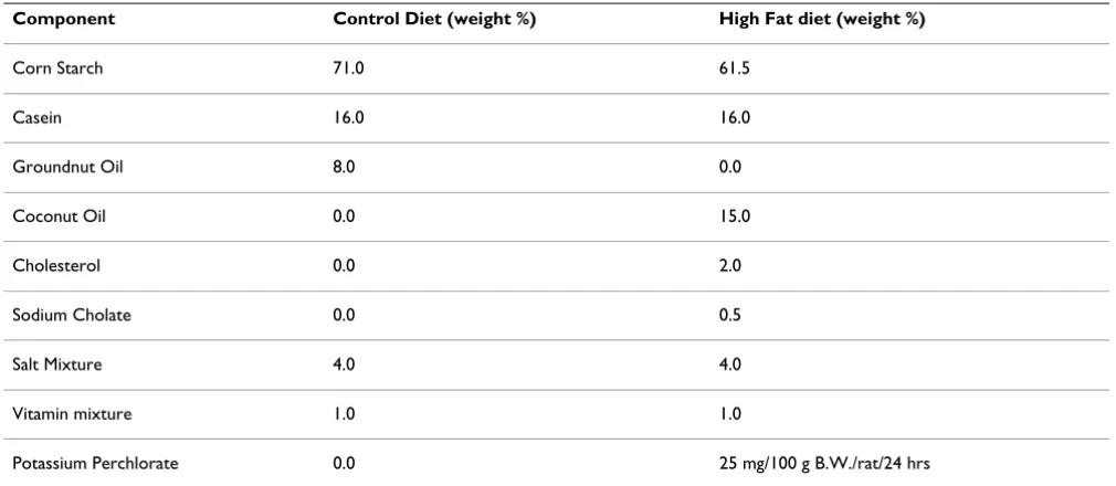
Casein based diets i.e. control diet, HFD was prepared in the laboratory itself according to the composition given by Abraham et al [59] Table 3.
To the group III animals i.e. HFD + Se group, 1 ppm sele-nium was given as sodium selenite and was administered along with the high fat diet. The selenium was so chosen that the level is in excess to adequate levels of selenium (0.2 ppm) but well below the sub toxic limits (2.0 ppm).
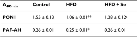
Table 2: PON1 and PAF-AH levels by ELISA after 4 months of control, HFD and HFD + Se feeding schedule
A405 nm Control HFD HFD + Se
PON1 1.55 ± 0.13 1.06 ± 0.01** 1.28 ± 0.12a
PAF-AH 0.26 ± 0.01 0.25 ± 0.01* 0.26 ± 0.01
Data is represented as Mean ± S.D. from 5 independent observations. Data is statistically analyzed by student's t-test. **p < 0.01,*p < 0.05 represent the comparison between control and HFD group. ap < 0.05
Selenium levels
Selenium levels were estimated in serum by fluorimetric method[60]. The assay is based on the principle that Se content in samples on acid digestion is converted to sele-nous acid. The reaction between selesele-nous acid and aro-matic-o-diamines such as 2, 3-diaminonapathalene leads to the formation of 4, 5- benzopiazselenol which displays brilliant lime-green fluorescence when excited at 366 nm in cyclohexane. Fluorescence emission in cyclohexane extract was read on fluorescence spectrophotometer using 366 nm as excitation and 520 nm as emission wave-lengths.
Lipid profile
Lipid profile analysis was done in serum using colorimet-ric kits to estimate the total cholesterol, triglycerides, HDL, LDL levels. Total Cholesterol level was estimated using CHOP-PAP based kit (Human Diagnostic Ger-many). Triglyceride levels were estimated using GPO based kit (Accurex Biomedical India). HDL and LDL cho-lesterol levels were estimated using (Fortress direct kit) enzymatic kit.
Reactive oxygen species (ROS) levels
Determination of ROS was based on the modified method of Driver et al [61]. Liver homogenates were pre-pared in ice-cold Locke's buffer (154 mM NaCl, 5.6 mM KCl, 3.6 mM NaHCO3, 2 mM CaCl2, 10 mM d-glucose and 5 mM HEPES pH 7.4). The homogenates were allowed to warm at 21°C for 5 min. The reaction mixture containing 10 μM DCFH-DA and 5 mg tissue/mL was incubated for 15 min at room temperature (21°C). After another 30 min of incubation, the conversion of DCFH to the fluorescent product 2, 7 dichloroflourescein (DCF)
was measured using fluorescence spectrophotometer with excitation at 485 nm and emission at 530 nm. Back-ground fluorescence (Conversion of DCFH-DH to DCF in the absence of homogenate) was corrected by inclusion of parallel blanks. The relative fluorescence intensity was taken as the measure of amount of ROS in different groups.
Measurement of PON1 activity
PON1 activity was assessed in serum by measuring the ini-tial rate of Paraoxon hydrolysis to yield p-Nitrophenol at 412 nm at 25°C. The basal assay mixture included 2 mM Paraoxon, 2 mM CaCl2 and 0.5 ml serum in 100 mM Tris/ Cl buffer. The extinction coefficient for the reaction is 18290 M-1 cm-1. Activity is expressed as μM Paraoxon
hydrolyzed/mg protein/min [62].
RNA isolation and mRNA expression of PON1 and PAF-AH using RT-PCR
Total RNA was isolated from fresh liver using Tri-reagent (Molecular research Centre, Inc Ohio, USA) and the qual-ity of isolated RNA was checked on 1.2% agarose gel elec-trophoresis. For reverse-transcriptase polymerase chain reaction (RT-PCR), primers for PON1, PAF-AH were designed with the aid of software and β- actin primer was taken from literature. The primer sequence for PON1 was Fav- 5'-TGGCATTGGCATTTCCCTTG-3', Rev- CAG-TAGCTTTCACTCCGGTAA-3' and for PAF-AH Fav- 5'-CTGATGACAAGACCCTCCGTG-3', Rev- 5'-CCGTAAC-CAGTGTGGTCCGGAT-3' and for β-actin Fav- 5'-AGAGCTATGAGCTGCCTGAC-3', Rev-3'-CTGCATCCT-GTCAGCCTACG-5'. After pilot experiments, it was found that PCR products for PON1, PAF-AH were progressively amplified till 35 cycles and hence 35 amplification cycles
Component Control Diet (weight %) High Fat diet (weight %)
Corn Starch 71.0 61.5
Casein 16.0 16.0
Groundnut Oil 8.0 0.0
Coconut Oil 0.0 15.0
Cholesterol 0.0 2.0
Sodium Cholate 0.0 0.5
Salt Mixture 4.0 4.0
Vitamin mixture 1.0 1.0
were performed. The RT-PCR reaction (Qiagen kit) used a template cDNA followed by PCR amplification with Accu Taq DNA polymerase in the same tube. PCR products were analysed by 1.5% agarose gel electrophoresis. Densi-tometric analysis from six independent observations was done by Image-J software.
PON1 and PAF-AH protein expression by ELISA
Wells were coated with 5 μg of sample for PON1 and PAF-AH in 100 μl of 0.05 M carbonate buffer (pH 9.6) and kept overnight at 4°C, in a moist chamber. Plates were flicked to remove the unbound antigen solution and wells were blocked with 1% BSA in 0.1 M phosphate buffer saline (pH 7.2) for1 hr at 37°C. Plates were flicked and wells were washed with 200 μl of PBS containing 0.05% (v/v) Tween-20. Wells were then incubated with anti-PON1 and anti-PAF-AH respectively, diluted in PBS (con-taining 0.05% Tween and 1% BSA) and kept for 2 hr at 37°C. Plates were washed again and incubated with anti-goat secondary antibody (peroxidase labeled) for PON1 (1:1000) and PAF-AH (1:1000) for 2 hr at 37°C. Wells were washed further thrice as described above and color was developed by addition of 2, 2'-azino-di (3-ethyl)-ben-zothiozolinsulphonic acid reagent and absorbance at 405 nm was measured by ELISA reader.
Statistical Analysis
Data is represented as mean ± S.D. Statistical analysis of the data was performed by student's T-test.
Abbreviations
A260: Absorption at 260 nm; A412: Absorption at 412 nm; B.W: Body Weight; LCAT: Lecithin Cholesterol Acyl Trans-ferase; LDL: Low Density Lipoproteins; OD: Optical Den-sity; oxLDL: Oxidized Low Density Lipoprotein; PAF: Platelet activating Factor; PAF-AH: Platelet activating Fac-tor-Acetyl Hydrolase; PON1: Paraoxonase1; Se: Selenium; SRB1: Scavenger receptor B (class) 1(type); VLDL: Very Low Density Lipoproteins.
Competing interests
The authors declare that they have no competing interests.
Authors' contributions
MPB designed the study and participated in drafting man-uscript and result analysis. HDK carried out all the exper-imental work, participated in statistical analysis and drafting the manuscript under the guidance of MPB. All authors read and approved the final manuscript.
Acknowledgements
Authors acknowledge the support given by Director: Prof. Tapas Mukho-padhyay (Department of human genomics, Panjab University, Chandigarh, 160014 India) for using various equipments.
References
1. Descamps OS, Gilbeau JP, Luwaert R, Heller FR: Impact of genetic defects on coronary atherosclerosis in patients' suspects of
having Familial Hypercholesterolemia. Eur J Clin Invest 2003,
33:1-9.
2. Seidel C, Deufel T, Jahreis G: Effects of fat-modified dairy prod-ucts on blood lipids in humans in comparisons with other
fats. Ann Nutr Metab 2005, 49:42-48.
3. Gordon DJ, Probstfield JL, Garrison RJ, Neaton JD, Castelli WP, Knoke JD, Jacobs DR Jr, Bangdiwala S, Tyroler HA: High density
lipoprotein cholesterol and cardiovascular diseases.
Circula-tion 1989:8-15.
4. Zuliani G, Volpato S, Blè A, Bandinelli S, Corsi AM, Lauretani F, Paolisso G, Fellin R, Ferrucci L: High interleukin-6 plasma levels are associated with low HDL-C levels in community-dwelling
older adults: the Inchianti study. Atherosclerosis 2007,
192(2):384-90.
5. Navab M, Hama-Levy S, Van Lenten BJ, Fonarow GC, Cardinez CJ, Castellani LW, Brennan ML, Lusis AJ, Fogelman AM, La Du BN: Mildly oxidized LDL induces an increased apolipoproteins J/
paraoxanase ratio. J Clin Invest 1997, 99:2005-19.
6. Mackness MI, Arrol S, Durrington PN: Paraoxanse prevents
accu-mulation of lipoperoxides in LDL. FEBS Lett 1991, 286:152-154.
7. Stafforini DM, Zimmerman GA, McIntyre TM, Prescott SM: The platelet activating factor acetylhydrolase from human
plasma prevents oxidation modification of LDL. Trans Assoc
Am Phys 1993, 105:44-63.
8. Navab M, Hama-Levy S, Van Lenten BJ, Fonarow GC, Cardinez CJ, Castellani LW, Brennan ML, Lusis AJ, Fogelman AM, La Du BN: Mildly oxidized LDL induces an increased apolipoproteins J/
paraoxanase ratio. J Clin Invest 1997, 99:2005-19.
9. Miller GJ, Miller NE: Plasma HDL concentration and
develop-ment of ischemic heart disease. Lancet 1975, 4(1(7897)):16-9.
10. Trudy M, Subbanagounder G, Berliner JA, Blanche PJ, Clermont AO, Jia Z, Oda MN, Krauss RM, Bielicki JK: Altered activities of anti-atherogenic enzymes LCAT, paraoxonase, and platelet acti-vating factor acetylhydrolase in atherosclerosis-susceptible
mice. J Lipid Res 2002, 43:477-485.
11. Shih DM, Gu L, Xia YR, Navab M, Li WF, Hama S, Castellani LW, Fur-long CE, Costa LG, Fogelman Na AM, Lusis AJ: Mice lacking serum paraoxonase are susceptible to organophosphate toxicity
and atherosclerosis. Nature 1998, 394(6690):284-7.
12. Fuhrman B, Volkova N, Aviram M: Oxidative stress increases the expression of the scavenger receptors and the cellular uptake of oxidized low density lipoproteins in macrophages from atherosclerotic mice protective role of antioxidants
and of paraoxonase. Atherosclerotic 2002, 161(2):307-16.
13. Navab M, Berliner JA, Watson AD, Hama SY, Territo MC, Lusis AJ, Shih DM, Van Lenten BJ, Frank JS, Demer LL, Edwards PA, Fogelman
AM: The yin and yang of oxidation in the development of the
fatty streak. A review based on the 1994 George Lyman Duff
Memorial Lecture. Arteriosclerosis Thromb Vasc Biol 1996,
16(7):831-42.
14. Aviram M, Rosenblatt M, Bisgaier CL, Newton RS, PrimoParma SL, LaDu BN: Paraoxanase inhibit high -density lipoproteins oxi-dation and preserves its functions. A possible peroxidative
role for paraoxanase. J Clin Invest 1998, 101(8):1581-90.
15. Rozenberg O, Rosenblat M, Coleman R, Shih DM, Aviram M: Paraoxanase (PON1) deficiency is associated with the increased macrophage oxidative stress: studies on
PON1-knockout mice. Free radical Biol Med 2003, 34(6):774-84.
16. Rosenberg O, Shih DM, Aviram M: Human serum paraoxanase 1 decreases macrophage cholesterol biosynthesis: possible role for its phospholipases-A2-like activity and
lysophos-phatidylcholine formation. Arteriosclerosis Thromb Vasc Biol 2005,
23(3):461-7.
17. Aviram M, Hardak : Human serum paraoxanase (PON1) Q and R selectively decrease lipid peroxides in human coronary and carotid atherosclerotic lesions; PON1 esterase and
peroxi-dase-like activities. Circulation 2000, 101(21):2510-17.
18. Mackness MI, Arrol S, Durrington PN: Paraoxanse prevents
accu-mulation of lipoperoxides in LDL. FEBS Lett 1991, 286:152-154.
19. Rosenblat M, Aviram M: Paraoxonases role in the prevention of
Acta 1995, 1254:231-24.
21. Stafforini DM, McIntyre TM, Zimmerman GA, Prescott SM: Platelet
activating factor-actylhydrolase. J Biol Chem 1997,
272:17895-17898.
22. Stafforini DM: Biology of platelet-activating factor acetylhy-drolase (PAF-AH, lipoprotein associated phospholipase A2).
Cardiovasc Drugs Ther 2009, 23(1):73-83.
23. Stemler KE, Stafforini DM, Prescott SM, McIntyre TM: Human plasma activating factor acetlyhydrolase. Oxidatively
frag-mented phospholipids as substrates. J Biol Chem 1991,
266:11095-111003.
24. Macphee CH, Moores KE, Boyd HF, Dhanak D, Ife RJ, Leach CA, Leake DS, Milliner KJ, Patterson RA, Suckling KE, Tew DG, Hickey
DM: The lipoproteins -associated phospholipases A2, platelet
activating factor acetylhydrolase, generates two bioactive products during the oxidation of low density lipoproteins:
use of a novel inhibiotor. Biochem J 1999, 338:479-478.
25. Macphee CH, Milliner K, Moores K, Tew DG: The involvement of
LDL-associated phospholipases A2 in atherogenesis. Pharmcol
Rev Commun 1996, 8:309-315.
26. Huang K, Liu H, Chen Z, Xu H: Role of selenium in cytoprotec-tion against cholesterol oxide induced vascular damage in
rats. Atherosclerosis 2002, 162:137-44.
27. Lee O, Moon J, Chung Y: The relationship between serum
sele-nium levels and lipid profiles in adult women. J Nutr Sci
Vitami-nol (Tokyo) 2003, 49:397-404.
28. Vijaya J, Subramanyam G, Sukhaveni V, Abdul SA, Latheef , Gupta SR, Sadhasivaiah G, Salam NM: Selenium levels in dilated
cardiomy-opathy. J Indian Med Assoc 2000, 98:166-179.
29. Beaglehole R, Jackson R, Watkinson J, Scragg R, Yee RL: Decreased
blood selenium and risk of myocardial infarction. Int J
Epide-miol 1990, 19:918-922.
30. Oster O, Prillwitz W: Selenium and cardiovascular diseases.
Biol Trace Ele Res 1990, 4:91-103.
31. Wojicicki J, Rosewicka L, Barcew-Wiszniewska B, Samochowiee L, Juwiak S, Kadlubowska D, Tustanowski S, Juzyszyn Z: Effects of sele-nium and Vitamin-E on the development of experimental
atherosclerosis in rabbits. Atherosclerosis 1991, 87:9-16.
32. Kang BP, Mehta U, Bansal MP: Hyperlipidemia and Type-I 5'-monodeiodinase activity: regulation by selenium
supple-mentation. Ind J Biochem Biophys 2000, 7:183-187.
33. Descamps OS, Gilbeau JP, Luwaert R, Heller FR: Impact of genetic defects on coronary atherosclerosis in patients' suspects of
having Familial Hypercholesterolemia. Eur J Clin Invest 2003,
33:1-9.
34. Castro C, Campistol JM, Baretinno D, Andres V: Transcriptional profiling of early onset diet-induced atherosclerosis in
apoli-poproteins-E deficient mice. Front Biosci 2005, 1:1932-1945.
35. Seidel C, Deufel T, Jahreis G: Effects of fat-modified dairy prod-ucts on blood lipids in humans in comparisons with other
fats. Ann Nutr Metab 2005, 49:42-48.
36. Hu FB, Manson JE, Willett WC: Types of Dietary fat and risk of
coronary heart disease: A critical review. J Am Col Nutr 2001,
20:5-19.
37. Rayman Margaret P: The importance of selenium to human
health. Lancet 2000, 356:233-41.
38. Mehta U, Kang BPS, Kukreja RS, Bansal MP: SEM of aorta after
high fat diet and selenium supplementation. Asia/pacific
micro-scopy and Analysis 2001, 22:9-10.
39. Palmer AM, Murphy N, Graham A: Triglyceride-rich lipoproteins inhibit cholesterol efflux to apo-lipoprotein (apo) A1 from
human macrophage foam cells. Atherosclerosis 2004, 173:27-28.
40. Koul D, Kukreja RS: Atherogenesis: Preventive action of
tri-flouroperazine. Atherosclerosis 1987, 64:211-214.
41. Wojicicki J, Rosewicka L, Barcew-Wiszniewska B, Samochowiee L, Juwiak S, Kadlubowska D, Tustanowski S, Juzyszyn Z: Effects of sele-nium and Vitamin-E on the development of experimental
atherosclerosis in rabbits. Atherosclerosis 1991, 87:9-16.
42. Abraham R, Kumar NS, Kumar GS, Sudhakaran PR, Kurub PA: Syn-thesis and secretion of Apo-B containing lipoproteins by pri-mary cultures of hepatocytes isolated from rats fed
atherogenic diet. Atherosclerosis 1993, 100:75-83.
mentation. Ind J Biochem Biophys 2000, 7:183-187.
44. Mabuchi H, Higashikata T, Kawashiri MA: Clinical applications of long-term LDL-apheresis on and beyond refractory
hyperc-holesterolemia. Transfus Apheresis Sci 2004, 30:233-243.
45. Hussein O, Roseblat M, Refael G, Aviram M: Dietary selenium increases cellular glutathione peroxidase activity and reduces the enhanced susceptibility to lipid peroxidation of plasma and low density lipoprotein in kidney transplant
recipients. Transplantation 1997, 63:679-685.
46. Gonca S, Ceylan S, Yardimoglu M, Dalcik H, Yumbul Z, Kokturk S, Filiz S: Protective effects of vitamin E and selenium on the
renal morphology in rats fed high -cholestreol diet.
Pathobiol-ogy 2000, 68:258-263.
47. Parthasarathy S, Barnett J, Fong LG: HDL inhibit the oxidative
modification of LDL. Biochem Biophy Acta 1990, 1044:275-283.
48. Navab M, Hama-Levy S, Van Lenten BJ, Fonarow GC, Cardinez CJ, Castellani LW, Brennan ML, Lusis AJ, Fogelman AM, La Du BN: Mildly oxidized LDL induces an increased apolipoprotein J/
paraoxonase ratio. J Clin Invest 1997, 99(8):2005-19.
49. Miller GJ, Miller NE: Plasma HDL concentration and
develop-ment of ischemic heart disease. Lancet 1975, 4(1(7897)):16-9.
50. Moya DeLa Llera, Atger MV, Paul JL, Fournier N, Moatti N, Giral P, Friday EK, Rothblat G: A cell culture system for screening human serum for ability to promote cellular cholesterol efflux. Relation between serum components and efflux
esterification and transfer. Arterioscler Thromb 1994,
14:1056-1065.
51. Kang BPS, Bansal MP, Mehta U: Selenium supplementation and diet induced hypercholesterolemia in the rat: changes in lipid levels, malonyldialdehyde production and the nitric
oxide synthetase activity. Gen Physiol Biophysics 1998, 17:71-78.
52. Mehta U, kang BPS, Bansal G, Bansal MP: Studies of apoptosis and bcl-2 in experimental atherosclerosis in rabbit and influence
of selenium supplementation. Gen Physiol Biophysics 2002,
21:15-29.
53. Kang BPS, Mehta U, Bansal MP: Selenium supplementation pro-tects from high fat diet-induced atherogenesis in rats: role of mitogen stimulated lymphocytes and macrophages NO
pro-duction. Ind J Expt Biol 2001, 39:793-797.
54. Stafforini DM, Zimmerman GA, McIntyre TM, Prescott SM: The platelet activating factor -acetylhydrolase from human plasma prevents oxidative modification of low density
lipo-proteins. Trans Assoc Am Physicians 1992, 105:44-63.
55. Ross R: Atherosclerosis -an inflammatory disease. N Engl J Med
1999, 340:115-126.
56. Steinberg D: Atherogenesis in perspective:
Hypercholestero-lemia and inflammation as partners in crime. Nat Med 2002,
8:1211-7.
57. Stafforini DM: Biology of platelet-activating factor acetylhy-drolase (PAF-AH, lipoprotein associated phospholipase A2).
Cardiovasc Drugs Ther 2009, 23(1):73-83.
58. Stemler KE, Stafforini DM: Human plasma platelet-activating factor acetylhydrolase. Oxidatively fragmented
phospholip-ids as substrates. J Biol Chem 1991, 266:11095-11103.
59. Abraham R, Kumar NS, Kumar GS, Sudhakaran PR, Kurub PA: Syn-thesis and secretion of Apo-B containing lipoproteins by pri-mary cultures of hepatocytes isolated from rats fed
atherogenic diet. Atherosclerosis 1993, 100:75-83.
60. Hasunama R, Ogawi T, Kawanishka Y: Flouremetric determina-tion of selenium in nanogram amounts in biological
materi-als using 2,3-diaminonapthalene. Anal Biochem 1982,
26:242-245.
61. Driver AS, Kodavanti PR, Mundy WR: Age related changes in Reactive oxygen Species production in rat brain
homoge-nate. Neurotoxicol Teratol 2000, 22(2):175-81.
62. Aviram M, Rosenblatt M: Paraoxanase inhibits high -density lipoproteins oxidation and preserves its functions. A possible
peroxidative role for paraoxanase. J Clin Invest 1998,