P R O C E E D I N G S
Open Access
Cell wall structure and function in lactic acid
bacteria
Marie-Pierre Chapot-Chartier
1,2*, Saulius Kulakauskas
1,2From
11th International Symposium on Lactic Acid Bacteria
Egmond aan Zee, the Netherlands. 31 August - 4 September 2014
Abstract
The cell wall of Gram-positive bacteria is a complex assemblage of glycopolymers and proteins. It consists of a thick peptidoglycan sacculus that surrounds the cytoplasmic membrane and that is decorated with teichoic acids, polysaccharides, and proteins. It plays a major role in bacterial physiology since it maintains cell shape and integrity during growth and division; in addition, it acts as the interface between the bacterium and its environment. Lactic acid bacteria (LAB) are traditionally and widely used to ferment food, and they are also the subject of more and more research because of their potential health-related benefits. It is now recognized that understanding the composition, structure, and properties of LAB cell walls is a crucial part of developing technological and health applications using these bacteria. In this review, we examine the different components of the Gram-positive cell wall: peptidoglycan, teichoic acids, polysaccharides, and proteins. We present recent findings regarding the structure and function of these complex compounds, results that have emerged thanks to the tandem development of structural analysis and whole genome sequencing. Although general structures and biosynthesis pathways are conserved among Gram-positive bacteria, studies have revealed that LAB cell walls demonstrate unique properties; these studies have yielded some notable, fundamental, and novel findings. Given the potential of this research to contribute to future applied strategies, in our discussion of the role played by cell wall components in LAB physiology, we pay special attention to the mechanisms controlling bacterial autolysis, bacterial sensitivity to bacteriophages and the mechanisms underlying interactions between probiotic bacteria and their hosts.
Introduction
The cell wall of Gram-positive bacteria is a complex arrangement of macromolecules. It consists of a pepti-doglycan (PG) sacculus that surrounds the cytoplasmic membrane and that is decorated with other glycopoly-mers, such as teichoic acids (TAs) or polysaccharides (PSs), and proteins. The cell wall has multiple functions during bacterial growth, including maintaining bacterial cell integrity and shape as well as resisting internal tur-gor pressure. Furthermore, it must remain flexible to accommodate the remodeling that is required for cell division and growth. Since it serves as the interface between the bacterial cell and its environment, the cell wall also mediates bacterial interactions with abiotic sur-faces, infecting bacteriophages, or eukaryotic host cells.
Lactic acid bacteria (LAB) are Gram-positive bacteria that belong to numerous genera, includingLactococcus, Enterococcus, Oenococcus, Pediococcus, Streptococcus, and
Lactobacillus[1-3]. These bacteria metabolize sugars,
mainly converting them to lactic acid, and are widely used as starters in the fermentation of food such as meat, vegetables, fruit, beverages, and milk. They play key roles in food preservation and contribute to the development of food texture and flavor [4,5]. Furthermore, LAB are present in the human gut microbiota. Certain natural LAB strains, lactobacilli strains in particular, are com-mercially sold as probiotics with health-promoting prop-erties [6]. Finally, due to their GRAS (generally recognized as safe) status, LAB may be suitable vectors for the delivery of therapeutic proteins or antigens to mucosal surfaces [7,8].
When it comes to the technological and health appli-cations of LAB, cell wall composition, structure, and * Correspondence: [email protected]
1INRA, UMR1319 Micalis, F-78350 Jouy-en-Josas, France Full list of author information is available at the end of the article
component organization play major roles. The LAB cell wall has been the subject of research because it contains receptors for bacteriophages that threaten milk fermen-tation [9,10]. Research has also focused on the need to favor LAB cell wall disruption to provoke autolysis, so that, during cheese ripening, bacteria release their cyto-plasmic content, which is rich in enzymes involved in the development of organoleptic properties [11]. It has also been suggested that increasing bacterial lysis by weakening the LAB cell wall can improve the efficiency of LAB as antigen-delivery vectors in immune system stimulation efforts [12]. More recently, it has been pro-posed that bacterial surface adhesins could favor the persistence of probiotic bacteria in the gastrointestinal tract [13]. Also, cell wall microbe-associated molecular patterns (MAMPs) identified in pathogens could play a role in the cross-talk that takes place between commen-sal or probiotic bacteria and their hosts [14,15]. As pre-dicted by Delcour et al. [16], the availability of whole genome sequences has boosted research on LAB cell wall structure and function over the last fifteen years.
Here, we review the current state of knowledge on the structure and function of the cell wall components (PG, TAs, PSs, and proteins) of the most investigated LAB, including Lactococcus lactis and several lactobacilli, mainlyLactobacillus plantarum, Lactobacillus casei, and
Lactobacillus rhamnosus.
Peptidoglycan
Chemical composition and structural analysis
PG is the main component of the Gram-positive cell wall. It consists of glycan chains made of alternating
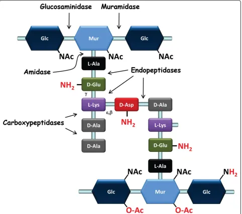
N-acetylglucosamine (GlcNAc) and N-acetylmuramic acid (MurNAc) that are linked viab-1,4 bonds (Figure 1). Peptidic chains are linked covalently through their N-terminus to the lactyl group of MurNAc. These pepti-dic chains vary in composition across species and can be cross-linked directly or indirectly, through short chains of one or more amino acids that generate a three-dimen-sional structure around the cell, which ensures bacterial integrity. In LAB, the amino acid sequence of the stem peptide is L-Ala-g-D-Glu-X-D-Ala, while the third amino acid (X) is a di-amino acid. It is most often L-Lys (e.g., in
L. lactisand most lactobacilli) but can also be
meso-diaminopimelic acid (mDAP) (e.g., inL. plantarum) or L-ornithine (e.g., inL. fermentum) [17]. Among LAB, D-Ala predominates at position five in newly synthesized PG; however, D-Lac residues are found in naturally vancomy-cin-resistant lactobacilli such asL. caseiandL. plan-tarum. Cross-linking between neighboring stem peptides takes place between the D-Ala in position four of one peptide chain and the diamino acid in position three (4-3 cross-link) of another chain. A direct cross-connection is seen in mDAP-type PG, which is typically found in
Gram-negative bacteria but which is also present in
L. plantarum. In other LAB, the Lys-type PG is found
and includes an interpeptide bridge made of one D-amino acid (e.g., D-Asp or D-Asn inL. lactis, L. casei, and most lactobacilli) (Figure 1) or several L-amino acids (e.g., L-Ala2 or L-Ala3 in Streptococcus thermophilus) [17]. PG peptide chains connected by 3-3 cross-links, which predominate inMycobacterium tuberculosis[18] and inClostridium difficile[19], have not been described in LAB to date.
Although a given bacterial species has a basic, charac-teristic PG structure, the PG layer remains in a dynamic state throughout a bacterium’s life, and PG structure is the result of complex biosynthetic, maturation, and degradation reactions, which will be described below. Structural analysis of PG muropeptides using HPLC and mass spectrometry has allowed the identification of the nature of peptide cross-bridges, the degree of cross-link-ing, and the frequency of maturation and hydrolysis events. It has also revealed the existence of covalent PG modifications, such asO-acetylation,N-deacetylation, or amidation; these modifications may play essential roles in bacterial physiology. Detailed PG structure has been ascertained for several LAB, including L. lactis [20],
L. casei [21],L. rhamnosus[22], andL. plantarum[23]. The first three species were found to have D-Ala4 -D-Asp/Asn-L-Lys3 cross-bridges, while the latter has a direct D-Ala4-mDAP3cross-bridge (Figure 1).
Biosynthesis as a multi-step process
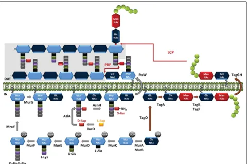
PG synthesis can be divided in three general steps: the first step takes place in the cytoplasm and leads to the synthesis of lipid II, the second step involves the transfer of lipid II to the extracellular side of the membrane, and the third step results in the polymerization of the synthe-sized subunits into a macromolecule [24] (Figure 2).
precursors that terminate with D-Lac, D-Ala-D-Ala-dipeptidase (Aad) eliminates D-Ala-D-Ala dipeptides that are produced by the Ddl ligase, thereby preventing their incorporation into the precursors [26]. PG precursors terminating with Lac instead of with D-Ala-D-Ala were successfully produced inL. lactiswhen theL. plantarumDdl ligase gene was heterologously expressed. Modification of the last residue of the stem peptides of PG precursors has been shown to result in significant changes to PG structure and cell morphology [27]. The UDP-MurNAc-pentapeptide is then attached with a pyr-ophosphate link to the lipid transporter, bactoprenol
(undecaprenyl-phosphate), by the membrane translocase MraY, a process that yields undecaprenyl-pyrophosphoryl-MurNAc-pentapeptide, or lipid I (Figure 2). Finally, the glycosyl-transferase MurG adds GlcNAc to lipid I, forming undecaprenyl-pyrophosphoryl-disaccharide-pentapeptide, or lipid II, which is the basic subunit used in PG assembly [28].
Another important enzymatic step that takes place in the cytoplasm is the assembly of peptide side chains that are added either to the nucleotide MurNAc-pentapeptide or the lipid precursors, depending on the species [29]. D-Asp, the amino acid most commonly included in LAB
side chains and that is found inL. lactisand in most lac-tobacilli, is added to the third amino acid (L-Lys) of the stem peptide by aspartate ligase (AslA) (Figure 2) [30]. This enzyme belongs to the ATP-Grasp family, which includes enzymes that catalyze ATP-dependent carboxy-late-amine ligation reactions and that use activated D-Asp–in the form ofb-aspartyl phosphate–as a substrate [31]. D-Asp is produced from L-Asp by the aspartate racemase encoded byracD, which is located in the same operon as theaslAgene inL. lactis[30,31]. The L-amino acids of the PG side chains are transferred from aminoa-cyl-tRNA by specific transferases, identified as BppA1 and BppA2 inEnterococcus faecalis[32], a species that has L-Ala-L-Ala cross-bridges likeS. thermophilus.
Lipid II (with or without a side chain) is then translo-cated outside the cytoplasmic membrane by a flippase (Figure 2). The integral membrane protein FtsW has been shown to transport lipid-linked PG precursors across the membrane and is proposed to act at the sep-tum level. The RodA homologous protein appears to be
involved in lateral PG synthesis during cell elongation in ovococci and bacilli [33].
In the last step of PG synthesis, PG monomer units are polymerized via transpeptidation and transglycosyla-tion reactransglycosyla-tions, which take place outside the cytoplasmic membrane (Figure 2). The major proteins involved in PG assembly are called penicillin-binding proteins (PBPs) because they are targets for penicillin and other beta-lactam antibiotics [34]. Class A PBPs contain both transglycosylation and transpeptidation domains located at the N- and C-terminals of the protein, respectively, whereas class B PBPs are exclusively involved in trans-peptidation. During transglycosylation, lipid II’s disac-charide is bound to the pre-existing PG chain; the bactoprenol loses one inorganic phosphate and is recycled to the inner side of the cytoplasmic membrane to initiate another round. To create a solid PG mesh around the bacterial cell, newly extended chains must be connected to neighboring chains by transpeptidation. A covalent bond is created between the carbonyl group
of the D-Ala in position four of one pentapeptide chain (donor chain) and the free amine of either the diamino acid in position three of a second peptide chain or the attached side-chain amino acid (acceptor chain). This step leads to the release of the C-terminal Ala or D-Lac of the donor chain. Alternative 3-3 cross-links require L,D-transpeptidases, which are not PBPs [19,35].
Analysis of the genome ofL. lactis, an ovococcus spe-cies, has revealed the presence of six PBPs: five high molecular weight (HMW) PBPs (PBP1a, PBP1b, PBP2a, PBP2b, and PBPx) and one low molecular weight (LMW) PBP (D-Ala-D-Ala-carboxypeptidase DacA) [36].
L. lactisalso possesses an L,D-carboxypeptidase (DacB), which cleaves the L-Lys3-D-Ala4 bonds of the stem pep-tides (Figure 1) [20]. Ovococci display both septal and peripheral growth, which results in the slight longitudi-nal expansion that generates their ovoid shape. It has been shown that lateral or septal growth is mediated by functionally different PG biosynthesis mechanisms, each under the control of a specific class B PBP: PBP2b and PBP2x, respectively. The other PBPs appear to have redundant functions, acting in both biosynthetic path-ways [36]. Furthermore, alteration of PBP2x and PBP2b activity has been proposed to directly affect the coccus-to-rod transition and further filamentation observed in
L. lactis during growth, both in planktonic conditions
and biofilms [37].
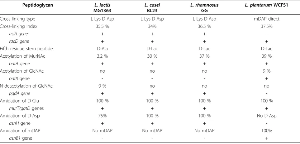
Only part of the PG stem peptides are connected by transpeptidation, and the degree of cross-linking is a PG characteristic. During the exponential growth phase, the cross-linking index has been estimated to be 35.5% in
L. lactis, 37.5% in L. plantarum, 34% in L. casei, and
36.5% in L. rhamnosus(Table 1). D,D-carboxypeptidase DacA and L,D-carboxypeptidase DacB participate in PG maturation and thus produce tetra-and and tripeptide chains in mature PG [20-23].
Another important feature of PG that likely influences PG architecture is glycan chain length. InL. lactis, long gly-can chains (chains with more than 50 disaccharides, which represent 50% of all chains) were detected after an amidase treatment [38]. PG nanoscale architecture was examined using atomic force microscopy (AFM) in livingL. lactis
cells. When a mutant without PSs on its surface was imaged, using a tip functionalized with the PG-binding LysM domain, PG was found to be organized in the form of cables running parallel to the short axis of the cells [39].
Peptidoglycan structural variations
In most bacterial species, PG basic structure is partially modified–either the glycan chains undergoN- deacetyla-tion orO-acetylation or the free carboxyl groups of the amino acids in the peptide chains are amidated (Figure 1) (Table 1) [40]. These structural modifications usually have functional consequences (Table 2); for instance, they may modulate the activity of endogenous PG hydro-lases (PGHs) as well as that of exogenous PGHs pro-duced by eukaryotic organisms, such as lysozyme. PG modifications have been shown to allow pathogenic bac-teria to escape from the host’s innate immune system [41]. Below, we will review PG modifications by chemical groups, given that wall TA or PS polymers that covalently attach to PG may also be considered to be modifications; they can even be linked to the same sites on PG (see text below).
Table 1 Peptidoglycan structural variations in selected LAB and genes involved in PG synthesis or modification.
Peptidoglycan L. lactis
MG1363
L. casei BL23
L. rhamnosus GG
L. plantarumWCFS1
Cross-linking type L-Lys-D-Asp L-Lys-D-Asp L-Lys-D-Asp mDAP direct
Cross-linking index 35.5 % 34% 36.5 % 37.5%
aslA gene + + +
-racD gene + + + +
Fifth residue stem peptide D-Ala D-Lac D-Lac D-Lac
Acetylation of MurNAc 3.2 % 30 % 37 % 39 %
oatAgene + + + +
Acetylation of GlcNAc no no no 9 %
oatBgene - - - +
N-deacetylation of GlcNAc 9 % no no no
pgdA gene + + +
-Amidation of D-Glu 100 % 100 % 100 % 100 %
murT/gatDgenes + + + +
Amidation of D-Asp 75% 100 % 100 % No D-Asp
asnHgene + + +
-Amidation of mDAP No mDAP No mDAP No mDAP 100%
O-Acetylation of glycan chains
In many Gram-positive pathogens, O-acetylation of MurNAc is associated with resistance against lysozyme [42]. A certain proportion of MurNAc residues have an extra acetyl group linked to their C6-OH that can be used to form a 2,6-N,O-diacetyl muramic acid (Figure 1). The first MurNAcO-acetyltransferase, named OatA, was identified inStaphylococcus aureus[43]. OatA is con-served among a large number of Gram-positive species, including LAB [23,44,45]. The enzyme is composed of two domains: the N-terminal domain contains 11 pre-dicted transmembrane helices, whereas the C-terminal domain appears to contain a catalytic acetyltransferase domain. The donor of the acetyl group is probably acetyl-CoA [46]. The acetyl group is likely added to the newly polymerized PG outside the cytoplasmic mem-brane since O-acetylation of lipid precursors has not been observed [43] and the OatA acetyltransferase domain is predicted to be located outside the membrane.
O-acetylation of MurNAc residues has been detected in
the different LAB species for which structural analysis of PG has been performed; estimated levels ofO-acetylation vary, from rather low inL. lactisMG1363 (3.2%) [20] to intermediate in lactobacilli:L. casei BL23 (30%) [21],
L. plantarumNZ7100 (39%) [23], andL. rhamnosusGG
(37%) (Table 1) [22].
InL. lactis, the oatAgene has been shown to be regu-lated at the transcriptional level in response to cell envelope stress, which may be provoked by lysozyme or other cell wall-targeting antimicrobials such as bacitra-cin, vancomybacitra-cin, and plantaricin [45,47]. It has been proposed that the first lactococcal response to treatment with lysozyme is the activation of the two-component system (TCS) CesSR, which then activates the transcrip-tion of several genes belonging to the cesSR regulon, among which is spxB[47], which belongs to the family of global transcriptional factors found in Gram-positive bacteria [48]. SpxB activates oatA expression; OatA activity increases PG resistance to lysozyme and thus counteracts cell wall stress [45]. Interestingly, while Table 2 Role of cell wall glycopolymers and their structural variations in LAB.
Role Organism* Ref.
PG variations
O-acetylation of MurNAc Resistance to lysozyme
Activation of autolysis by amidase LytH
Ll, Lc, Lp Lp
[23,45]
O-acetylation of GlcNAc
Inhibition of major autolysin Acm2 (glucosaminidase)
Lp [23]
N-deacetylation of GlcNAc
Resistance to lysozyme Inhibition of autolysis
Inhibition of AcmA major autolysin (glucosaminidase)
Ll Ll Ll
[54]
Amidation of D-Asp
Resistance to lysozyme Inhibition of autolysis Resistance to nisin.
Ll Ll Ll
[56]
Amidation of mDAP
Essential for growth Control of septation
Increase DacB L,D-carboxypeptidase activity
Lp Lp Lp
[57]
Teichoic acids
WTA Bacterial morphogenesis and division Lp [120]
LTA Immunomodulatory properties La [127]
TA alanylation Cell morphology
UV stress response Protein secretion Resistance to nisin Bacteriophage receptor Adhesion to epithelial cells
Decrease of anti-inflammatory properties Colonization of mouse gastrointestinal tract
Lp, Lrh Ll Ll Ll, Lp
Ld Lj Lp, Lrh
Lr
[85,112] [114] [113] [52,85,111]
[122] [123] [100,126]
[124] Polysaccharides
Cell division and morphology Bacteriophage receptor Protection against phagocytosis
Ll Ll Ll
[131] [131,133,144]
[131]
Immunosuppressive function Lc, Lp [136,142]
Decrease adhesion and biofilm formation Protection against antimicrobial peptides (LL-37)
Lrh Lrh
[137] [147]
increased PGO-acetylation makes L. lactismore resis-tant to the PGH activity of lysozyme, it has been shown that thecesSR regulon is induced by overexpression of membrane-anchored proteins [49,50] or by bacterioph-age infection [51]. The regulon has also been shown to be induced in anL. lactismutant resistant to the bacter-iocin nisin [52].
TheO-acetylation of GlcNAc, never before described in bacteria, was discovered in L. plantarum; both GlcNAc and MurNAc are acetylated in this species [23]. In this bacterium, around 9% of GlcNAc residues areO -acetylated. The addition of the acetyl group to GlcNAc is performed by a second, specificO-acetyltransferase– OatB–that shares a similar two-domain structure with
L. plantarum OatA but has a rather low amino acid
sequence identity (21%). It is noteworthy that, until now, the presence of two Oat proteins has only been found in a very limited number of bacterial species, including two other LAB species, Lactobacillus sakei
andWeissella paramesenteroides[23].
PG O-acetylation has an impact on L. plantarum
autolysis. The O-acetylation of GlcNAc inhibits the
N-acetylglucosaminidase Acm2, the major autolysin of
L. plantarum. In contrast, in this species,O-acetylation of MurNAc has been shown to activate autolysis through the activity of the putative N -acetylmuramoyl-L-alanine amidase LytH [23]. Thus, bothO -acetyltrans-ferases, OatA and OatB, which co-occur in L.
plan-tarum, play antagonistic roles when modulating the
activity of endogenous autolysins. In contrast to the O -acetylation of MurNAc, the O-acetylation of GlcNAc does not inhibit lysozyme activity [23].
N-Deacetylation of glycan chains
TheN-deacetylation of GlcNAc, which leads to the pre-sence of glucosamine (GlcNH2on Figure 1) in the PG backbone, is performed by PG-deacetylase PgdA, which was first identified inStreptococcus pneumoniaethanks to its sequence homology with chitin deacetylases [53]. GlcNAc deacetylation has been found to occur at a level of around 9% inL. lactis; in this species, it protects PG from hydrolysis by AcmA autolysin [54] and increase resistance to lysozyme [45]. In contrast, theN -deacetyla-tion of GlcNAc has not been observed in L. casei,
L. rhamnosus, orL. plantarumunder laboratory growth
conditions. ApgdAhomolog is present in the L. casei
BL23 genome, while no homolog exists in theL.
plan-tarumgenome (Table 1). Deacetylated MurNAc residues
were found inBacillus anthracisPG [40] and, recently, a MurNAc-deacetylase was discovered inBacillus subtilis
[55]; neither have been found in LAB to date.
Amidation of amino acids
The free carboxyl groups of PG-forming amino acids can be amidated; these amino acids include D-Glu and mDAP found on stem peptides and D-Asp on side
chains or cross-bridges (Figure 1). These modifications are catalyzed by specific enzymes and take place intra-cellularly; PG precursors, either UDP-MurNAc-penta-peptide or lipid intermediates, are amidated before the molecules are translocated through the cytoplasmic membrane [29]. Amidation of D-Asp cross-bridges has been observed in L. lactis [20]. D-Asn and D-iso-Asn are not substrates for aspartate ligase, as has been shown inL. lactisand Enterococcus faecium, a species that also has D-Asp cross-bridges [30,31]. As a result, amidation of the alpha-carboxyl group of D-Asp takes place after D-Asp has been added to the PG precursor and is performed by an asparagine synthase (AsnH), which was identified in L. lactis [56] (Figure 2). In
L. lactis, amidation of the D-Asp cross-bridge during
the exponential phase is partial (75%); in contrast, in
L. casei, it is almost complete (near 100%) during all growth phases. PGH activity is affected by D-Asp ami-dation. Indeed, anL. lactis asnHmutant with PG that contained exclusively D-Asp bridges exhibited a higher autolysis rate than the wild-type strain, as well as increased sensitivity to lysozyme. D-Asp amidation also decreases L. lactissensitivity to cationic antimicrobials such as nisin, which may be explained by a decrease in the negative charge inside the cell wall [56].
InL. plantarum, almost all the mDAP side chains are amidated. In this bacterium, amidation has also been shown to be mediated in the cytoplasm by an amido-transferase named AsnB1, the first enzyme to be asso-ciated with such activity [57]. Interestingly, theasnB1
gene co-localizes withmurE, which encodes the ligase catalyzing the addition of mDAP to the PG precursor UDP-N-muramoyl-L-Ala-D-Glu. TheasnB1 gene has been found to play an essential role inL. plantarum. In a mutant strain with a mDAP amidation defect, growth and cell morphology were strongly affected; filamentation and long-chain formation were observed, suggesting that mDAP amidation may play a critical role in controlling the septation process. In addition, L-D-carboxypeptidase DacB activity requires mDAP amidation to reach optimal levels [57].
The D-Glu on the PG stem peptide has an amidated
a-carbonyl group (which transforms it into an iso-Gln) in several bacterial species, including LAB. The level of amidation is close to 100% in all four of the LAB species studied [20-23]. The genes responsible for D-Glu amida-tion have been identified inS. aureus [58,59]. The con-version of iso-Glu to iso-Gln is catalyzed by the glutamine amidotransferase GatD and the Mur ligase homolog MurT. Lipid precursors, but not soluble UDP-MurNAc-pentapeptide precursors, are substrates for this enzymatic complex [59]. ThemurTandgatDgenes are grouped in an operon and play an essential role in
amidation results in a markedly reduced bacterial growth rate, which suggests that amidated PG may serve as a better substrate for proteins that catalyze PG bio-synthesis and cell division; furthermore, resistance to beta-lactam antibiotics and increased sensitivity to lyso-zyme have been observed [58]. Homologs of co-localized
gatDand murTgenes are present in LAB with amidated D-Glu PG including L. lactis and L. plantarum; in
L. caseiandL. rhamnosus, both genes can be found but at different places in the genome sequences (Table 1).
Degradation by PGHs
PGHs are enzymes that can hydrolyze specific bonds in bacterial cell wall PG. Among them are bacterial autoly-sins and phage endolyautoly-sins. Autolyautoly-sins are endogenous bacterial PGHs whose activity may lead to autolysis, in particular when cells experience stressful conditions. Moreover, the cleavage of PG strands is required to insert newly synthesized PG subunits during bacterial cell growth and to separate daughter cells following cell division [60,61]. Bacteriophage genomes encode PGHs called endolysins that, in association with holins, are responsible for host cell destruction after the viral parti-cles have multiplied during phage dissemination [62]. They may also encode PGHs that are tail-associated lysins involved in phage entry into the host bacteria [63]. From a technological cheese making point of view, highly focused, applied studies have sought to under-stand and control LAB lysis, with the aim of being able to release the intracellular pool of enzymes of starter bacteria to improve cheese flavor development [11,64].
Different classes of PGHs can be defined on the basis of their hydrolytic specificity for different bonds (Figure 1): (i)N-acetylmuramidases (muramidases) hydrolyze the
b1-4 bond between MurNAc and GlcNAc–among the known muramidases are lysozymes that result in a pro-duct with a terminal-reducing MurNAc residue and lytic transglycosylases that yield anhydromuropeptides as a result of the formation of a 1,6-anhydro ring inside Mur-NAc; (ii)N-acetylglucosaminidases (glucosaminidases) hydrolyze theb1-4 bond between GlcNAc and MurNAc; (iii)N-acetylmuramyl-L-Ala amidases (amidases) hydro-lyze the bond between the lactyl group of MurNAc and thea-amino group of L-Ala, which is the first amino acid of the lateral peptidic chain; and (iv) peptidases, including endopeptidases and carboxypeptidases, hydrolyze a vari-ety of PG bonds.
Bacterial PGHs as well as phage endolysins usually exhibit modular organization and have a catalytic domain associated with a cell wall binding domain (CWBD). The catalytic domain determines hydrolytic specificity for the PG molecule, whereas the CWBD, because it specifically recognizes a cell wall component, influences localization, target bacteria specificity, and/or
PGH catalytic efficiency. CWBDs, such as the LysM domain, the SH3 domain, or the Lc-LysBD domain, that are found in PGHs but also possibly in other cell wall proteins are described in more detail in the text below.
The catalytic domains present in LAB PGHs are charac-teristic of the following enzyme families: the glucosamini-dases (glycoside hydrolase family 73, PF01832), the muramidases (glyco_hydro_25, PF01183), the lytic trans-glycosylases (transglycosylase-like, PF06737), the amidases (two domains: Amidase_2, PF01510 and Amidase_3, PF01520), the CHAP-domain enzymes with amidase or endopeptidase specificity (the cysteine, histidine-depen-dent amidohydrolase/peptidase domain) (PF05257), and theg-glutamyl-diamino-acid endopeptidases (NlpC_P60, PF00877). Furthermore, the Peptidase_S11 domain (PF00768) is present in D,D-carboxypeptidases (DacA) and the VanY domain (PF02557) is found in L,D-carboxy-peptidase (DacB).
The availability of complete genome sequences allows the full PGH complement of a given bacterial species to be analyzed and identified using amino acid sequence similarity searches that employ representative sequences of all known classes of PGHs. Most Gram-positive bac-teria possess a complex PGH complement that includes a variable number of PGHs. Generally, a given bacterial species produces several PGHs that have various hydroly-tic specificities, although not necessarily all the specifici-ties listed above. In LAB, sequence analyses have revealed that rather complex PGH systems exist; 12 PGHs were identified inL. casei[21], 9 inL. helveticus[65], and 12 inL. plantarum[66]. Five PGHs were initially identified inL. lactis, before the description of the CHAP domain [67]; re-examination of the genome sequence ofL. lactis
MG1363 has allowed us to identify 4 additional putative PGHs that contain a CHAP domain (unpublished results).
Before whole genome sequencing, the first LAB PGH characterized at the molecular level was the major autoly-sin AcmA inL. lactis[68]. AcmA has a modular structure; its N-terminal catalytic domain demonstratesN -acetylglu-cosaminidase specificity [69], and its C-terminal domain is made up of three LysM sequences. The LysM repeats have been shown to bind to PG, and binding appears to be hin-dered by other cell wall constituents, which results in loca-lized binding of AcmA to the cellular septum [70]. In
L. plantarum, the major autolysin Acm2 is also anN -acet-ylglucosaminidase, but its modular structure differs from that of AcmA. In addition to its catalytic domain, it has three SH3 domains and an N-terminal Ala/Ser/Thr (AST)-rich domain [66,71]. Two other major PGHs with
g-D-Glu-L-Lys-endopeptidase activity, Msp1 (p75) and Lc-p75, have been characterized inL. rhamnosus[22] and
L-endopeptidase activity and mediates cleavage of the PG cross-bridge [72]. It is worth noting that these enzymes are, respectively, the major autolysins of the aforemen-tioned bacterial species and they are involved in daughter cell separation. They illustrate the diversity that exists among bacterial species in cell-separating enzymes, a point highlighted in past research [73]. In each species, inactivation of the corresponding genes led to defects in daughter cell separation and long-chain formation. In agreement with their role, all these PGHs were located at the cell septum. Also, AcmA and Acm2 are the major autolysins involved in the bacterial cell autolysis that is observed during the stationary phase or after bacteria are transferred to buffer solution. Other PGHs have been characterized inL. lactis: three other glucosaminidases, one with a LysM domain (AcmD) and two without LysM domains (AcmB and AcmC) as well as oneg -D-Glu-L-Lys-endopeptidase, YjgB [74]. AcmB and AcmD contri-bute to autolysis, in tandem with AcmA [75,76] and AcmD but not AcmB, is involved in cell separation [76]. InL. plantarum, LytA with putativeg -D-Glu-L-Lys-endo-peptidase activity, appears to be required for cell shape maintenance and cell wall integrity [66].
Interestingly, several LAB PGHs have recently been shown to beO-glycosylated [71,77,78]. Their sugar resi-dues are covalently linked to low complexity domains rich in Ala, Ser, and Thr (AST domains). L. rhamnosus
p75 (Msp1) has been found to be glycosylated with hexoses, and probably mannose, given that they are recognized by the lectin concanavalin A.O-glycosylation of p75 (Msp1) appears to confer protection against pro-teolytic degradation [77]. Furthermore, L. plantarum
Acm2 contains more than 20 bound N-acetyl-hexosa-mines: most are probably GlcNAc residues given that they are recognized by wheat germ agglutinin [78]. In this species,O-glycosylation has been shown to modu-late Acm2 PG-degradation activity (see section 1.5) [71]. Very recently, the glycosyltransferases involved in Acm2
O-glycosylation were identified [79].
Factors controlling PG hydrolysis
A strong PG mesh is needed to maintain cell shape and to counteract both high turgor pressure and cell wall stress related to environmental factors. At the same time, the growth and separation of bacterial cells also require a high degree of PG elasticity. These two oppos-ing demands require the coordinated and balanced action of PG synthetic and degradation enzymes. The loss of this equilibrium may cause growth arrest and cell lysis. In bacteria, such equilibrium is achieved mostly by regulating activities of potentially lethal autolytic enzymes that are PGHs. PGH regulation can take place at the transcriptional level but may also be mediated by mechanisms involving post-transcriptional modifications
of PGHs or modification of their substrate, PG [60,61,80].
Control of PGH activity
One of the factors that affects autolysin activity is pro-teolytic degradation. It has been shown that the main lactococcal autolysin, AcmA, is degraded by extracellular proteinase PrtP and that the autolysis of L. lactis
MG1363 depends on the expression of the plasmid encoded cell wall-anchored proteinases PrtPI and PrtPIII [81]. Also, the cell wall-housekeeping protease HtrA has been shown to process lactococcal autolysin AcmA [82]. The activity of a given PGH can also be affected by its specific location in the bacterial cell. Depending on their role in bacterial physiology, PGHs may be distributed all along the cell periphery or located at the septum, as has been observed for PGHs involved in daughter cell separation. By immunofluorescent labeling, the major LAB autolysins (L. lactis AcmA, L. casei Lc-p75,
L. rhamnosus p75, S. thermophilus Cse, and L.
plan-tarum Acm2) were found to be localized in the septal
zone of dividing cells [21,22,70-72].
Secondary cell wall polymers (TAs or PSs) can modulate autolytic activity by shielding PG [83]. InL. lactis, second-ary cell wall polymers can hinder the binding of AcmA LysM domain to PG, which results in the localized binding of AcmA [70]. Furthermore, the autolysis of LAB strains can be influenced by the level of D-alanylation of TAs. An
L. lactis dltDmutant, deficient in LTA alanylation, exhib-ited increased autolysis, which was tied to the decreased degradation of AcmA by HtrA protease [84]. The absence of D-Ala on LTAs inL. plantarumincreases autolysis, caused, at least partially, by the autolysin Acm2 [85].
Also, as described above in the text above, structural variations in the PG substrate, such as the O-acetylation of glycan chains and the amidation of peptide chains, can contribute to the modulation of PGH activity.
Finally, glycosylation of the autolysin Acm2 has recently been shown to control the enzyme’s activity [71]. The N-terminal AST-rich domain of Acm2 is glycosy-lated; this domain bears 21 mono-GlcNAc that are linked to Ser or Thr residues. When the AST domain is notO -glycosylated, Acm2 enzymatic activity significantly increases. In the model that has been proposed, the access of the Acm2 catalytic domain to its substrate may be hindered by the AST domain;O-glycosylation could change the domain conformation and/or mediate inter-domain interactions [71].
Regulation at the transcriptional level
instead of affecting the transcription of genes that encode endogenous PGHs, the lactococcal TCS that responds to cell envelope stress, CesSR, increases expression of the
oatAgene, and this gene encodes PGO-acetyltransferase, whose activity increases PG resistance to the PGH lyso-zyme [45] (see text above).
PG and PGH as mediators of bacteria-host interactions PG and certain of its fragments are known MAMPs that are recognized by host pattern-recognition receptors (PRRs), such as Nod receptors or Toll-like receptors (TLR) [89]. Nod receptors are intracellular receptors expressed by both epithelial cells and immune cells, such as dendritic cells. The minimum ligand recognized by Nod1 is the dipeptide D-Glu-mDAP, which is present in most Gram-negative bacteria, and the minimum ligand recognized by Nod2 is MurNAc-L-Ala-D-Glu, which is present in most bacteria [90]. In addition to the activity of host PGHs such as lysozyme and certain PG-recognition proteins (PGRP), endogenous bacterial PGHs may contribute to the release of PG fragments that can modulate host response [80]. For example, the PG of an L. caseimutant contained less disaccharide-dipeptide (GlcNAc-MurNAc-L-Ala-D-Gln), a known Nod2 agonist; this mutant lacked the major PGH Lc-p75, which demonstrates g-D-Glu-L-Lys-endopeptidase activity [21], a fact that possibly affected Nod2 signaling. When the PG structures of two Lactobacillus strains with different inflammation profiles were compared, the presence of the muropeptide MurNAc-L-Ala-D-Glu-L-Lys (M-tri-MurNAc-L-Ala-D-Glu-L-Lys) in the PG structure and the anti-inflam-matory properties of Lactobacillus salivariusLs33 were found to be correlated [91]. The corresponding synthetic muropeptide has been shown to have a protective effect in a mouse model of intestinal inflammation (Nod2-dependent). These results show that PG originating from probiotic or commensal LAB may play an active role in the gut’s immune balance.
Furthermore, in the well-documented probiotic
L. rhamnosus GG, two secreted PGHs, Msp1 (p75) and
Msp2 (p40), were found to promote the survival and growth of epithelial cells under pro-inflammatory condi-tions [92]. Furthermore, p40 has been shown to prevent and treat colonic epithelial cell injury and inflammation in mouse models of colitis through a mechanism that is dependent on the epidermal growth factor (EGF) recep-tor [93]. The non-catalytic N-terminal domain, which does not contain any characterized functional domains, appears to be responsible for the beneficial effects [94].
Teichoic acids
Structures of teichoic acids
The cell wall of most Gram-positive bacteria contains TAs, which are anionic polymers made of
alditol-phosphate repeating units [95]. They are classified into two groups: wall teichoic acids (WTAs), which are cova-lently linked to the PG molecule, and lipoteichoic acids (LTAs), which are anchored in the cytoplasmic mem-brane with a glycolipid moiety. WTAs may constitute up to half of cell wall total dry weight in certain bacter-ial species [96].
The most common WTA structures are poly-glycero-phosphate [poly(Gro-P)] or poly-ribitolpoly-glycero-phosphate [poly (Rbo-P)] chains. They are covalently attached to PG by a phosphodiester linkage to the C6-hydroxyl of Mur-NAc, via a linkage unit usually consisting of a disacchar-ide, N-acetylmannosaminyl-b(1-4)-N-acetylglucosamine, and a Gro-P unit (Figure 3). The typical LTA structure consists of a poly(Gro-P) chain linked to a glycolipid anchor (Figure 3). The free hydroxyl groups on the Gro-or Rbo-alditol units are partly decGro-orated with D-Ala Gro-or monosaccharides, such as Glc, Gal or GlcNAc. The length of the poly(Gro-P) or poly(Rbo-P) chains varies between species and strains, as does the substitution level. In most Gram-positive bacteria, LTAs and WTAs coexist, but certain bacterial species, including L. casei
and L. rhamnosus, appear to contain only LTAs.
Remarkably, inL. plantarum(depending on the strain), the two types of WTAs–with either poly(Gro-P) or poly (Rbo-P) chains–have been found [97]; in addition, cer-tain strains concer-tain the genes needed to synthesize the two types of WTAs [98]. In anL. plantarum mutant in which the Gro-P type WTA synthesis was abolished, an alternative ribitol-type WTA was synthesized instead of the wild-type Gro-P type WTA [99]. LTA purified from
L. rhamnosusand L. plantarumwere analyzed by NMR;
they were found to have poly(Gro-P) backbones contain-ing an average of 50 and 22 Gro-P repeatcontain-ing units (for each species, respectively), with D-Ala being the only detectable substituent (74% and 42% D-Ala/GroP, respectively) [100,101].L. lactis was found to have poly (Gro-P) LTAs with D-Ala and Gal substituents [52].
Biosynthesis of WTAs and attachment to PG
MnaA. The synthesis of the linkage unit is finished when TagB primase sequentially attaches one Gro-P unit taken from CDP-glycerol. CDP-glycerol is formed from CTP and glycerol by TagD. WTA polymerization is catalyzed by TagF, which adds Gro-P taken from CDP-glycerol to the nascent WTA chain. Up to 60 aldi-tol-phosphate groups may be successively added. After the intracellular steps are completed, the WTA chain is translocated to the extracellular side of the membrane by the ABC transporter TagGH; subsequently, the chain is covalently linked to PG–on the C6-OH of MurNAc. Three transferases (TagTUV) belonging to the LytR-CpsA-Psr (LCP) family involved in the transfer of the
bactoprenol-linked neo-synthesized WTAs to PG have been identified in B. subtilis [104]. Homologous enzymes are also found in LAB such as L. lactis, but their role has yet to be investigated.
Biosynthesis of LTAs and their anchoring in the cytoplasmic membrane
LTAs are linked to the bacterial cell by a glycolipid inserted in the outer layer of the membrane (Figure 3). This glycolipid is a diglucosyldiacylglycerol synthesized from diacylglycerol by the successive addition of two Glc from UDP-Glc by YpfP [105]. LtaA then transfers the diglucosyldiacylglycerol from the inner to the outer
side of the membrane. Once outside the membrane, the diglucosyl portion of the lipid anchor is elongated by LtaS via a polymerization process that adds, in most species, Gro-1-P units; the resulting LTA chain can con-tain up to fifty such units. In cercon-tain species, the first unit is added by a specific LTA-primase (LtaP), the role of which is to initiate elongation. The donor of Gro-P is a phosphatidyldiacylglycerol molecule. The diacylgly-cerol that is released can be recycled to synthesize another LTA molecule.
Modifications of teichoic acids
As mentioned above, the free hydroxyl groups of the aldi-tol-phosphate chains of both the WTAs and the LTAs may be replaced by different sugars (e.g., Glc, Gal, or GlcNAc) or by D-Ala. InB. subtilis, a glycosyltransferase named TagE adds Glc to WTAs [106]. The D-alanylation process is the best characterized and involves the
dltABCDoperon [107]. The first step is the activation of D-Ala, which consumes ATP, and the alanylation of the DltC carrier by DltA. Two models have been proposed for the next steps involving DltB and DltD [108,109]. In the first model [110], DltB transfers D-Ala from DltC to undecaprenol-phosphate lipid (C55-P) and is then flipped outside the membrane. DltD is then involved in transfer of D-Ala to LTA. In the second model [107], DltD would rather act in the cytoplasm by promoting transfer of D-Ala to DltC. The DltC-D-Ala is then transported through the membrane by the protein DltB. DltC would be the only protein required for the transfer of D-Ala to LTA. Recent data rather substantiates the first model although a lipid-linked intermediate has not been detected until now [109]. It seems that D-Ala residues are then free to move along a single alditol-phosphate chain or between different chains, which allows the D-alanylation of WTAs [107]. D-alanyl substituents can modulate the net nega-tive charge of teichoic acids by providing protonated amino groups that serve as counterions to the negatively charged phosphate groups. These modifications widely contribute to TA functionality (see text below).
Adltoperon has been identified in the different LAB that have been studied. InL. plantarum, thedltoperon contains two supplementary genes,pbpX2anddltX, that encode, respectively, a protein whose sequence is similar to that of a low molecular weight PBP endowed with D, D-carboxypeptidase activity and a small protein of 49 amino acids in length whose function is unknown [85]. When thedltoperon is inactivated, D-Ala substituents on teichoic acids are completely absent or strongly reduced in number, a result that has been observed in
L. lactis, L. rhamnosus, andL. plantarum[84,85,111,112]. Unexpectedly, D-Ala-depleted LTAs in theL. plantarum
NCIMB8826dltBmutant contained high levels of Glc subtituents that were absent from wild-type LTAs and
that were threefold longer than wild-type LTAs [100]. Also, inL. rhamnosusGG, LTA extracted from adltD
mutant revealed longer fatty acid chains of the glycolipid anchor, and shorter chain of Gro-P compared to the wild-type LTA [112].
Functions of teichoic acids
WTAs and LTAs contribute significantly to cell wall functionality (Table 2). The various roles attributed to TAs are, at least in part, related to their anionic charac-ter or their distribution within the baccharac-terial cell wall. The level of D-Ala substituents, which modify the global and local charge of TAs, also has a major impact on their functionality [107].
In general, TAs provide a reservoir of ions close to the cell wall that may be required for enzymes to function properly. Due to their anionic character, they can bind both cations, such as Mg2+, and protons, thus creating a pH gradient across the cell wall [107]. They play other roles: they control autolysins, maintain bacterial cell mor-phology, recognize bacteriophages, interact with the host immune system, and are involved in host colonization. These roles are detailed below. InL. lactis, LTA D-alanyla-tion also has an impact on the efficiency of protein secre-tion [113], UV stress resistance [114], and resistance to the cationic antimicrobial peptide nisin [52].
TA and autolysis control
TAs and their substituents have long been considered to play a role in the control of bacterial autolysis in certain Gram-positive bacterial species; they are thought to act using several proposed mechanisms. LTAs were initially considered to be autolysin inhibitors. By determining the number of binding sites for cationic autolysins, their degree of D-alanylation has been also proposed to be a means of regulating autolysis [107]. Finally, WTAs/LTAs have been shown to prevent autolysin binding on the bacterial surface, except to the cell septum, where these molecules are presumably absent [115].
In LAB, L. lactis, L. plantarum, andL. rhamnosus dlt
mutants had faster autolysis rates than did wild-type strains, as a result of the activity of the major autolysins AcmA, Acm2, and Msp1, respectively [84,85,112]. In
L. lactis, this phenotype was associated with a decreased degradation of AcmA by HtrA, cell wall-housekeeping protease [84].
Role of TAs in bacterial cell morphogenesis
by the fact that viable mutants lacking WTAs were obtained in B. subtilisandS. aureus by inactivating the
tagOandtarOgenes, respectively; they each encode the first enzyme of the biosynthesis pathway. As a result, WTAs are no longer considered as essential in B. subti-lis, although their absence severely alters cell morphol-ogy and growth [117]. The role of teichoic acids in cell division and morphogenesis has been investigated in
B. subtilis, and it appears that WTAs are involved in
bacterial elongation, while LTAs participate in cellular division [118]. The concurrent absence of WTAs and LTAs is lethal forB. subtilis, which suggests that anionic polymers are a necessary component of Gram-positive cell walls. In S. aureus, WTAs have been described as acting as temporal and spatial regulators of PG cross-linking [119].
InL. plantarum, atagO deletion mutant revealed that while WTAs are not essential for survival, they are required for proper cell elongation and cell division [120]. Atomic force microscopy (AFM) imaging of the bacterial cell surface combined with fluorescent labeling with lectin probes has revealed that WTAs exhibit a polarized distribution across the cell surface and that they are absent from the cell’s poles. In addition, it appears that the polarized distribution of WTAs plays a key role in controlling cell morphogenesis (surface roughness, cell shape, and elongation and division) [120]. Furthermore, in L. plantarum, D-alanylation of LTAs plays an important role in cell morphology: the longer bacterial cells observed in thedltDmutant indi-cate that its elongation process is altered [85].
LTAs as bacteriophage receptors
LTAs have been shown to be receptor components for the bacteriophage LL-H that infectsLactobacillus
del-bruekii subsp. lactis ATCC15808 [121]. Moreover,
D-Ala anda-Glc substituents of the poly(Gro-P) LTA backbone affect phage adsorption. A high degree of D-alanylation decreased adsorption, whereas Glc substi-tuents were required for efficient binding, indicating that these LTA structural modifications affect how well the anti-receptor protein of the phage tail binds to LTAs [122].
Role of LTAs in bacteria-host cross-talk
LTAs appear to play a prominent role in host-lactoba-cilli interactions [101]. First, LTAs have been reported to be major players inLactobacillus johnsoniiLa1 adhe-sion to human intestinal epithelial cells (Caco-2), possi-bly via hydrophobic interactions [123]. Also, TA D-Ala depletion can result in impaired colonization of the mouse gastrointestinal tract byL. reuteri[124].
Moreover, LTAs are MAMPs that bind to Toll-like receptor 2 (TLR2), a PRR that is present on the surface of epithelial and antigen-presenting cells and that, after being stimulated, can activate cytokine release [89]. It
has been reported that LTAs purified from L. caseiYIT 9029 andL. fermentum YIT 0159 significantly induce TNF-a secretion from murine macrophages, via a TLR2-mediated strain-dependent mechanism [125]. A
dltmutant of L. plantarum NCIMB8826–whose LTA D-alanylation was substantially reduced–exhibited anti-inflammatory properties, which contrasted with the properties demonstrated by the parental strain. In the mutant as compared to the wild type, the secretion of pro-inflammatory cytokines, such as TNFaand IL12, by peripheral blood monocyte-derived cells (PBMCs) was dramatically reduced and IL10 secretion was concur-rently increased [100]. Moreover, thedlt mutant con-ferred protection against inflammation in a murine model of trinitrobenzene sulfonic acid (TNBS)-induced colitis. These results were confirmed using highly puri-fied LTAs, which stimulated TLR2-dependent pro-inflammatory cytokine production [100]. In contrast, in
in vitro studies, an L. rhamnosus GG dltD mutant,
whose LTAs lacked D-alanyl esters, did not demonstrate significantly changed cytokine production [112]; how-ever, it was associated with improvement in some colitic parameters in moderate to severe DSS-induced colitis in a murine model [126]. Furthermore, in an Lactobacillus
acidophilus ltaS mutant deficient in LTAs, IL12 and
TNF-asecretion in bone marrow-derived dendritic cells (DCs) was downregulated, while IL10 secretion and the expression of costimulatory molecules on the surfaces of the DCs were significantly enhanced. When mice with DSS-established colitis were treated with the ltaS
mutant, their condition improved as a result of a mechanism involving IL10 and CD4+ FoxP3+ T-regula-tory cells [127].
Wall polysaccharides
Structure and biosynthesis
extensive research because of their role in adding texture to fermented milk products; they have been covered by other reviews [128,129]. We will only examine the PSs that have been found to be associated with bacterial cells. The genes encoding molecules involved in PS biosynth-esis are typically organized in clusters of 8 to 25 genes, which are located in the chromosome or plasmids. These clusters contain genes that encode glycosyltransferases and genes that are responsible for export, and regulation [130]. The PS synthesis pathway may overlap with those responsible for generating other cell wall polymers, such as PG or WTAs, since it may also involve undecaprenyl phosphate as a lipid carrier (Figure 2).
In L. lactisMG1363, a WPS was discovered at the
bacterial surface and subsequently characterized [131]. AFM as well as complementary transmission electron microscopy observations have shown that this WPS type forms a compact outer layer that surrounds the cell, named the pellicle. Its structure was established by NMR and is distinct from those of other bacterial PSs, including the previously characterizedL. lactisEPS. The PS chains are composed of hexasaccharide-phosphate repeating units that contain rhamnose (Rha). They are likely covalently attached to the cell wall since they were only able to be extracted using a harsh acid treatment.
In L. lactis MG1363, the molecules involved in PS
synthesis are encoded by a single large cluster of genes. This gene cluster is found in many different L. lactis
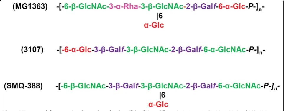
strains but exhibits a rather high level of genetic diver-sity, which suggests that there are structural variations in the WPSs synthesized by different L. lactis strains [132]. The structures of WPSs purified from two other
L. lactis strains, 3107 and SMQ-388, were recently
described and confirmed the PS structural diversity between L. lactisstrains. Like the WPSs making up the
MG1363 PS pellicle, these WPSs are acidic PSs made of oligosaccharide repeating units that are linked by phos-phodiester bonds; however, the structure of the oligosac-charide repeating units differs among the three strains (Figure 4) [133,134].
HeteroPSs, which are composed of different sugar moieties (Glc, Gal, Rha, GlcNAc, and GalNAc), and other residues, such as glucuronic acid and Gro-3-P, have also been found to be associated with the cell sur-faces of lactobacilli [135]. Notably, the L. plantarum
WCFS1 genome contains four gene clusters associated with surface PS production [136]. The structure of these different PSs has not yet been determined. InL.
rham-nosus GG, a long galactose-rich PS (named EPS–
extra-cellular polysaccharide–by the authors) was detected at the bacterial surface using AFM [137,138]. It likely cor-responds to a previously described PS structure [139].
L. rhamnosusstrain GG and other strains show genetic
differences in the gene cluster that encodes PS biosynth-esis, differences that are linked to variation in PS com-position [140]. Exploration of theL. rhamnosus GG cell surface using AFM revealed that its morphology is rough and characterized by wave-like structures [138]. In contrast, the cell surface of a PS-negative mutant was found to be much smoother, which suggests that the wave-like structures reflect PS production. Furthermore, single molecule force spectroscopy with lectin-modified tips revealed that cell surface PS chains had heteroge-neous structures: there were PSs rich in mannose (or glucose) that had moderate extensions and PSs rich in galactose that had much longer extensions [138]. A lec-tin microarray developed to compare the surface gly-comes of L. caseistrains revealed that different strains had different profiles, which suggests that their WPSs are different [135]. In probiotic L. casei Shirota, two
types of WPSs were found: longer, high molecular mass PS-1 and shorter low molecular mass PS-2. The struc-ture of PS-1 has been described [141], and the gene cluster encoding proteins involved in PS-1 biosynthesis has been identified [142]. Furthermore, it was recently observed that Lactobacillus helveticus strains differ in WPS structure, and it has been hypothesized that these differences may partially explain variation in autolytic properties among the strains studied [143]. In conclu-sion, WPSs are omnipresent components of LAB cell surfaces, and it is likely that they differ structurally among strains of the same species.
Functions of WPSs
A number of roles have already been assigned to WPSs in LAB in bacterial physiology as well as in interactions with bacteriophages or eukaryotic hosts (Table 2).
WPSs as bacteriophage receptors in L. lactis
In L. lactis, WPSs are now considered to be receptors
for bacteriophages belonging to the 936 and P445 families, which means that they allow bacteriophage adsorption at the cell surface. An L. lactis MG1363 mutant that lacked a PS pellicle made of hexasaccharide subunits linked through phosphodiester bonds was shown to be resistant to the 936-bacteriophage sk1, which strongly suggests that this WPS could be the sk1 phage receptor [131]. Indeed, previous studies using transposon random mutagenesis mapped the genes required for the adsorption of two 936-type bacterio-phages in their respective host strains; these genes were found inside gene clusters potentially implicated in WPS biosynthesis inL. lactisIL1403 and Wg2 [144] and that are homologous to the cluster that encodes the PS pelli-cle in MG1363. Recent research revealed a correlation between the pellicle genotype of a given L. lactisstrain and the host range of these 936-type phages [132]. The findings support the PS pellicle’s proposed role as a 936-phage receptor and suggest that variation in PS pelli-cle structure among strains could explain the narrow host range of this phage group. On the basis of bioinfor-matic analysis of the PS-encoding gene cluster, three major groups ofL. lactisstrains were distinguished (types A, B and C) [132]; more recently five subtypes (C1 to C5) could be identified in the C-group on the basis of differ-ences in the variable region present in the C-type PS bio-synthesis locus [133]. When genes from the variable region of the C2 subtype strain 3107 were expressed in a mutant ofL. lactisNZ9000 of subtype C1 deficient in WPS synthesis, the resulting recombinant NZ9000 strain synthesizes WPS with the structure of subtype C2. In addition, by challenging the recombinant strain with bac-teriophages infectingL. lactis3107, it was shown that WPS is the host cell surface receptor of the tested phages
from 936 and P335 groups [133]. At the phage level, receptor-binding proteins (RBPs; also named anti-recep-tors) located at the tip of phage tail are involved in phage adsorption: they specifically recognize receptors on the bacterial surface. The 3D structures of RBPs in different lactococcal phages have been established, which means that the recognition mechanism that mediates interac-tions between RBPs and the PS pellicule can now be explored, with a view to understanding the molecular mechanisms underlying recognition specificity [145]. As a first step, surface-plasmon resonance experiments have demonstrated that bacteriophage p2 RBPs bind to PS pel-licle purified from the phage’s host strain–MG1363 [146].
Other functions of WPSs
AnL. lactisMG1363 mutant that lacked surface WPSs
produced long chains of unseparated cells that showed some morphological defects [131]. These observations suggest that WPSs are required for normal cell mor-phology and that they play a role in cell division.
Additionally, surface-exposed PSs are involved in a wide range of bacterial properties and functions, includ-ing adhesion to abiotic surfaces and biofilm formation; they also participate in interactions with other microor-ganisms and host cells. Inactivation of the glycosyltrans-ferasewelEgene in L. rhamnosusGG greatly reduced levels of high molecular mass, galactose-rich WPSs [137]. ThewelEmutant exhibited increased adherence and a greater capacity to form biofilms, possibly because sur-face adhesins, such as pili structures, were more exposed.
Bacterial CPSs have been shown to be potent immuno-modulating molecules; they have largely been character-ized in pathogenic species [130] and are considered to be virulence factors that act by preventing phagocytosis. TheL. lactisPS pellicle has also been shown to protect bacteria against phagocytosis by murine macrophagesin
vitro[131], which suggests that WPSs may shield other
Cell wall proteins
Different modes of attachment to the cell wall
After being synthesized in the cytoplasm, 5-10% of bac-terial proteins are released outside the cytoplasmic membrane [148]. In Gram-positive bacteria, most of these proteins are secreted by the universally conserved and essential Sec pathway. This pathway has been exten-sively studied in E. coli, and genome analyses have revealed that homologs exist in other bacteria, including LAB [149]. Almost all proteins that are targeted by this secretory pathway have an N-terminal signal peptide composed of approximately 30 amino acids. Once the proteins have been translocated across the cytoplasmic membrane, this signal peptide is cleaved off by the appropriate signal peptidase. Then, the protein is either released into the extracellular medium or, alternatively, it is retained in the cell envelope, if it contains a specific sequence ensuring its attachment to the cytoplasmic membrane or the components of the cell wall in addi-tion to the signal peptide. In LAB, surface-associated proteins make up around 80% of predicted secreted pro-teins [148]. Secreted propro-teins can be covalently attached to the cell surface by sortase-mediated reactions or non-covalently attached via i) transmembrane anchors; ii) lipid anchors; or iii) different cell wall binding domains (CWBD) [150,151]. We will review here LAB proteins which are linked to cell wall components through cova-lent or non covacova-lent binding.
PG-anchored proteins
A portion of a given cell wall protein is covalently bound to PG by a transpeptidation mechanism that is catalyzed by sortase A (SrtA, also called housekeeping sortase). In addition to an N-terminal signal peptide, they also contain, at their C-terminal, a conserved LPXTG motif that is followed by a stretch of hydropho-bic residues and a positively charged tail [149,152,153]. Transpeptidase SrtA, which is located in the membrane, cleaves the Thr-Gly bond of the LPXTG motif and links the Thr carboxyl group to the free amino group of the side chain of the lipid II PG precursor. The presence of SrtA and LPXTG-containing proteins is well documen-ted in pathogens such as S. aureus [154], E. faecalis, E. faecium[155], andL. monocytogenes[156]. This SrtA-specific mode of protein attachment to PG is character-istic of all Gram-positive bacteria, including LAB [157]. Inactivation ofsrtAinL. lactisIL1403 has demonstrated that this gene is responsible for the cell wall anchoring of at least five LPXTG-containing proteins [158].
One remarkable family of LPXTG proteins found in LAB is the one of mucus-binding proteins. These pro-teins contain mucus-binding domains (MUB or MucBP) that are thought to play an important role in the adhe-sion of LAB to the mucus layer that covers intestinal
epithelial cells [159]. Other functionally important LPXTG proteins are the pilins, which are the structural components of pili. Pili (or fimbriae) are long filamen-tous structures that extend from the surfaces of various Gram-negative and Gram-positive bacteria. Most studies on pili in Gram-positive bacteria have been conducted on pathogenic species, including streptococci, entero-cocci, corynebacteria, and bacilli [160-162]. Pili have been shown to be involved in adhesion to host cells and tissues and are thus considered to promote host coloni-zation and invasion [162]. In Gram-positive bacteria, the sortase-dependent pili (Spa-type for sortase-mediated pilus assembly) are composed of a major backbone pilin, whose subunits are covalently assembled by sortase C, and of one or two accessory pilins. The minor pilins are located at the base and the tip of the pilus and are pos-sibly also dispersed along the shaft. The pili structures are anchored to PG by housekeeping sortase A [161]. The presence of pili in LAB and in bifidobacteria has also been described and has been linked to the ability of these bacteria to colonize the guts of their hosts and persist in their gastrointestinal tracts [163,164].L. rham-nosusGG cells have been found to contain multiple pili (an average of 10-50 per cell) with lengths of up to 1
μm that are predominantly located near the cell poles [165]. More recently,L. lactissurface pili were visualized using electron microscopy and AFM [166,167]. In a nat-uralL. lactis isolate, a plasmid-encoded pilin gene clus-ter that encodes sortase-dependent pili was shown to be responsible for the assembly of surface pili [167]. This strain produces thin pili that are rather short (averaging 350 nm length).
Proteins that are noncovalently bound to the cell wall
These proteins contain specific CWBDs that have been described in several reviews [151,168]. Here we focus on CWBDs that are found in LAB and their bacteriophages.
LysM domain
The LysM sequence (Lys motif, PF01476)is about 40 amino acid residues long and is present in more than 2,000 eukaryotic and prokaryotic proteins. Several LysM sequences linked by intervening sequences constitute a LysM domain [70,168,169]. Studies examining the bind-ing patterns of different PG chemotypes have found that LysM non-covalently binds to the GlcNAc moiety of glycan chains [70]. However, binding is not disrupted by
N-deacetylation of GlcNAc [54] or byO-acetylation of MurNAc [170].
are involved in cell growth, cell separation, and autolysis. The main lactococcal autolysin AcmA, which is one of the best studied PGHs, has a modular structure and a C-terminal LysM domain that contains three LysM sequences and an N-terminal N-acetyl-glucosaminidase catalytic domain [68]. All three LysM sequences are required for AcmA to function optimally [69], but a sin-gle LysM suffices for PG binding [172]. AcmA also binds to PG in other bacteria, and even to the cells of different Gram-positive species in mixed communities [173].
It has been shown that, in L. lactis, the AcmA LysM domain binds near the cell poles and the cell septum [70]. At the surface of Gram-positive bacteria, the bind-ing of LysM-containbind-ing proteins may be hindered by CW polymers, such as WPSs or WTAs, which results in localization of PGHs in the septal region of the cell [169]. In the case of the lactococcal autolysin AcmA, it has been proposed that attachment to the cell wall can be hindered by CW constituents; LTAs are suggested candidates [70,174]. Another possible candidate is the surface PS pellicle: using AFM and employing tips coated with the AcmA-derived LysM domain,L. lactis
PS pellicle was found to be capable of blocking the binding of LysM to PG [39,175].
Bacterial SH3b domain (including the subfamilies SH3_3, SH3_4, and SH3_5)
This domain is the bacterial equivalent of the well-char-acterized SH3 domain that is found in eukaryotes and viruses. Conflicting results have been obtained when it comes to the PG motif recognized by this domain. In staphylococci with a five-Gly PG crossbridge, the length and amino acid composition of the cross-bridge have been found to have a significant impact on the binding of the SH3-containing homolog of lysostaphin ALE-1 [176]. Also, it has been proposed that the C-terminal domain of lysostaphin, which contains the SH3_5 domain, directs the enzyme to the cross-linked PG [177]. However, more recently, single-molecule AFM experiments using tips functionalized with the L.
plan-tarumAcm2 (which contains five SH3_5 domains) have
found that SH3b domains bind to PG glycan chains and that the binding site contains GlcNAc [178].
WxL domain
This domain was initially identified based onin silico
analysis of gene clusters that encode the cell surface proteins of lactobacilli, enterococcoci, and listeria spe-cies [179]. Proteins containing the WxL domain have been experimentally shown to non-covalently bind to PG in E. faecalis [180]. Proteins containing the WxL domain are present inL. plantarum (19), L. sakei(15),
L. lactis (7),L. casei, andLactobacillus coryniformis (1) [180]; however, their functions have not yet been identified.
Lc-LysBD domain
This domain was recently discovered in the C-terminal of the endolysins (Lc-Lys and Lc-Lys2) of prophages found in the complete genome sequence of L. casei
BL23 [181]. It does not share amino acid sequence simi-larity with any known CWBDs. The domain can bind to PG and can specifically recognize the amidated D-Asp cross-bridge that occurs in L. casei PG (Figure 1). Remarkably, it does not bind to PG molecules with non-amidated D-Asp cross-bridges or PG molecules with dif-ferent types of cross-bridges, such as the L-Ala-L-Ala/L-Ser bridge. This domain is also present in the endolysins of otherL. caseiphages–A2 and PL-1–and in the endo-lysin ofL. lactisphage 949 [182].
SLH-domains
The surface (S) layer entirely coats the bacterial surface and is composed of (glyco)proteins that intrinsically form a two-dimensional paracrystalline structure. Most prokaryotic S-layer proteins possess a signal peptide. These proteins bind non-covalently via their N- or C-terminus to PG or secondary cell wall polymers. The attachment is mediated by S-layer homologous domains (SLHDs), which can also be found in other enzymes of Gram-positive bacteria [149,183]. Most often, S-layer proteins possess three SLHDs, each consisting of 50-70 amino acids. S-layer proteins are present in lactobacilli, and their structure and functions have already been extensively reviewed [184]. The cell wall ligands of the S-layer proteins isolated from different Lactobacillus
species have been proposed to be carbohydrates either teichoic acids or neutral polysaccharides [184].
Role and applications of cell wall proteins
Role of cell wall proteins in bacteria-host interactions