R E S E A R C H
Open Access
The impacts of different embolization
techniques on splenic artery embolization
for blunt splenic injury: a systematic review
and meta-analysis
Jing-Jing Rong
1, Dan Liu
1, Ming Liang
1, Qing-Hua Wang
2, Jing-Yang Sun
1, Quan-Yu Zhang
1, Cheng-Fei Peng
1,
Feng-Qi Xuan
1, Li-Jun Zhao
1, Xiao-Xiang Tian
1and Ya-Ling Han
1*Abstract
Background:Splenic artery embolization (SAE) has been an effective adjunct to the Non-operative management
(NOM) for blunt splenic injury (BSI). However, the optimal embolization techniques are still inconclusive. To further understand the roles of different embolization locations and embolic materials in SAE, we conducted this system review and meta-analyses.
Methods:Clinical studies related to SAE for adult patients were researched in electronic databases, included PubMed, Embase, ScienceDirect and Google Scholar Search (between October 1991 and March 2013), and relevant information was extracted. To eliminate the heterogeneity, a sensitivity analysis was conducted on two reduced study sets. Then, the pooled outcomes were compared and the quality assessments were performed using Newcastle-Ottawa Scale (NOS). The SAE success rate, incidences of life-threatening complications of different embolization techniques were compared byχ2test in 1st study set. Associations between different embolization techniques and clinical outcomes were evaluated by fixed-effects model in 2nd study set.
Results:Twenty-three studies were included in 1st study set. And then, 13 of them were excluded, because lack of the necessary details of SAE. The remaining 10 studies comprised 2nd study set, and quality assessments were performed using NOS. In 1st set, the primary success rate is 90.1% and the incidence of life-threatening complications is 20.4%, though the cases which required surgical intervention are very few (6.4%). For different embolization locations, there was no obvious association between primary success rate and embolization location in both 1st and 2nd study sets (P> 0.05). But in 2nd study set, it indicated that proximal embolization reduced severe complications and complications needed surgical management. As for the embolic materials, the success rate between coil and gelfoam is not significant. However, coil is associated with a lower risk of life-threatening complications, as well as less complications requiring surgical management.
Conclusions:Different embolization techniques affect the clinical outcomes of SAE. The proximal embolization is the best option due to the less life-threatening complications. For commonly embolic material, coil is superior to gelfoam for fewer severe complications and less further surgery management.
Keywords:Blunt splenic injury, Embolization, Location, Material, Clinical outcome
* Correspondence:[email protected]
1Department of Cardiology, General Hospital of Shenyang Military Region, Shenyang 110016, China
Full list of author information is available at the end of the article
Background
Splenic injury is the most common injury following blunt abdominal trauma in daily life and can lead to high mortality because of massive blood loss [1–5]. In the early 1990s, splenectomy was the only choice for splenic rupture. Because overwhelming postsplenectomy infec-tion (OPSI) has occurred in 0.5% of all splenectomies in trauma patients and in over 20% of elective splenecto-mies [6], along with its associated high mortality (8–10%) and high postoperative infectious complications rate (45–49%), including pneumonia, bacteremia, urinary tract infections, abscesses, wound infections and so on [7–9]. So surgeons make every effort to preserve the spleen using various surgical and nonsurgical approaches [10]. Over the past few decades, nonoperative management (NOM) of blunt splenic injuries (BSI) in hemodynamically stable patients with or without splenic artery embolization (SAE) has been widely accepted and became the standard care currently [11–14]. There are growing evidences suggesting that SAE improves splenic salvage [15–19], as well as preserves the immunologic function of injured spleen [20–22]. However, the choices of different tech-niques (including embolization locations and materials) used in SAE were still at the interventional radiologists’ discretion or experience, and the superiority of different specific techniques is debated [23–28]. This situation may partly due to a vague scene of the effect and out-comes with different embolization techniques. Until now, no one prospective randomized study comparing the association between different embolization tech-niques with effects and outcomes of SAE is available. And in most published studies, the sample size of pa-tients and the details of SAE were too limited to ana-lysis and make a meaningful study. To improve the therapeutic efficacy and outcome, it is very necessary to further clarify the role of different embolization techniques of SAE.
Methods Search strategy
Electronic databases of PubMed, Embase, ScienceDirect and Google Scholar Search were used to search pub-lished studies between October 1991 and March 2013 which related to the use of SAE as an adjunct to the NOM in patients of BSI. The Mesh terms used for search were“splenic” (“spleen”),“trauma” (“injury”), and
“embolization”(“splenic artery embolization”or“SAE”).
Inclusion criteria
Retrospective study evaluating BSI adult patients who underwent SAE were included and the following data were required: 1) basic demographics of patients; 2) indi-cations for SAE; 3) the details of SAE techniques were given, including embolization locations (proximal, distal
or combination) and/or embolic materials (coil or gel-foam); 4) the number of severe complications which were life-threatening or complications which need fur-ther surgical management.
Exclusion criteria
The exclusion criteria were as follows: 1) single case re-port, reviews or editor letters; 2) studies involved only open injury, multiple trauma, pediatric patients or caused by iatrogenic injury; 3) non English language publications; 4) studies with insufficient or unconfirmed information.
Study selection
The results of literature searching were screened prelim-inarily by two reviewers (ML and DL) using titles and abstracts. Then, the full texts of potentially appropriate literatures were searched for further screening.
Data extraction
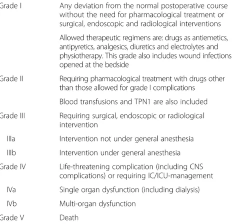
Data including the first author, publication year, number of patients, age, gender, indications for embolization, grades of American Association for the Surgery of Trauma-Organ Injury Scale (AAST-OIS), locations of embolization, embolic materials were extracted from studies, as well as the clinical outcomes including the number of successfully treated patients, severe complica-tions which were life-threatening (rebleeding, infarction, abscess, cyst and contrast-induced renal insufficiency) and Dindo-Clavien classification of morbidity for complication III (DC III, see Table 1) which need further surgical man-agement after SAE [7, 11, 15, 25, 26, 28–32].
Table 1Clavien-Dindo classification of morbidity for complication
Grade I Any deviation from the normal postoperative course without the need for pharmacological treatment or surgical, endoscopic and radiological interventions
Allowed therapeutic regimens are: drugs as antiemetics, antipyretics, analgesics, diuretics and electrolytes and physiotherapy. This grade also includes wound infections opened at the bedside
Grade II Requiring pharmacological treatment with drugs other than those allowed for grade I complications
Blood transfusions and TPN1 are also included
Grade III Requiring surgical, endoscopic or radiological intervention
IIIa Intervention not under general anesthesia
IIIb Intervention under general anesthesia
Grade IV Life-threatening complication (including CNS complications) or requiring IC/ICU-management
IVa Single organ dysfunction (including dialysis)
IVb Multi-organ dysfunction
Statistical analysis
The grades of AAST-OIS of patients among the included studies were compared using one-way ANOVA and then sensitivity analysis was performed on two reduced sets of studies to assess the impact on outcomes as a result of het-erogeneity. First, all included studies were analyzed as 1st study set. Sequentially, studies were reanalyzed excluding those that lacked the necessary details of SAE (embolization locations, embolic materials and complications) and those without detail data of AAST-OIS grade or the outliers. The remaining studies were included in 2nd study set, and qual-ity assessments were performed using Newcastle-Ottawa Scale (NOS). The total score over 6 was judged to be of good quality, otherwise, of poor quality.
SAE success was defined as spleen in situ after embolic treatment. The pooled rates of SAE success rate, incidences of life-threatening complications and DC III were compared among patients who treated by different embolization techniques by χ2 test. Associations between different embolization techniques and clinical outcomes (including SAE success rate, incidences of severe complications and incidences of DC III) were evaluated by odds ratio (OR) and 95% confidence interval (CI) in 2nd study set, respectively.
Heterogeneity in 2nd study set was assessed using Q test,PandI2value.P> 0.05 for theQ-test indicated a lack of heterogeneity across studies, allowing to use the fixed-effects model (the Mantel-Haenszel method) [33–35]. The funnel plot and Begg’s test were used to examine the publication bias [36]. The P value was two-sided and of less than or equal to 0.05 was considered statistically significant. All statistical analyses were performed using Review Manager 5.0 and Stata 12.0 software. To ensure reliability and accuracy of the results, the data was ana-lyzed by two researchers (JJR and QHW) independently and reached a consensus.
Results
Study selection and characteristics
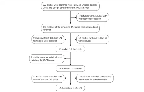
Two hundred and twenty-two researches were found from literature search. After review of titles and ab-stracts, 44 studies were preliminarily identified for fur-ther evaluation. Of the 44 studies, 12 were excluded without follow-up [1, 12, 15, 17, 20–23, 37–40], and 9 studies were excluded because of the lack of detailed data about SAE techniques [19, 29, 31, 41–46]. Finally, 23 relevant studies [7, 10, 11, 16, 25–28, 30, 32, 47–59] were included as 1st study set. Then, 13 studies in 1st study set were excluded with significant differences of grades of AAST-OIS and the remaining 10 studies were included in 2nd study set (Flow diagram shown in Fig. 1). The NOS score of studies in 2nd study set was all over 6 and judged as good quality (Table 2).
Overall, 876 patients who sustained blunt mechanism of injury and then received embolization treatment were
included (Table 3). The average age in included studies was 38.8 ± 5.4 years (range 16–89) and most of the patients were male (72.4%, range 57.9–92.9%). The con-trast blush (38.4%) or pseudoaneurysm (8.2%), high-grade (AAST III-V) splenic injury (50.1%), and large hemoperitoneum (18.0%) were widely considered as major indications for embolization after splenic injury [4, 7, 10, 11, 25–27, 30, 32, 47–51, 53–55, 57]. More than one indication per patient could occur and the dis-tribution of these individual indications for embolization differed significantly in studies (Table 3). However, AAST-OIS is a quantitative and widely accepted grading scale for solid organ injuries which based on computed tomography (CT) image. The overall mean AAST-OIS grade of splenic injury was 3.4 ± 0.4 and differed signifi-cantly among studies in 1st study sets (range 2.9–4.5;P< 0.05, analysis of variance; Table 3). But within the 2nd study set, the differences were not significant (average 3.32 ± 0.20; range 3.1–3.7;P= 0.305, analysis of variance).
The overall primary success rate of SAE in 1st study set was 90.1% (range 72.7–100%; Table 4). Proximal embolization was performed more often than distal and combined significantly (52.1% vs 24.8% vs 5.5%). Exclu-sively proximal embolization, distal embolization and combination were performed in 5 studies [44, 48, 50, 52, 54], 1 study [10] and 10 studies [7, 11, 27, 28, 30, 47, 53, 55, 56, 58] (Table 4). On embolization materials, coil and gelfoam were commonly used materials in included studies. And only coil was used in 5 studies [11, 48, 50–52], gelfoam was exclusively used in 1 study [55].
Data synthesis of different embolization locations
Success rate
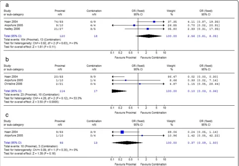
Due to the most common way, the proximal embolization (P) was compared with distal (D) and combination (P + D) on success rate. Within both 1st and 2nd study sets, the success rates of proximal embolization are all higher than distal embolization and combination (Table 5), but all these trends did not reach statistical significance (Figs. 2a and 3a).
Incidence of the severe complications and the Clavien-Dindo classification III
In 2nd study set, the incidence of severe complications of proximal embolization was lower than distal (18.2%vs 28.7%) and combination (20.2%vs58.8%, Table 5). Com-pared with proximal embolization, distal embolization increased the overall incidence of severe life-threatening complications [P= 0.05, OR= 0.51, 95%CI 0.26–1.00, I2
= 0%, Fig. 2b], as well as combination embolization
[P= 0.0005, OR= 0.10, 95%CI 0.03–0.36, I2= 53.3%, Fig. 3b]. The incidence of DC III of proximal embolization was lower than distal (9.9% vs 20.0%, Table 5) and combination (10.8% vs 23.1%, Table 5). Although the association was lost [Proximal vs Distal: P= 0.07,OR= 0.49, 95%CI 0.22–1.06, I2= 0%; Proximal vs Combination: P= 0.16, OR= 0.37, 95%CI 0.09–1.50, I2=
Table 2Quality assessment of studies in 2nd study set for meta-analyses using Newcastle-Ottawa Scale
Author Year Selection Compatibility Outcomes Total
1 2 3 4 5 6 7 8
Liu et al.[10] 2004 ▲ ▲ ▲ - ▲▲ ▲ ▲ ▲ 8
Franco et al.[25] 2011 ▲ ▲ ▲ - ▲ ▲ ▲ ▲ 7
Wu et al. [26] 2011 ▲ ▲ ▲ - ▲ ▲ ▲ ▲ 7
Smith et al.[27] 2006 ▲ ▲ ▲ - ▲ ▲ ▲ ▲ 7
Haan et al.[28] 2004 ▲ ▲ ▲ - ▲ ▲ ▲ ▲ 7
Ekeh et al.[30] 2005 ▲ ▲ ▲ - ▲ ▲ ▲ ▲ 7
Wu et al.[32] 2008 ▲ ▲ ▲ - ▲ ▲ ▲ ▲ 7
Killeen et al.[53] 2001 ▲ ▲ ▲ - ▲ ▲ ▲ ▲ 7
Gaarder et al.[56] 2006 ▲ ▲ ▲ - ▲▲ ▲ ▲ ▲ 8
Cooney et al.[57] 2005 ▲ ▲ ▲ - ▲▲ ▲ ▲ ▲ 8
▲one point ▲▲two points
0%], there were trends that the more proximal embolization used, the less DC III occurred (Figs. 2c and 3c).
Data synthesis of different embolic materials
Success rate
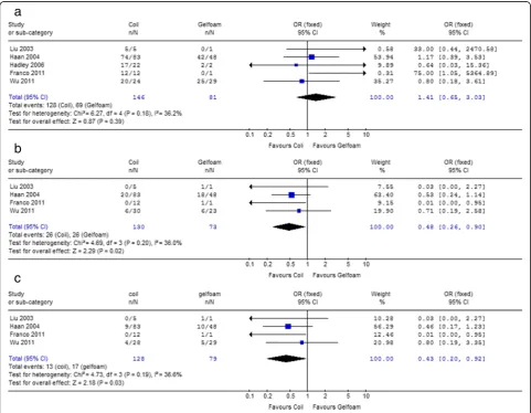
As the most widely used materials, the therapeutic effects and clinical outcomes between coil and gelfoam were compared. The results of our study indicated that coil showed higher success rate than gelfoam (92.4% vs 83.9%) and the difference was significant (P= 0.006) in 1st study set (Table 6). However, within the five studies in 2nd study set, there was no association between em-bolic material and primary success rate [P= 0.39, OR= 1.41, 95%CI 0.65–3.03,I2= 36.2%, Fig. 4a].
Incidence of the severe complications and the Clavien-Dindo classification III
In 1st study set, the average incidence of the life-threatening complications was significantly lower by using coil than gelfoam (12.5% vs 41.6%,P< 0.0001), as
well as the incidence of DC III (7.3% vs 20.9%, P< 0.0001, Table 6).
Within the 2nd study set, strong associations were found between embolic material and life-threatening complication [P= 0.02,OR= 0.48, 95%CI 0.26–0.90,I2= 36%] or DC III [P= 0.03,OR= 0.43, 95%CI 0.20–0.92, I2 = 36.6%]. The severe complication of coil was less than gelfoam significantly (20.0% vs 34.2%, P= 0.02, Fig. 4b). Our results also showed that the more the coil used, the lower incidence of DC III (coil vs gelfoam = 10.2% vs 21.5%,P= 0.03, Fig. 4c).
Heterogeneity analysis and publication bias analysis For both embolization locations and embolic materials study subgroups, there was no significant heterogeneity in each comparison (P> 0.05).
Discussion
In our study, SAE represented an effective adjunct to NOM for adult patients of blunt splenic injury, and the
Table 3Demographic characteristics of included studies
Author n Average age
(year)
Male/ Female
AAST Indication [n(%)]
Contrast blush Pseudoaneurysm Large hemoperitoneum AAST III-V
Niloy et al.[7] 45 48.0 28/17 3.0 27 (60.0) 7 (15.6) 0 (0) 31 (68.9)
Liu et al.a[10] 6 43.8 4/2 3.7 2 (33.3) 0 (0) 3 (50.0) 6 (100.0)
Ekeh et al. [11] 88 37.8 59/29 3.4 17 (19.3) 21 (23.9) 0 (0) 79 (89.8)
Liu et al. [16] 15 - - 3.4 - - -
-Franco et al.a[25] 14 44.8 13/1 3.1 8 (57.1) - - 6 (42.9)
Wu et al.a[26] 53 37.5 33/20 3.3 33 (62.3) 11 (20.8) 0 (0)
-Smith et al.a[27] 41 - - 3.1 - - - 27 (65.9)
Haan et al.a[28] 140 33.0 106/34 3.5 107 (76.4) 0 (0) 9 (6.4) 87 (62.1)
Ekeh et al.a[30] 15 36.0 11/4 3.5 8 (53.3) - - 14 (93.3)
Wu et al.a[32] 19 46.5 11/8 3.5 8 (42.1) 1 (5.3) 10 (52.6) 19 (100.0)
Edmund et al. [47] 8 35.1 7/1 4.5 5 (62.5) 0 (0) 0 (0) 8 (100.0)
Ashraf et al [48] 109 - - -
-Kaseje et al. [49] 11 32.7 - - 11 (100.0) 0 (0) 0 (0)
-Haan et al. [50] 32 - - - 0 (0) 32 (100.0) 0 (0)
-Wu et al. [51] 10 - - - 8 (80.0) 0 (0) 2 (20.0)
-Bessoud et al. [52] 37 40.0 28/9 3.7 14 (37.8) 0 (0) 34 (91.9)
-Killeen et al.a[53] 53 37.6 - 3.2 - - - 42 (79.2)
Sclafani et al. [54] 60 33.9 45/15 2.9 - - - 38 (63.3)
Sclafani et al. [55] 18 - - - 18 (100.0) 0 (0) 0 (0)
-Gaarder et al.a[56] 27 31.0 21/6 3.1 14 (51.9) 0 (0) 11 (40.7) 7 (25.9)
Cooney et al.a[57] 9 39.0 6/3 3.2 6 (66.7) 0 (0) 0 (0) 9 (100.0)
Hagiwara et al. [58] 15 36.0 11/4 4.0 15 (100.0) 0 (0) 6 (40.0) 15 (100.0)
Wei et al. [59] 51 47.0 - 3.8 35 (68.6) 0 (0) 23 (45.1) 51 (100.0)
Total 876 38.8 634/242 3.4 336 (38.4) 72 (8.2) 158 (18.0) 439 (50.1)
a
Study in 2nd study set
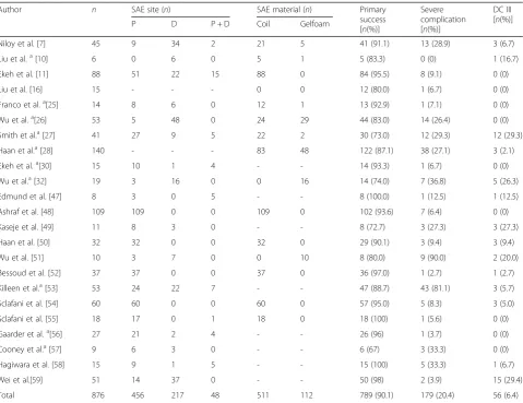
Table 4Details of the included studies
Author n SAE site (n) SAE material (n) Primary
success [n(%)]
Severe complication [n(%)]
DC III [n(%)]
P D P + D Coil Gelfoam
Niloy et al. [7] 45 9 34 2 21 5 41 (91.1) 13 (28.9) 3 (6.7)
Liu et al.a[10] 6 0 6 0 5 1 5 (83.3) 0 (0) 1 (16.7)
Ekeh et al. [11] 88 51 22 15 88 0 84 (95.5) 8 (9.1) 0 (0)
Liu et al. [16] 15 - - - 0 0 12 (80.0) 1 (6.7) 0 (0)
Franco et al.a[25] 14 8 6 0 12 1 13 (92.9) 1 (7.1) 0 (0)
Wu et al.a[26] 53 5 48 0 24 29 44 (83.0) 14 (26.4) 0 (0)
Smith et al.a[27] 41 27 9 5 22 2 30 (73.0) 12 (29.3) 12 (29.3)
Haan et al.a[28] 140 - - - 83 48 122 (87.1) 38 (27.1) 3 (2.1)
Ekeh et al.a[30] 15 10 1 4 - - 14 (93.3) 1 (6.7) 0 (0)
Wu et al.a[32] 19 3 16 0 0 16 14 (74.0) 7 (36.8) 5 (26.3)
Edmund et al. [47] 8 3 0 5 - - 8 (100.0) 1 (12.5) 1 (12.5)
Ashraf et al. [48] 109 109 0 0 109 0 102 (93.6) 7 (6.4) 0 (0)
Kaseje et al. [49] 11 8 3 0 - - 8 (72.7) 3 (27.3) 3 (27.3)
Haan et al. [50] 32 32 0 0 32 0 29 (90.1) 3 (9.4) 3 (9.4)
Wu et al. [51] 10 3 7 0 0 10 8 (80.0) 9 (90.0) 2 (20.0)
Bessoud et al. [52] 37 37 0 0 37 0 36 (97.0) 1 (2.7) 1 (2.7)
Killeen et al.a[53] 53 24 22 7 - - 47 (88.7) 43 (81.1) 3 (5.7)
Sclafani et al. [54] 60 60 0 0 60 0 57 (95.0) 5 (8.3) 3 (5.0)
Sclafani et al. [55] 18 17 0 1 18 0 18 (100) 1 (5.6) 0 (0)
Gaarder et al.a[56] 27 21 2 4 - - 26 (96) 1 (3.7) 0 (0)
Cooney et al.a[57] 9 6 3 0 - - 6 (67) 3 (33.3) 0 (0)
Hagiwara et al. [58] 15 9 1 5 - - 15 (100) 5 (33.3) 1 (6.7)
Wei et al.[59] 51 14 37 0 - - 50 (98) 2 (3.9) 15 (29.4)
Total 876 456 217 48 511 112 789 (90.1) 179 (20.4) 56 (6.4)
a
Study in 2nd study set; SAE. Splenic artery embolization; P. Proximal splenic artery embolization; D. Distal splenic artery embolization; P + D: Combination of proximal and distal splenic artery embolization; DC III: Clavien-Dindo classification of morbidity for complication III;“-”Not mentioned
Table 5Comparisons of clinical outcomes between proximalvsdistal and combined embolization in 1st and 2nd study sets
Outcome Study set Location Percentage (%) Pvalue OR(95% CI)
Success rate 1st PvsD 91.4 (444/486)vs87.7 (213/243) 0.11a 1.49 (0.91–2.45)
PvsP + D 91.4 (444/486)vs86.4 (38/44) 0.27a 1.67 (0.67–4.18)
2nd PvsD 85.0 (122/142)vs82.3 (108/131) 0.52a 1.28 (0.61–2.67)
PvsP + D 86.7 (104/120) vs 72.2 (13/18) 0.11a 2.56 (0.81–8.05)
Severe complication 1st PvsD 10.7 (50/466) vs 30.7 (67/218) <0.01a 0.27 (0.18–0.41)
PvsP + D 10.7 (50/466) vs 35.6 (16/45) <0.01a 0.22 (0.11
–0.43)
2nd PvsD 18.2 (24/132) vs 28.7 (31/108) 0.05 0.51 (0.26–1.00)
PvsP + D 20.2 (23/114) vs 58.8 (10/17) 0.00 0.10 (0.03–0.36)
DC III 1st PvsD 7.3 (32/438) vs 13.0 (28/216) 0.02a 0.53 (0.31
–0.90) PvsP + D 7.3 (32/438) vs 10.3 (4/39) 0.52b 0.69 (0.23
–2.06)
2nd PvsD 9.9 (13/131) vs 20.0 (21/105) 0.07 0.49 (0.22–1.06)
PvsP + D 10.8 (10/93) vs 23.1 (3/13) 0.16 0.37 (0.09–1.50)
P: Proximal splenic artery embolization; D: Distal splenic artery embolization; P + D: Combination of proximal and distal splenic artery embolization; DC III: Clavien-Dindo classification of morbidity for complication III
a Chi-square b
therapeutic effect of SAE is prominent (primary success rate of SAE = 90.1%). The overall incidence of severe com-plications after SAE is 20.4%, though the cases which really required further surgical intervention is relatively fewer (incidence of DC III = 6.4%). For different embolization locations, there was no obvious association between primary success rate and embolization location in both 1st and 2nd study sets. However, our study sug-gests that proximal SAE reduced the risk of clinical ad-verse events than distal and combination, especially the life-threatening complications. As for the embolic mate-rials, the using of coil is associated with a higher success rate and with a lower risk of developing life-threatening complication and DC III than using of gelfoam.
SAE may increase the nonoperative salvage rate in pa-tients with splenic injury [10, 60, 61]. However, no clear
on two reduced sets of included studies. By doing this, the differences in the grades of splenic injury should be elimi-nated in 2nd study set. And the main conclusions of this study are based on the analysis results of 2nd study set.
Embolization locations
The choice of methods will have an effect on angioem-bolization efficacy and results [31, 42]. Emangioem-bolization may be performed proximal (main splenic artery), distal (small arterial branches within the splenic parenchyma), or combination. Several researches over the past few years have reported conflicting clinical outcomes of dif-ferent embolization locations [7, 10, 11, 23, 25–28, 32, 48, 53, 56, 57]. Proximal SAE achieves hemostasis by re-ducing pulse pressure and decreasing the distal flow of the splenic parenchyma. It is beneficial for clot formation rather than stop the hemorrhage directly [4, 23, 40, 47, 48], and it is advantageous to control multiple splenic injuries [23, 28]. Meanwhile, the rich network of collateral cir-culation from the left gastric, gastroepiploic arteries, pancreatic and omental branches enters the spleen to minimize the risk of infarction and preserve the
function [12, 15, 20, 23, 25, 38, 39, 47, 48, 58]. In contrast, distal subselection embolization may not be feasible in these situations. Distal embolization oc-cludes bleeding segmental vessels but may lead to wedge infarctions or increase the risk of abscess forma-tion. And bleeding segmental vessels may be overlooked because of vasospasm caused by hematoma, potentially increasing the risk of rebleeding [7, 11, 28, 68]. In addition, proximal embolization is to simply occlude the main trunk of the splenic artery, which was approximately 2-3cm beyond the origin of the dorsal pancreatic and proximal to the first great pancreatic artery (arteria pancreatica magna) [15, 47, 54]. However, distal embolization is more technically challenging and has to occlude beyond the splenic hilum and distal to any major potential collateral pathways [15]. Therefore, proximal embolization is more technically simple and will be performed in less time and used less contrast agent. The reasons above could explain the results of our study that proximal SAE reduced the risk of life-threatening complications (including rebleeding, infarc-tion, abscess and contrast nephropathy).
Embolic materials
In the early 1980s, Sclafani et al. [40, 54] firstly introduced the concept of “angiographic hemostasis” using gelfoam, vasopressor and steel-wool coil injected into the splenic artery to treat blunt splenic trauma. Until now, coil and gelfoam still remain the most common agents of SAE. Coil is a kind of permanent and radio-opaque embolic
agent [25], and can be injected from catheterization to the predetermined embolization location of injured splenic ar-tery to block or decrease blood flow for clot formation and durable hemostasis. However, migration is a compli-cation of coil often occurred during an embolization pro-cedure and causes rebleeding or more infraction of spleen parenchyma [7, 11, 28, 30, 52]. Gelfoam is a kind of
water-Fig. 4Forest plot of different embolic materials (CoilvsGelfoam) associated with success rate (a), severe complications (b) and the incidence of DC III (c) in 2nd study set
Table 6Comparisons of clinical outcomes between coil and gelfoam
Outcome Study set Coil (%) Gelfoam (%) Pvalue OR(95% CI)
Success rate 1st 92.4 (391/423) 83.9 (94/112) 0.0060a 2.34 (1.26–4.35)
2nd 87.7 (128/146) 85.2 (69/81) 0.39 1.41 (0.66–3.03)
Severe complication 1st 12.5 (61/487) 41.6 (42/101) <0.0001a 0.20 (0.12–0.32)
2nd 20.0 (26/130) 34.2 (25/73) 0.02 0.48 (0.26–0.90)
DC III 1st 7.3 (36/493) 20.9 (23/110) <0.0001a 0.30 (0.17–0.53)
2nd 10.2 (13/128) 21.5 (17/79) 0.03 0.43 (0.20–0.92)
DC IIIClavien-Dindo classification of morbidity for complication III a
insoluble hemostatic agent prepared from purified skin gelatin. The first angiographic embolization used gelfoam was performed for hemostatic purpose prior splenectomy [29, 40]. Once injected, gelfoam follows the arterial blood stream and occludes the vessels, thus stopping the bleeding [25]. In addition, its hemostatic proper-ties are also the result of entrapping the platelets in the porous and hastening the development and providing structural support to the thrombus [24]. One of the attractive characteristics of gelfoam is that it can be rapidly absorbed by macrophages and re-storing vessel patency within days or a few weeks [25]. Instead, several papers report an increased risk of rebleeding before final hemostasis when gelfoam is used as embolic agent [10, 25, 27, 69, 70]. On the other side, once injected into the artery, the gelfoam particles would not only occlude the embolization location, but also occlude splenic vessels distal to col-lateral circulation with the flow, and splenic infarction was noted by ultrasound or CT follow-up [51, 54]. In addition, due to the capacity of retaining air bubbles that might give a chance for aerobic organisms to grow, the gelfoam eventually may lead to infection and abscess [24].
Our study indicated that the primary success rate of SAE was irrelevant to the embolic material. This result is consistent with Wu et al. [26] and Haan et al. [28]. At present, the association between embolization material and severe complication still remains controversial. It has been reported that the use of gelfoam resulted in the higher severe complication rate than coil [10, 25, 27, 32].
However, some other studies suggested that the
difference was not significant [7, 26, 28]. According to our study, strong associations were found between different embolic materials and adverse clinical events. Coils should be the preferred embolic material used
in SAE. Due to the higher incidence of
life-threatening complications and DC III, an exclusively gelfoam embolization should be considered only as an urgent preoperative maneuver to deal with refractory shock and to enable patient transfer to the operating room [54].
Conclusions
The studies in 2nd study set was judged as good quality (NOS > 6), and the results were reliable. This study dem-onstrated that the use of different embolization tech-niques would have significantly effects on clinical outcomes of SAE. Proximal embolization was the best option due to the less life-threatening complications than distal and combination. In addition, embolic mater-ial was another key factor which affected the out-comes of SAE. The use of coil in SAE surpassed using gelfoam, due to less adverse clinical outcomes.
And more effective and reliable embolic materials would be further developed to improve the thera-peutic effects of SAE.
Abbreviations
AAST-OIS:American Association for the Surgery of Trauma-Organ Injury Scale; BSI: Blunt splenic injury; CI: Confidence interval; CT: Computed tomography; D: Distal embolization; DC III: Clavien-Dindo classification of morbidity for complication III; NOM: Non-operative management; NOS: Newcastle-Ottawa Scale; OPSI: Overwhelming postsplenectomy infection; OR: Odds ratio; P: Proximal embolization; P + D: Combination embolization; SAE: Splenic artery embolization
Acknowledgements Not applicable.
Funding
This work was supported by grant from the Innovation Project of Military Medicine (No. 16CXZ006).
Availability of data and material
The databases used in this study are from the electronic databases: PubMed: https://www.ncbi.nlm.nih.gov/pubmed;
Embase: https://www.elsevier.com/solutions/embase-biomedical-research; ScienceDirect: http://www.sciencedirect.com/;
Google Scholar Search: http://scholar.google.com/
The statistical analysis tools and methods used in this study are as following: The grades of AAST-OIS of patients among the included studies were com-pared using one-way ANOVA and then sensitivity analysis was performed on two reduced sets of studies to assess the impact on outcomes as a result of heterogeneity. First, all included studies were analyzed as 1st study set. Sequentially, studies were reanalyzed excluding those that lacked the necessary details of SAE (embolization locations, embolic materials and complications) and those without detail data of AAST-OIS grade or the outliers. The remaining studies were included in 2nd study set, and quality assessments were performed using Newcastle-Ottawa Scale (NOS). The total score over 6 was judged to be of good quality, otherwise, of poor quality.
SAE success was defined as spleen in situ after embolic treatment. The pooled rates of SAE success rate, incidences of life-threatening complica-tions and DC III were compared among patients who treated by different embolization techniques byχ2test. Associations between different embolization techniques and clinical outcomes (including SAE success rate, incidences of se-vere complications and incidences of DC III) were evaluated by odds ratio (OR) and 95% confidence interval (CI) in 2nd study set, respectively.
Heterogeneity in 2nd study set was assessed usingQtest,PandI2value.P> 0.05 for theQ-test indicated a lack of heterogeneity across studies, allowing to use the fixed-effects model (the Mantel-Haenszel method) [33–35]. The funnel plot and Begg’s test were used to examine the publication bias [36]. ThePvalue was two-sided and of less than or equal to 0.05 was considered statistically significant. All statistical analyses were performed using Review Manager 5.0 and Stata 12.0 software. To ensure reliability and accuracy of the results, the data was analyzed by two researchers independently and reached a consensus.
Authors’contributions
JJR participated in the design of the study, collected the data, performed the statistical analysis and drafted the manuscript. DL, ML and JYS participated in the design of the study and as adviser. QHW,QYZ and CFP helped to collect data and perform statistical analysis. FQX, LJZ and XXT participated in data collection and were responsible for the results of statistical analysis. YLH participated in the design of the study and the writing of the manuscript. All authors read and approved of the final manuscript.
Competing interests
The manuscript has been read and approved by all the authors and that the criteria for authorship have been met. In addition, this study has no conflicts of interest and financial disclosures.
Ethics approval and consent to participate Not applicable.
Author details
1Department of Cardiology, General Hospital of Shenyang Military Region, Shenyang 110016, China.2Department of Cardiology, Xinqiao Hospital of Third Military Medical University, Chongqing 400038, China.
Received: 13 July 2016 Accepted: 10 May 2017
References
1. Banerjee A, Duane TM, Wilson SP, Haney S, O’Neill PJ, Evans HL, et al. Trauma center variation in splenic artery embolization and spleen salvage: A multicenter analysis. J Trauma. 2013;75:69–75.
2. Peitzman AB, Heil B, Rivera L, Federle MB, Harbrecht BG, Clancy KD, et al. Blunt splenic injury in adults: multi-institutional study of the Eastern Association for the Surgery of Trauma. J Trauma. 2000;49:177–89.
3. Cogbill TH, Moore EE, Jurkovich GJ, Morris JA, Mucha JRP, Shackford SR, et al. Nonoperative management of blunt splenic trauma: a multicenter experience. J Trauma. 1989;29:1312–7.
4. van der Vlies CH, Hoekstra J, Ponsen KJ, Reekers JA, van Delden OM, Goslings JC. Impact of splenic artery embolization on the success rate of nonoperative management for blunt splenic injury. Cardiovasc Intervent Radiol. 2012;35:76–81.
5. Smith J, Caldwell E, D’Amours S, Jalaludin B, Sugrue M. Abdominal trauma: a disease in evolution. ANZ J Surg. 2005;75:790–4.
6. Uranüs S, Pfeifer J. Nonoperative treatment of blunt splenic injury. World J Surg. 2001;25:1405–7.
7. Dasgupta N, Matsumoto AH, Arslan B, Turba UC, Sabri S, Angle JF. Embolization therapy for traumatic splenic lacerations. Cardiovasc Intervent Radiol. 2012;35: 795–806.
8. Gauer JM, Gerber-Paulet S, Seiler C, Schweizer WP. Twenty years of splenic preservation in trauma: lower early infection rate than in splenectomy. World J Surg. 2008;32:2730–5.
9. Wiseman J, Brown CV, Weng J, Salim A, Rhee P, Demetriades D. Splenectomy for trauma increases the rate of early postoperative infections. Am Surgeon. 2006;72:947–50.
10. Liu PP, Lee WC, Cheng YF, Hsieh PM, Hsieh YM, Tan BL, et al. Use of splenic artery embolization as an adjunct to nonsurgical management of blunt splenic injury. J Trauma. 2004;56:768–73.
11. Ekeh AP, Khalaf S, Ilyas S, Kauffman S, Walusimbi M, McCarthy MC. Complications arising from splenic artery embolization: a review of an 11-year experience. Am J Surg. 2013;205:250–4.
12. Pirasteh A, Snyder LL, Lin R, Rosenblum D, Reed S, Sattar A, et al. Temporal assessment of splenic function in patients who have undergone percutaneous image-guided splenic artery embolization in the setting of trauma. J Vasc Interv Radiol. 2012;23:80–2.
13. Schnüriger B, Inaba K, Konstantinidis A, Lustenberger T, Chan LS, Demetriades D. Outcomes of proximal versus distal splenic artery embolization after trauma: a systematic review and meta-analysis. J Trauma. 2011;70:252–60.
14. Requarth JA, D’Agostino Jr RB, Miller PR. Nonoperative management of adult blunt splenic injury with and without splenic artery embolotherapy: a meta-analysis. J Trauma. 2011;71:898–903.
15. Imbrogno BF, Ray CE. Splenic Artery Embolization in Blunt Trauma. Semin Interv Radiol. 2012;29(2):147–9.
16. Liu PP, Liu HT, Hsieh TM, Huang CY, Ko SF. Nonsurgical management of delayed splenic rupture after blunt trauma. J Trauma. 2012;72:1019–23. 17. Malhotra AK, Carter RF, Lebman DA, Carter DS, Riaz OJ, Aboutanos MB, et al.
Preservation of splenic immunocompetence after splenic artery angioembolization for blunt splenic injury. J Trauma. 2010;69:1126–30. 18. Jeremitsky E, Kao A, Carlton C, Rodriguez A, Ong A. Does splenic embolization
and grade of splenic injury impact nonoperative management in patients sustaining blunt splenic trauma? Am Surgeon. 2011;77:215–20.
19. Chen IC, Wang SC, Shih HC, Wang CY, Liu CC, Wen YS, et al. Spleen artery embolization increases the success of nonoperative management following blunt splenic injury. J Chin Med Assoc. 2011;74:341–4.
20. Walusimbi MS, Dominguez KM, Sands JM, Markert RJ, McCarthy MC. Circulating cellular and humoral elements of immune function following splenic arterial embolisation or splenectomy in trauma patients. Injury. 2012;43:180–3.
21. Skattum J, Titze TL, Dormagen JB, Aaberge IS, Bechensteen AG, Gaarder PL, et al. Preserved splenic function after angioembolisation of high grade injury. Injury. 2012;43:62–6.
22. Shih HC, Wang CY, Wen YS, Wu JK, Huang MS, Huang CI, et al. Spleen artery embolization aggravates endotoxin hyporesponse of peripheral blood mononuclear cells in patients with spleen injury. J Trauma. 2010;68:532–7. 23. Requarth JA, Miller PR. The splenic artery stump pressure is affected by arterial
anatomy after proximal embolotherapy in blunt splenic injury. J Trauma Acute Care Surg. 2012;73:1221–4.
24. Abada HT, Golzarian J. Gelatine sponge particles: handling characteristics for endovascular use. Tech Vasc Interv Radiol. 2007;10:257–60.
25. Franco F, Monaco D, Volpi A, Marcato C, Larini P, Rossi C. The role of arterial embolization in blunt splenic injury. Radiol Med. 2011;116:454–65. 26. Wu SC, Fu CY, Chen RJ, Chen YF, Wang YC, Chung PK, et al. Higher incidence
of major complications after splenic embolization for blunt splenic injuries in elderly patients. Am J Emerg Med. 2011;29:135–40.
27. Smith HE, Biffl WL, Majercik SD, Jednacz J, Lambiase R, Cioffi WG. Splenic artery embolization: have we gone too far? J Trauma. 2006;61:541–6. 28. Haan JM, Biffl W, Knudson MM, Davis KA, Oka T, Majercik S, et al. Splenic
embolization revisited: a multicenter review. J Trauma. 2004;56:542–7. 29. Moog R, Mefat L, Kauffmann I, Becmeur F. Non-operative management of
splenic trauma. Arch Pediatr. 2005;12:219–23.
30. Ekeh AP, McCarthy MC, Woods RJ, Haley E. Complications arising from splenic embolization after blunt splenic trauma. Am J Surg. 2005;189:335–9. 31. Ekeh AP, Izu B, Ryan M, McCarthy MC. The impact of splenic artery embolization on
the management of splenic trauma: an 8-year review. Am J Surg. 2009;197:337–41. 32. Wu SC, Chen RJ, Yang AD, Tung CC, Lee KH. Complications associated with
embolization in the treatment of blunt splenic injury. World J Surg. 2008;32: 476–82.
33. Liu D, Guo H, Li Y, Xu X, Yang K, Bai Y. Association between polymorphisms in the promoter regions of matrix metalloproteinases (MMPs) and risk of cancer metastasis: a meta-analysis. PLoS One. 2012;7:e31251.
34. Mantel N, Haenszel W. Statistical aspects of the analysis of data from retrospective studies of disease. J Natl Cancer Inst. 1959;22:719–48.
35. DerSimonian R, Laird N. Meta-analysis in clinical trials. Control Clin Trials. 1986;7:177–88.
36. Egger M, Davey Smith G, Schneider M, Minder C. Bias in meta-analysis detected by a simple, graphical test. BMJ. 1997;315:629–34.
37. Bhullar IS, Frykberg ER, Siragusa D, Chesire D, Paul J, Tepas 3rd JJ, et al. Age does not affect outcomes of nonoperative management of blunt splenic trauma. J Am Coll Surg. 2012;214:958–64.
38. Requarth JA. Distal splenic artery hemodynamic changes during transient proximal splenic artery occlusion in blunt splenic injury patients: a mechanism of delayed splenic hemorrhage. J Trauma. 2010;69:1423–6.
39. Krohmer SJ, Hoffer EK, Burchard KW. Transcatheter embolization for delayed hemorrhage caused by blunt splenic trauma. Cardiovasc Intervent Radiol. 2010;33:861–5.
40. Sclafani SJ. The role of angiographic hemostasis in salvage of the injured spleen. Radiology. 1981;141:645–50.
41. Paul DB, Opalek JM. Proximal splenic arterial embolization may also result in pancreatic necrosis. J Trauma. 2011;71:268–9.
42. Harbrecht BG, Ko SH, Watson GA, Forsythe RM, Rosengart MR, Peitzman AB. Angiography for blunt splenic trauma does not improve the success rate of nonoperative management. J Trauma. 2007;63:44–9.
43. Rajani RR, Claridge JA, Yowler CJ, Patrick P, Wiant A, Summers JI, et al. Improved outcome of adult blunt splenic injury: a cohort analysis. Surgery. 2006;140:625–32.
44. Bessoud B, Denys A, Calmes JM, Madoff D, Qanadli S, Schnyder P, et al. Nonoperative management of traumatic splenic injuries: is there a role for proximal splenic artery embolization? AJR Am J Roentgenol. 2006;186:779–85. 45. Bessoud B, Denys A. Main splenic artery embolization using coils in blunt splenic
injuries: effects on the intrasplenic blood pressure. Eur Radiol. 2004;14:1718–9. 46. Elliott JA, Millward SF, Kribs SW. Use of computed tomographic scanning and embolization to improve the nonoperative management of splenic trauma: critically appraised topic. Can Assoc Radiol J. 2003;54:183–4. 47. Ng EH, Comin J, David E, Pugash R, Annamalai G. AMPLATZER Vascular Plug
4 for proximal splenic artery embolization in blunt trauma. J Vasc Intrev Radiol. 2012;23:976–9.
49. Kaseje N, Agarwal S, Burch M, Glantz A, Emhoff T, Burke P, et al. Short-term outcomes of splenectomy avoidance in trauma patients. Am J Surg. 2008; 196:213–7.
50. Haan JM, Marmery H, Shanmuganathan K, Mirvis SE, Scalea TM. Experience with splenic main coil embolization and significance of new or persistent pseudoaneurysm: reembolize, operate, or observe. J Trauma. 2007;63:615–9. 51. Wu SC, Chow KC, Lee KH, Tung CC, Yang AD, Lo CJ. Early selective
angioembolization improves success of nonoperative management of blunt splenic injury. Am Surgeon. 2007;73:897–902.
52. Bessoud B, Duchosal MA, Siegrist CA, Schlegel S, Doenz F, Calmes JM, et al. Proximal splenic artery embolization for blunt splenic injury: clinical, immunologic, and ultrasound-Doppler follow-up. J Trauma. 2007;62:1481–6. 53. Killeen KL, Shanmuganathan K, Boyd-Kranis R, Scalea TM, Mirvis SE. CT
findings after embolization for blunt splenic trauma. J Vasc Intrev Radiol. 2001;12:209–14.
54. Sclafani SJ, Shaftan GW, Scalea TM, Patterson LA, Kohl L, Kantor A, et al. Nonoperative salvage of computed tomography–diagnosed splenic injuries: utilization of angiography for triage and embolization for hemostasis. J Trauma. 1995;39:818–27.
55. Sclafani SJ, Weisberg A, Scalea TM, Phillips TF, Duncan AO. Blunt splenic injuries: nonsurgical treatment with CT, arteriography, and transcatheter arterial embolization of the splenic artery. Radiology. 1991;181:189–96. 56. Gaarder C, Dormagen JB, Eken T, Skaga NO, Klow NE, Pillgram-Larsen J, et al.
Nonoperative management of splenic injuries: improved results with angioembolization. J Trauma. 2006;61:192–8.
57. Cooney R, Ku J, Cherry R, Maish 3rd GO, Carney D, Scorza LB, et al. Limitations of splenic angioembolization in treating blunt splenic injury. J Trauma. 2005;59: 926–32.
58. Hagiwara A, Fukushima H, Murata A, Matsuda H, Shimazaki S. Blunt splenic injury: usefulness of transcatheter arterial embolization in patients with a transient response to fluid resuscitation. Radiology. 2005;235:57–64. 59. Wei B, Hemmila MR, Arbabi S, Taheri PA, Wahl WL. Angioembolization
reduces operative intervention for blunt splenic injury. J Trauma. 2008;64: 1472–7.
60. Haan J, Scott J, Boyd-Kranis RL, Ho S, Kramer M, Scalea TM. Admission angiography for blunt splenic injury: advantages and pitfalls. J Trauma. 2001;51:1161–5. 61. Dent D, Alsabrook G, Erickson BA, Myers J, Wholey M, Stewart R, et al. Blunt
splenic injuries: high nonoperative management rate can be achieved with selective embolization. J Trauma. 2004;56:1063–7.
62. Stassen NA, Bhullar I, Cheng JD, Crandall ML, Friese RS, Guillamondegui OD, et al. Selective nonoperative management of blunt splenic injury: an Eastern Association for the Surgery of Trauma practice management guideline. J Trauma Acute Care Surg. 2012;73:S294–300.
63. Harbrecht BG, Peitzman AB, Rivera L, Heil B, Croce M, Morris JA, et al. Contribution of age and gender to outcome of blunt splenic injury in adults: multicenter study of the eastern association for the surgery of trauma. J Trauma. 2001;51:887–95. 64. Albrecht RM, Schermer CR, Morris A. Nonoperative management of blunt
splenic injuries: factors influencing success in age > 55 years. Am Surgeon. 2002;68:227–30.
65. Knudson MM, Maull KI. Nonoperative management of solid organ injuries: past, present, and future. Surg Clin N Am. 1999;79:1357–71.
66. Gaunt WT, McCarthy MC, Lambert CS, Anderson GL. Traditional criteria for observation of splenic trauma should be challenged. Am Surgeon. 1999;65: 689–91.
67. Siriratsivawong K, Zenati M, Watson GA, Harbrecht BG. Nonoperative management of blunt splenic trauma in the elderly: does age play a role? Am Surg. 2007;73:585–9. 68. Davis KA, Fabian TC, Croce MA, Gavant ML, Flick PA, Minard G, et al. Improved
success in nonoperative management of blunt splenic injuries: embolization of splenic artery pseudoaneurysms. J Trauma. 1998;44:1008–13.
69. Jander HP, Russinovich NA. Transcatheter gelfoam embolization in abdominal, retroperitoneal, and pelvic hemorrhage. Radiology. 1980;136:337–44. 70. Omert LA, Salyer D, Dunham CM, Porter J, Silva A, Protetch J. Implications of
the“contrast blush”finding on computed tomographic scan of the spleen in trauma. J Trauma. 2001;51:272–8.
• We accept pre-submission inquiries
• Our selector tool helps you to find the most relevant journal
• We provide round the clock customer support
• Convenient online submission
• Thorough peer review
• Inclusion in PubMed and all major indexing services
• Maximum visibility for your research
Submit your manuscript at www.biomedcentral.com/submit