
R E V I E W
Open Access
From bench to bedside a comprehensive
review of pancreatic cancer
immunotherapy
Paul R. Kunk
1, Todd W. Bauer
2, Craig L. Slingluff
3and Osama E. Rahma
1*Abstract
The incidence of pancreatic cancer has been increasing while its 5-year survival rate has not changed in decades. In
the era of personalized medicine, immunotherapy has emerged as a promising treatment modality in a variety of
malignancies, including pancreatic cancer. This review will discuss the unique pancreatic tumor microenvironment,
including the cells and receptors that transform the pancreas from its normal architecture into a complex mix of
suppressor immune cells and dense extracellular matrix that allows for the unrestricted growth of cancer cells. Next,
we will highlight the recently completed immunotherapy clinical trials in pancreatic cancer. Finally, we will explore
the on-going immunotherapy clinical trials and future directions of this engaging and changing field.
Keywords:
Pancreatic cancer, Immunotherapy, Cancer vaccine, Immune checkpoint, Tumor Immunology
Background
Despite intensive research efforts to better understand
its tumor microenvironment, the prognosis of pancreatic
cancer remains dismal [1, 2]. The Pancreatic Cancer
Action Network estimates that deaths from pancreatic
cancer will be second only to lung cancer by 2020 [3].
Accordingly, novel treatment strategies for pancreatic
cancer are desperately needed.
Immunotherapy is one of these novel strategies that
has been under investigation in a variety of cancers. This
review will focus on pancreatic cancer from an immune
perspective, describing its immune microenvironment
and the completed and ongoing clinical trials in this area.
Review
Pancreatic cancer from an immune perspective
Pancreatic cancer is unique from an immunological
per-spective. First, intratumoral effector T-cells are rare, in
contrast to many other solid tumors for which
infiltra-tion of effector T-cells is often prominent [4, 5]. Second,
the
RAS
oncogene drives an inflammatory program that
establishes immune privilege in the pancreatic tumor
microenvironment (PTME) [6]. Third, pancreatic cancer
is associated with a massive infiltration of
immunosup-pressive leukocytes into the tumor microenvironment
[4, 5]. Fourth, the development of pancreatic cancer is
associated with a strong desmoplastic reaction that
consists of multiple cell types, molecular factors, and
extracellular matrix [7]. This dense desmoplastic stromal
reaction is one of the hallmarks of pancreatic cancer and
plays a vital role in promoting angiogenesis while evading
from immune cells [4, 8, 9]. Studies have uncovered a rich
communication between stellate cells (fibroblasts) and
cancer cells [4, 8, 9]. The abundance of PDGF (platelet
derived growth factor), fibronectin, proteoglycans and
hyaluronic acid distorts the normal pancreatic
architec-ture and transforms it into a complex, abnormal
configur-ation of seemingly impenetrable walls [7]. Accordingly,
this extensive stroma is not only a passive barrier for the
immune system but rather interacts with cancer cells and
participates in its progression and invasion [7].
It is useful to examine the immune cells and receptors
in pancreatic cancer based on their role in the
develop-ment of an immune response and their correlation with
prognosis. There are two immunological processes that
determine the immune response against cancer cells: the
effector process and the suppressor process. These cell
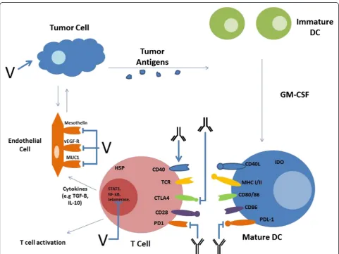
types are summarized in Table 1 and Fig. 1.
* Correspondence:[email protected]
1Department of Medicine, Division of Hematology-Oncology, University of Virginia Health System, UVA Box 800716, Charlottesville, VA 22908, USA Full list of author information is available at the end of the article
Table 1
Cellular Microenvironment of Pancreatic Cancer
Cell Role in pancreatic cancer Relationship to outcome Reference
NK deactivated ↑tumor stage and↓survival [11–13]
CD8+ T-Cell deactivated ↑tumor stage and↓survival [5,8,13]
CD4+ Th1-Cell ↓ ↑tumor stage and↓survival [12–14]
TAM (M1) ↓ ↑tumor stage [4,12–14]
DC deactivated ↑survival [8,13,17,18]
MDSC ↑ ↓survival [5,12,16,24]
Mast Cell ↑ ↑metastases [9,13]
T-regs ↑ ↓survival [4,5,12,22,23]
TAM (M2) ↑ ↑stage and↓survival [4,12–14]
Fibroblast ↑ ↑stage and↓survival [12,13,25,27]
CD4 + Th2 Cell ↑ ↑stage and↓survival [5,9,12–14,16,25]
↑increase,↓decrease,DCdendritic cell,MDSCmyeloid derived suppressor cell,NKnatural killer cell,TAMtumor associated macrophage
Effector immune cells
Natural killer cells (NK)
An increased number of NK
cells have been shown to be associated with a better
prognosis in a small set of 13 patients with pancreatic
cancer [10], presumably due to their role in recognition
and elimination of cancer cells. However, NK cells are
typically found in a limited number in pancreatic cancer
and often in a deactivated form due to the lack of
NKG2D, a cell surface receptor found to be upregulated
in activated NK cells [11
–
13].
CD8 Cytotoxic and CD4 helper T-cells or tumor
infiltrating lymphocytes (TILs)
The presence of TILs in
pancreatic cancer has been well described and it may
rep-resent the most important element in PTME [8, 9, 14, 5].
Among these TILs, the memory (CD45RO) CD8 T-cells
are thought to be the major anti-tumor effector cells and
their density in resected pancreatic tumors was found to
correlate with survival [8, 9]. On the other hand, the role
of CD4 T-cells is more complex. The Th1, the effector
form, activates antigen presenting cells (APC) such as
dendritic cells (DCs) while the ineffective form (Th2) plays
a major role in tumor tolerance [13, 5]. Although the
pres-ence of both CD8 and CD4 T-cells correlates with a better
prognosis [4, 8, 15], they are found in small numbers in
the PTME, possibly due to effect of stroma and
sup-pressor immune cells [15]. In addition, the number of
CD8 effector T-cells decreases during the malignant
transformation of pre-cancer cells [5, 8]. Studies have
shown that Th1 cells are found in disproportionately
lower concentrations among pancreatic cancer cells
than Th2, suggesting an ineffective immune response
against tumor cells [5, 16].
Dendritic cells (DCs)
The presence of DCs in the
PTIM is essential in order to generate an anti-tumor
im-mune response and, similar to TILs, is associated with a
better prognosis in pancreatic cancer [8, 17, 18].
How-ever, DCs are present in low numbers in the PTME and
often in an immature form [8, 17, 18], thus likely limiting
their ability to present foreign antigens to T-cells.
Co-stimulatory receptors and ligands
CD40
Is a co-stimulatory molecule that is expressed on
T-cells and binds to its ligand (CD40L) on APCs, leading
to the activation of lymphocytes [13]. In a retrospective
analysis of patients with pancreatic cancer, Unek et al.,
showed that the expression of CD40 in pancreatic cancer
tissue samples correlates with a trend towards improved
progression-free survival (PFS) and overall survival (OS)
[19]. Accordingly, CD40 represents a promising target in
pancreatic cancer as described below.
OX-40 (tumor necrosis factor receptor superfamily
member 4)
Is a member of the TNF receptor family
found on T- cells and acts as a secondary co-stimulatory
molecule as it requires other co-stimulatory molecules
to be expressed first prior to its activation. The
expres-sion of OX-40 is found in high concentrations on
acti-vated T-cells. In pancreatic cancer increased levels of
OX-40 was reported to correlate with better survival but
this data needs to be validated on a larger scale [10].
4-1BB (tumor necrosis factor receptor superfamily
member 9)
Is also a member of the TNF receptor family
and found on T-cells and NK cells. Upon interaction
with its ligand 4-1BBL, it promotes T cell activation,
par-ticularly CD8 lymphocyte. 4-1BB also acts as a
second-ary co-stimulatory molecule, similar to OX-40 [20, 21].
However, there is currently no data available regarding
the role of 4-1BB in the PTIM.
Suppressor immune cells
Tumor associated macrophages (TAMs)
These cells
express the program cell death ligand (PD-L1) which is
involved in immune suppression and T-cell apoptosis as
described below. In PTIM, the presence of TAMs and it
is association with poor outcomes and more frequent
metastases has been well described [4, 13].
T-regulatory cells (CD4 + CD25 + FoxP3+) (T-regs)
T-regs are other subsets of TILs known for their
immuno-suppressive activity through the release of cytokines
including TGF-
β
and IL-10. Based on few retrospective
analyses, pancreatic tumors with low numbers of
T-regs were found to have a significantly better survival
compared to tumors with high numbers [4, 22, 23].
Myeloid derived suppressor Cell (MDSCs)
In
pancre-atic cancer, these cells were found in pre-malignant
growths and increased in concentration as cancer cells
grow suggesting a direct role in immune suppression
and unrestricted cancer cell growth [5, 12, 16, 24].
Inter-estingly, high concentration of MDSCs in the peripheral
blood was associated with poor outcomes in patients
with pancreatic cancer [16]. Whether this correlation
ex-ists in the PTIM remains to be determined.
[26, 27]. Therefore, the role of fibroblasts in the PTME
is currently under intense investigation.
Mast cells
The role of mast cells in cancer has not been
well-defined [9, 28, 29]. In pancreatic cancer, it has been
suggested that low concentration of mast cells in the
PTME correlates with increased survival [28] while
in-creased concentration is associated with inin-creased lymph
node metastases based on retrospective analyses in small
subsets of patients [29].
Co-Inhibitory Receptors and Ligands
Program-death (PD-1)
Is an inhibitory receptor that
belongs to the B7-receptor family and interacts with its
ligand PD-L1 (B7-H1) to down regulate signals by
T-cells, leading to the induction of apoptosis in activated
T-cells [13]. PD-1 is expressed on progenitor T cells,
ac-tivated T- and B-lymphocytes, NK cells, and myeloid
cells [13]. Patients with pancreatic cancer with PD-L1
positive tumors have a significantly worse prognosis than
patients with PD-L1 negative tumors [27]. The PD-1/
PD-L1 pathway is currently one of the most targeted
pathways in cancer.
Cytotoxic T-lymphocyte associated protein-4 (CTLA-4)
Is a co-inhibitory molecule found on T- lymphocytes
that deactivates these cells to induce apoptosis in
re-sponse to interaction with APCs. This negative feedback
loop is a key to normal immune function to prevent
over-stimulation of T-cells and damage to healthy cells
but is stimulated inappropriately in pancreatic cancer to
create a microenvironment that promotes cancer growth
rather than its recognition. CTLA-4 is overexpressed in
pancreatic cancer cells and its overexpression was found
to correlate negatively with survival in patients who
underwent surgical resection [30].
CXCL12 (C-X-C motif chemokine ligand 12)
Is a
che-mokine that is found in high concentration in pancreatic
cancer and is involved in fibroblast migration and
prolif-eration. The increased concentration of CXCL12 in the
cancer microenvironment creates a network of dense
stroma restricting immune cells migration and recognition
of cancer antigens. Feig et al., showed that the inhibition
of this chemokine resulted in increased T-cell infiltration
into pancreatic cancer in murine models [27].
T-cell immunoglobulin and mucin-domain containing
molecule 3 (TIM-3)
Is a transmembrane protein that is
involved in the regulation of Th1 lymphocytes. The
interaction between TIM-3 and CD4 Th1 cells has been
studied in pancreatic cancer patients and found to
cor-relate with tumor vascular invasion [31]. However, the
role of TIM-3 needs to be further characterized in
pancreatic cancer and other malignancies.
Soluble lymphocyte activation gene-3 (LAG-3)
Is an
important T-cell regulator that interacts with MHC class
II molecules expressed on APCs. This interaction
pro-motes activation and maturation of DCs but negatively
regulates effector T-cells. LAG-3 has been shown to be
necessary for T-regulatory cell activity and represents a
novel target for therapy in pancreatic cancer [32].
Indoleamine 2,3-dioxygenase (IDO)
Is an enzyme
in-volved in cleaving tryptophan into kynurenine.
Trypto-phan is required for T-cell activation and kynurenine
leads to T-regs differentiation and chemotaxis [33].
In-creased levels of IDO, as seen in pancreatic cancer,
creates a microenvironment devoid of effector T-cells
but rich in immunosuppressive T-regs [34]. IDO inhibitors
are currently under investigation in pancreatic cancer and
other malignancies (NCT02048709 and NCT02077881).
Galectins (Gal-1, Gal-3 and Gal-9)
Are immune
modulating glycoproteins that are overexpressed in
pan-creatic cancer cells and thought to be involved in T-cell
homeostasis. These glycoproteins have been shown to
promote immune suppression in pancreatic cancer by
promoting Th2 and T-reg transformation, restricting DC
maturation and stimulating stellate cells [35, 36]. Their
association with survival in pancreatic cancer has been
conflicting, with several studies showing increased
con-centration associated with decreased survival [37, 38],
while others showed an association with improved
out-comes [39].
B7-H3
Is a member of the B7 ligand family that can be
induced in activated dendritic cells, monocytes, and T
cells leading to decrease Th1 type response and cytokine
production. B7-H3 expression was found to correlate
with lymph node metastases and advanced pathologic
stage in patients with pancreatic cancer [40]. The
inhib-ition of B7-H3 increased CD8+ TILs and inhibited
tumor growth in mice [40].
Pancreatic cancer vaccines
Cancer vaccines aim to stimulate the immune system
against tumor cells by generating humoral and/or
cellu-lar immune responses. Many forms of cancer vaccines
exist but generally they can be divided into synthetic and
cellular-based vaccines.
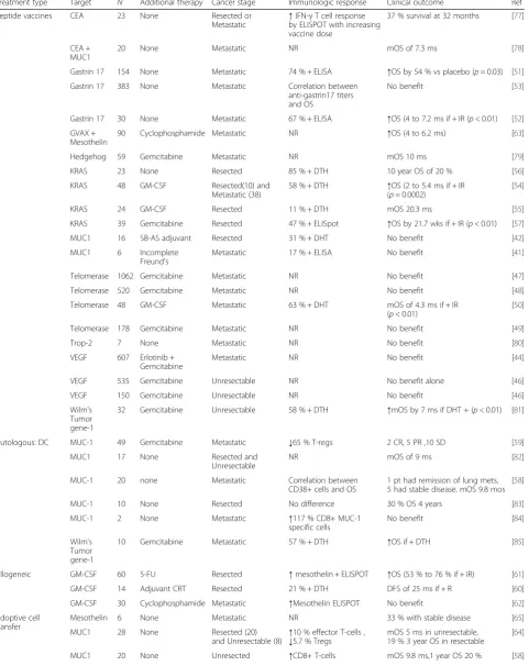
Synthetic vaccines (summarized in Table 2)
Table 2
Completed immunotherapy clinical trials
Treatment type Target N Additional therapy Cancer stage Immunologic response Clinical outcome Ref Peptide vaccines CEA 23 None Resected or
Metastatic ↑
IFN-γT cell response by ELISPOT with increasing vaccine dose
37 % survival at 32 months [77]
CEA + MUC1
20 None Metastatic NR mOS of 7.3 ms [78]
Gastrin 17 154 None Metastatic 74 % + ELISA ↑OS by 54 % vs placebo (p= 0.03) [51]
Gastrin 17 383 None Metastatic Correlation between anti-gastrin17 titers and OS
No benefit [53]
Gastrin 17 30 None Metastatic 67 % + ELISA ↑OS (4 to 7.2 ms if + IR (p< 0.01) [52]
GVAX + Mesothelin
90 Cyclophosphamide Metastatic NR ↑OS (4 to 6.2 ms) [63]
Hedgehog 59 Gemcitabine Metastatic NR mOS 10 ms [79]
KRAS 23 None Resected 85 % + DTH 10 year OS of 20 % [56]
KRAS 48 GM-CSF Resected(10) and Metastatic (38)
58 % + DTH ↑OS (2 to 5.4 ms if + IR (p= 0.0002)
[54]
KRAS 24 GM-CSF Resected 11 % + DTH mOS 20.3 ms [55]
KRAS 39 Gemcitabine Resected 47 % + ELISpot ↑OS by 21.7 wks if + IR (p< 0.01) [57]
MUC1 16 SB-AS adjuvant Resected 31 % + DHT No benefit [42]
MUC1 6 Incomplete Freund's
Metastatic 17 % + ELISA No benefit [41]
Telomerase 1062 Gemcitabine Metastatic NR No benefit [47]
Telomerase 520 Gemcitabine Metastatic NR No benefit [48]
Telomerase 48 GM-CSF Metastatic 63 % + DHT mOS of 4.3 ms if + IR (p< 0.01)
[50]
Telomerase 178 Gemcitabine Metastatic NR No benefit [49]
Trop-2 7 None Metastatic NR No benefit [80]
VEGF 607 Erlotinib + Gemcitabine
Metastatic NR No benefit [44]
VEGF 535 Gemcitabine Unresectable NR No benefit alone [46]
VEGF 150 Gemcitabine Unresectable NR No benefit [46]
Wilm's Tumor gene-1
32 Gemcitabine Unresectable 58 % + DTH ↑mOS by 7 ms if DHT + (p< 0.01) [81]
Autologous: DC MUC-1 49 Gemcitabine Metastatic ↓65 % T-regs 2 CR, 5 PR ,10 SD [59]
MUC1 17 None Resected and
Unresectable
NR mOS of 9 ms [82]
MUC-1 20 none Metastatic Correlation between CD38+ cells and OS
1 pt had remission of lung mets, 5 had stable disease. mOS 9.8 mos
[58]
MUC-1 10 None Resected No difference 30 % OS 4 years [83]
MUC-1 2 None Metastatic ↑117 % CD8+ MUC-1 specific cells
No benefit [84]
Wilm's Tumor gene-1
10 Gemcitabine Metastatic 57 % + DTH ↑OS if + DTH [85]
Allogeneic GM-CSF 60 5-FU Resected ↑mesothelin + ELISPOT ↑OS (53 % to 76 % if + IR) [61]
GM-CSF 14 Adjuvant CRT Resected 21 % + DTH DFS of 25 ms if + R [60]
GM-CSF 30 Cyclophosphamide Metastatic ↑Mesothelin ELISPOT No benefit [62]
Adoptive cell transfer
Mesothelin 6 None Metastatic NR 33 % with stable disease [65]
MUC1 28 None Resected (20) and Unresectable (8)
↑10 % effector T-cells , ↓5.7 % Tregs
mOS 5 ms in unresectable, 19 % 3 year OS in resectable
[64]
targeting MUC1 [41
–
43], VEGF [44
–
46], telomerase
[47
–
50] and gastrin-17 [51
–
53], none have shown a
meaningful survival benefit. These trials, however, were
able to show significant immune responses to the
tar-geted antigens. Some trials using mutated RAS peptide
vaccine alone [54
–
56] or in combination with
gemcita-bine [57] have shown clinical benefit. Importantly, this
benefit was mainly seen in patients who demonstrated
positive immune responses [54, 56, 57].
Cellular-based vaccines (summarized in Table 2)
Cellular-based vaccines use cancer cells (either whole
cells or cell lysates) as the source of the antigens,
allow-ing the immune system to utilize multiple antigens
ra-ther than a single epitope. Overall the results of these
trials are encouraging; however, each included a small
number of patients making it difficult for meaningful
in-terpretation. Cellular based vaccines can be divided into
autologous or allogeneic vaccines based on the source of
the cells.
Autologous vaccines
In this process, the patient
’
s own
dendritic cells are isolated and pulsed with a specific
antigen before being re-infused back to the patient.
Mul-tiple studies have used this vaccination approach
target-ing variety of antigens. MUC1 is one of the most
targeted antigens given its overexpression in pancreatic
cancer and its association with tumor invasion and
me-tastasis. The clinical outcome of this vaccination method
was not impressive; however, an interesting correlation
with immune biomarkers was identified such as
in-creased CD38 (a marker for activated lymphocytes) [58]
and decreased T-regs [59].
Allogeneic vaccine
In this method of vaccination, a
pancreatic cancer cell line is stimulated, usually with
GM-CSF, in order to elicit an immune response when
administered to patients with pancreatic cancer. A group
at John Hopkins used this form of cancer vaccine
(GVAX) in combination with 5-FU or chemoradiation in
the adjuvant setting and showed an increase in PFS and
OS in patients who developed an immune response
against mesothelin [60, 61]. The same group also
investi-gated GVAX in the metastatic setting in combination
with low dose cyclophosphamide, to deplete T-regs,
demonstrating an increase in mesothelin specific T-cell
response with no survival benefit [62]. More recently,
GVAX was combined with a Listeria vaccine that
ex-presses mesothelin in 90 patients with metastatic
dis-ease. The combination of these 2 vaccines showed an
improved OS compared to the allogeneic vaccine alone
(6.1 vs 3.9 months,
p
= 0.01). Interestingly, patients who
derived the most benefit of this combination were
pa-tients who received over 3 doses of the vaccine (9.7 vs
4.6 months,
p
= 0.01) and patients who received at least
2 previous chemotherapy regimens (5.1 vs 3.7 months,
p
= 0.001) [63]. This combination has currently a
break-through designation by FDA while being investigated
in patients with metastatic disease in a large ongoing
clinical trial.
Adoptive T- cell transfer (summarized in Table 2)
In this approach the patient
’
s T-cells are expanded and
activated
ex vivo
then re-infused back to the patient.
Based on the source and the method used for T-cell
activation, adoptive T- cell transfer could be classified
into: Tumor infiltrating lymphocytes (TILs),
engi-neered T- cells that express a specific cancer T-cell
re-ceptor (TCR), and T- cells that express a chimeric
antigen receptor (CAR). This methodology of
im-munotherapy had gained a lot of attention recently
due to promising clinical outcomes in hematological
malignancies. However, the efficacy of adoptive T- cell
transfer remains to be determined in solid tumors
in-cluding pancreatic cancer. Kawaoka et al., investigated
MUC1-specific cytotoxic T lymphocytes (CTLs) in 28
patients and showed 19 % 3-year survival in patients
with resectable disease along with increased effector
lymphocytes and decreased T-regs [64]. More recently,
the University of Pennsylvania group presented their
Table 2
Completed immunotherapy clinical trials
(Continued)
Immune checkpoint inhibitor
CD40 21 Gemcitabine Unresected (7) and Metastatic (20)
N/A ↑mOS by 1.7 ms vs gemcitabine alone, 1 patient had complete resolution of hepatic metasteses
[68]
CTLA-4 30 GVAX Unresectable or metastatic
N/A ↑1 year OS by 20 % compared to GVAX alone
[75]
CTLA-4 27 none Unresected (7) and Metastatic (20)
N/A 1 patient delayed regression of hepatic metasteses
[70]
PD-L1 14 none Metastatic N/A No benefit [69]
↑increase,↓decrease,CRcomplete response,CRTchemoradiation,CTLA-4Cytotoxic T-lymphocyte associated protein-4,DCdendritic cell,DTHdelayed typed hypersensitivity,GM-CSFgranulocyte macrophage colony stimulating factor,MDSCmyeloid derived suppressor cell,mOSmedian overall survival,Msmonths,N/A
not applicable,NKnatural killer cell,NRnot reported,OSoverall survival,PD-L1, programmed cell death ligand-1,PRpartial response,Rresponse,SDstable disease,
experience using autologous T- cells modified with a
chimeric antigen receptor (CAR) that recognizes
mesothelin in pancreatic cancer patients with
refrac-tory metastatic disease. Of the 6 patients treated 2 had
stable disease with one patient had a decreased PET
avidity of hepatic metastases. Overall, the treatment
was well tolerated [65]. The carcinoembryonic antigen
(CEA) is another attractive target in pancreatic cancer
that is currently been explored in clinical trials
(NCT01723306, NCT00004178 and NCT01212887). New
generations of CAR therapies are under investigation with
a focus on increasing their activities and specificities and
decreasing their toxicities. The CAR T-cells efficacy could
be enhanced by engineering the intracellular domain to
contain co-stimulatory molecules such as 41BB and
OX40 or combining CAR T-cells therapy with immune
modulators such as cyclophosphamide in order to
de-plete T-regulatory cells (NCT02465983) or immune
checkpoint inhibitors such as CTLA-4 and anti-PD1
antibodies.
Immune checkpoint inhibitors and co-stimulatory
agonists (summarized in Table 2)
Immune checkpoint inhibitors represent a paradigm
shift in cancer treatment due to their promising clinical
activities in melanoma and other malignancies [66, 67].
However, a limited number of studies targeting these
immune checkpoints have been completed in pancreatic
cancer. The
first
study
to
investigate
the
co-stimulatory agonists in pancreatic cancer used CD40
agonist
in
combination
with
gemcitabine
in
21
patients with locally advanced or metastatic disease.
This study demonstrated promising outcomes with an
improvement of median OS compared to gemcitabine
alone and one patient having a complete resolution of
his liver metastases [68]. In the area of immune
checkpoint inhibitors, both CTLA-4 and PD-L1
inhib-itors were investigated in patients with locally
ad-vanced or metastatic pancreatic cancer in 2 clinical
trials. The clinical outcomes were disappointing,
al-though, only small number of patients were treated
on both trials [69, 70]. To date, the only immune
checkpoint inhibitor to show activity in pancreatic
cancer is MEDI4736 (anti-PD-L1), which showed a
PR rate of 8 % in a preliminary analysis of this going
trial [71]. While this suggests a response can be
achieved with single immunotherapy, it remains
dis-mal and maybe improved by combination therapy.
This approach is currently being investigated in few
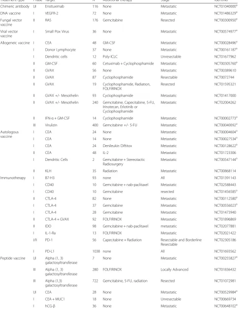
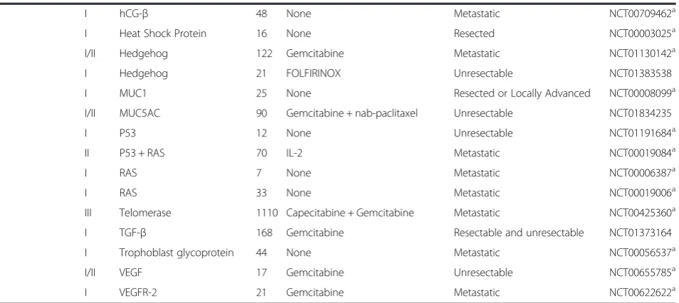
ongoing trials as detailed in Table 3.
Combination therapy
Due to the lack of meaningful clinical benefits of cancer
vaccines, the potential positive immunological effect of
chemotherapy and radiation therapy, and the promising
outcomes of immune checkpoint inhibitors, the focus
has shifted towards combining these modalities.
Gemci-tabine, a standard chemotherapy that is used
tradition-ally to treat pancreatic cancer, has been found to
mediate immunological effects such as tumor associated
antigen cross presentation by dendritic cells and the
in-duction and expansion of cytotoxic T cells responses in
addition to reduce the number of myeloid suppressor
cells [72, 73]. Radiation therapy can also increase the
im-munogenic properties of tumor cells by enhancing MHC
class I expression, thereby increasing their vulnerability to
CTLs. Another frequent effect of DNA damage inflicted
by radiotherapy or chemotherapy is the increase in the
ex-pression of death receptors (in particular Fas/CD95 and
TNF-related apoptosis-inducing ligand [TRAIL] receptors,
enabling lysis of the tumor cells by Fas/CD95 ligand and
TRAIL-positive immune effectors [74]. As detailed in
Table 3, the majority of ongoing trials investigate a
com-bination strategy of the immunotherapy with
chemother-apy, radiation or both. Of interest are multiple trials
targeting mesothelin and/or GVAX with chemoradiation
and multiple immune checkpoint inhibitors combined
with chemotherapy. Our group is currently investigating
the immunological effect of the combination of
chemora-diation and anti-PD-1 as a neoadjuvant treatment in
pa-tients with resectable or borderline resectable pancreatic
cancer compared to neoadjuvant chemoradiation alone
(NCT02305186). This neoadjuvant setting will allow
in-vestigators to study the effect of combination therapy on
the tumor microenvironment. Another promising
com-bination by the Hopkins group combined GVAX with
anti-CTLA-4 and demonstrated a 1-year improvement in
OS by 20 % compared to GVAX and cyclophosphamide
alone [75].
Conclusion
Table 3
On-going immunotherapy clinical trials
Treatment type Phase Target N Additional therapy Stage Identifier
Chimeric antibody I,II Ensituximab 116 None Metastatic NCT01040000a
DNA vaccine I VEGFR-2 72 None Metastatic NCT01486329a
Fungal vector vaccine
II RAS 176 Gemcitabine Resected NCT00300950a
Viral vector vaccine
I Small Pox Virus 36 None Metastatic NCT00574977a
Allogeneic vaccine I CEA 48 GM-CSF Metastatic NCT00028496a
I Donor Lymphocyte 37 None Metastatic NCT00161187a
I Dendritic cells 12 Poly-ICLC Unresectable NCT01677962
II GM-CSF 60 Cetuximab + Cyclophosphamide Metastatic NCT00305760a
II GVAX 56 None Metastatic NCT00389610
II GVAX 87 Cyclophosphamide Resectable NCT0072744
II GVAX 19 Cyclophosphamide, Radiation, FOLFIRINOX
Resected NCT01595321
II GVAX +/- Mesothelin 93 Cyclophosphamide Metastatic NCT01417000
II GVAX +/- Mesothelin 240 Gemcitabine, Capecitabine, 5-FU, Irinotecan, Erlotinib or
Cyclophosphamide
Metastatic NCT02004262
II IFN-α+ GM-CSF 14 Cyclophosphamide Metastatic NCT00002773a
III Virulizin 400 Gemcitabine +/- 5-FU Metastatic NCT00040092a
Autologous vaccine
I CEA 24 None Metastatic NCT00004604a
I CEA 14 None Metastatic NCT00027534a
I CEA 24 Denileukin Diftitox Metastatic NCT00128622a
II CEA 48 IL-2 Metastatic NCT01723306
I Dendritic Cells 2 Gemcitabine + Stereotactic Radiosurgery
Metastatic NCT00547144a
II KLH 35 Radiation Metastatic NCT00868114
Immunotherapy I B7-H3 93 none All NCT01391143
I CD40 10 Gemcitabine + nab-paclitaxel Metastatic NCT02588443
I CD40 10 Gemcitabine resected NCT01456585a
II CTLA-4 82 None Metastatic NCT00112580a
I CTLA-4 37 Gemcitabine Metastatic NCT00556023a
I CTLA-4 28 Gemcitabine Metastatic NCT01473940
II CTLA-4 + GVAX 92 FOLFIRINOX Metastatic NCT01896869
II IDO 98 Gemcitabine + nab-paclitaxel metastatic NCT02077881
I IL-1-Ra 13 FOLFIRINOX Metastatic NCT02021422
I/II PD-1 56 Capecitabine + Radiation Resectable and Borderline Resectable
NCT02305186
I PD-L1 1038 none All NCT01693562
Peptide vaccine I,II Alpha (1, 3) galactosyltransferase
7 None Metastatic NCT00255827a
III Alpha (1, 3) galactosyltransferase
280 FOLFIRINOX Locally Advanced NCT01836432
III Alpha (1,3) galactosyltransferase
722 Gemcitabine, 5-FU, radiation Resected NCT01072981
I,II CEA 28 None Metastatic NCT00529984a
I CEA + MUC1 18 None Unresectable NCT00669734
immunosuppressive cells and receptors; and
under-standing the role of the stromal reaction and its
inter-action with pancreatic cancer immune microenvironment.
Indeed, the recent encouraging data of patients with
mismatch-repair deficient colorectal cancer responding to
pembrolizumab requires further investigation, particularly
as it may be relevant for a small number of patients with
pancreatic cancer [76]. Improved response rates and
survival benefits may be achieved by using combination
therapies; identifying novel biomarkers in order to select
the group of patients who may drive the most benefit of
cancer immunotherapy; and implementing novel clinical
trials designs that allow for tumor samples collection in
order to understand the mechanism of action and
resist-ance of pancreatic cresist-ancer to immunotherapy.
Abbreviations
↑:increase;↓: decrease; 4-1BB: Tumor Necrosis Factor Receptor Superfamily Member 9; APC: antigen presenting cell; CD: cell differentiation; CR: complete response; CRT: chemoradiation; CTLA-4: Cytotoxic T-lymphocyte associated protein-4; CXCL12: C-X-C motif chemokine 12; DC: dendritic cell; DTH: delayed typed hypersensitivity; ELISA: enzyme linked immunosorbent assay; FOLFIRINOX: folinic acid, fluorouracil, irinotecan, oxaliplatin; Gal: Galectins; GM-CSF: granulocyte macrophage colony stimulating factor; HSP: heat shock protein; IDO: Indoleamine 2,3-dioxygenase; IFN-γ: interferon-gamma; IL: interleukin; LAG-3: Soluble Lymphocyte Activation Gene-3; MDSC: myeloid derived suppressor cell; MHC: major histocompatibility complex; mOS: median overall survival; ms: months; NA: not applicable; NK: natural killer cell; NR: not reported; OS: overall survival; OX-40: Tumor Necrosis Factor Receptor Superfamily Member 4; PD-1: programmed cell death-1; PDGF: platelet derived growth factor; PD-L1: programmed cell death-ligand 1; PR: partial response; PTME: pancreatic tumor microenvironment; R: response; SD: stable disease; TAM: tumor associated macrophages; TCR: T-cell receptor; TGF: tumor growth factor; TIL: tumor infiltrating lymphocyte; TIM-3: T-cell Immunoglobulin and mucin-domain containing molecule 3;
vEGF: vascular epithelial growth factor; vEGF-R: vascular epithelial growth factor-receptor.
Competing interests
Paul Kunk has no conflicts of interest to disclose.
Osama Rahma is supported in part by a research grant from Investigatory-Initiated Studies Program of Merck, Sharp & Dohme Corp.
Todd Bauer is has equity ownership of iTi Health, Inc. and is a consultant for Hemoshear, LLC.
Craig Slingluff receives funding from NIH but not related to the topic of this review.
Authors’contributions
PK collected the data and drafted the manuscript. OR revised the manuscript. TB and CS provided additional revisions for the manuscript. All authors read and approved the final manuscript.
Authors’information
PK is currently an Oncology Fellow at the University of Virginia (UVA) with interest in gastrointestinal malignancy. Awarded the Farrow Oncology Fellowship Award, he is currently studying the immune profile of cholangiocarcinoma and is gaining expertise in the field of hepatobiliary cancer and immunotherapy. OR is an Assistant Professor in the Division of Hematology/Oncology at UVA. During his previous training at the National Cancer Institute, he was involved in conducting cancer vaccine clinical trials and the development of immune checkpoint inhibitors. As a member of the Emily Couric Cancer Center and in collaboration with other investigators, his main focus is to study hepatobiliary cancers and to identify potential targets and immune biomarkers that may correlate with outcomes. He is currently a Principal Investigator on many immunotherapy clinical trials in GI cancers and is the lead physician of the GI Oncology team. TB is an Associate Professor of Surgery in the Division of Surgical Oncology, with emphasis on hepatobiliary tumors. Receipt of several awards for clinical and research excellence, his research focus is the immune relationship to pancreatic cancer and early detection of pancreatic cancer. He is the director of the High-Risk Pancreatic Cancer Clinic and a Principal Investigator in many clinical trials in GI cancers. CS is a Professor of Surgery in the Division of Surgical Oncology and a leading expert in the immune profile and immunotherapy of melanoma. He is a Principal Investigator in several clinical trials in melanoma.
Table 3
On-going immunotherapy clinical trials
(Continued)
I hCG-β 48 None Metastatic NCT00709462a
I Heat Shock Protein 16 None Resected NCT00003025a
I/II Hedgehog 122 Gemcitabine Metastatic NCT01130142a
I Hedgehog 21 FOLFIRINOX Unresectable NCT01383538
I MUC1 25 None Resected or Locally Advanced NCT00008099a
I/II MUC5AC 90 Gemcitabine + nab-paclitaxel Unresectable NCT01834235
I P53 12 None Unresectable NCT01191684a
II P53 + RAS 70 IL-2 Metastatic NCT00019084a
I RAS 7 None Metastatic NCT00006387a
I RAS 33 None Metastatic NCT00019006a
III Telomerase 1110 Capecitabine + Gemcitabine Metastatic NCT00425360a
I TGF-β 168 Gemcitabine Resectable and unresectable NCT01373164
I Trophoblast glycoprotein 44 None Metastatic NCT00056537a
I/II VEGF 17 Gemcitabine Unresectable NCT00655785a
I VEGFR-2 21 Gemcitabine Metastatic NCT00622622a
5-FU5-flurouracil,CEACarcinoembryonic antigen,CTLA-4Cytotoxic T-lymphocyte associated protein-4,FOLFIRINOXfolinic acid, fluorouracil, irinotecan, oxaliplatin,
GM-CSFgranulocyte macrophage colony stimulating factor,hCGHuman chorionic gonadotropin,IDOindoleamine 2,3-dioxygenase,IFNinterferon,IL-2interleukin-2,KLH keyhole limpet hemocyanin,MUCmucin,Nnumber,PDprogrammed death,PD-Lprogrammed death ligand,Poly-ICLCcarboxymethylcellulose, polyinosinic-polycytidylic acid, and poly-L-lysine double-stranded RNA,TGFTransforming growth factor,VEGFVascular endothelial growth factor,VEGF-RVascular endothelial growth factor receptor
a
Acknowledgements
No additional acknowledgements other than those listed as authors. There were no sources of funding for this review.
Author details
1Department of Medicine, Division of Hematology-Oncology, University of Virginia Health System, UVA Box 800716, Charlottesville, VA 22908, USA. 2
Department of Surgery, Division of Hepatobiliary Surgery, University of Virginia Health System, Charlottesville, VA, USA.3Department of Surgery, Division of Surgical Oncology, University of Virginia Health System, Charlottesville, VA, USA.
Received: 22 October 2015 Accepted: 16 February 2016
References
1. Jemal A, Siegel R, Xu J, Ward E. Cancer statistics, 2010. CA Cancer J Clin. 2010;60(5):277–300.
2. Yadav D, Lowenfels AB. The epidemiology of pancreatitis and pancreatic cancer. Gastroenterology. 2013;144(6):1252–61.
3. Rahib L, Smith BD, Aizenberg R, Rosenzweig AB, Fleshman JM, Matrisian LM. Projecting cancer incidence and deaths to 2030: the unexpected burden of thyroid, liver, and pancreas cancers in the United States. Cancer Res. 2014; 74(11):2913–21.
4. Ino Y, Yamazaki-Itoh R, Shimada K, Iwasaki M, Kosuge T, Kanai Y, et al. Immune cell infiltration as an indicator of the immune microenvironment of pancreatic cancer. Br J Cancer. 2013;108(4):914–23.
5. Fridman WH, Pages F, Sautes-Fridman C, Galon J. The immune contexture in human tumours: impact on clinical outcome. Nat Rev Cancer. 2012;12(4): 298–306.
6. Collins MA, Bednar F, Zhang Y, Brisset JC, Galban S, Galban CJ, et al. Oncogenic Kras is required for both the initiation and maintenance of pancreatic cancer in mice. J Clin Invest. 2012;122(2):639–53. 7. Feig C, Gopinathan A, Neesse A, Chan DS, Cook N, Tuveson DA. The
pancreas cancer microenvironment. Clin Cancer Res. 2012;18(16):4266–76. 8. Fukunaga A, Miyamoto M, Cho Y, Murakami S, Kawarada Y, Oshikiri T, et al.
CD8+ tumor-infiltrating lymphocytes together with CD4+ tumor-infiltrating lymphocytes and dendritic cells improve the prognosis of patients with pancreatic adenocarcinoma. Pancreas. 2004;28(1):e26–31.
9. Protti MP, De Monte L. Immune infiltrates as predictive markers of survival in pancreatic cancer patients. Front Physiol. 2013;4:210.
10. Davis M, Conlon K, Bohac GC, Barcenas J, Leslie W, Watkins L, et al. Effect of pemetrexed on innate immune killer cells and adaptive immune T cells in subjects with adenocarcinoma of the pancreas. J Immunother. 2012;35(8):629–40. 11. Duan X, Deng L, Chen X, Lu Y, Zhang Q, Zhang K, et al. Clinical significance of
the immunostimulatory MHC class I chain-related molecule A and NKG2D receptor on NK cells in pancreatic cancer. Med Oncol. 2011;28(2):466–74. 12. Sideras K, Braat H, Kwekkeboom J, van Eijck CH, Peppelenbosch MP, Sleijfer S,
et al. Role of the immune system in pancreatic cancer progression and immune modulating treatment strategies. Cancer Treat Rev. 2014;40(4):513–22. 13. Wachsmann MB, Pop LM, Vitetta ES. Pancreatic ductal adenocarcinoma: a
review of immunologic aspects. J Investig Med. 2012;60(4):643–63. 14. Disis ML. Immune regulation of cancer. J Clin Oncol. 2010;28(29):4531–8. 15. Vonderheide RH, Bayne LJ. Inflammatory networks and immune surveillance
of pancreatic carcinoma. Curr Opin Immunol. 2013;25(2):200–5. 16. Gabitass RF, Annels NE, Stocken DD, Pandha HA, Middleton GW. Elevated
myeloid-derived suppressor cells in pancreatic, esophageal and gastric cancer are an independent prognostic factor and are associated with significant elevation of the Th2 cytokine interleukin-13. Cancer Immunol Immunother. 2011;60(10):1419–30.
17. Yamamoto T, Yanagimoto H, Satoi S, Toyokawa H, Yamao J, Kim S, et al. Circulating myeloid dendritic cells as prognostic factors in patients with pancreatic cancer who have undergone surgical resection. J Surg Res. 2012; 173(2):299–308.
18. Tjomsland V, Spangeus A, Sandstrom P, Borch K, Messmer D, Larsson M. Semi mature blood dendritic cells exist in patients with ductal pancreatic adenocarcinoma owing to inflammatory factors released from the tumor. PLoS One. 2010;5(10):e13441.
19. Unek T, Unek IT, Agalar AA, Sagol O, Ellidokuz H, Ertener O, et al. CD40 expression in pancreatic cancer. Hepatogastroenterology. 2013;60(128): 2085–93.
20. Moran AE, Kovacsovics-Bankowski M, Weinberg AD. The TNFRs OX40, 4-1BB, and CD40 as targets for cancer immunotherapy. Curr Opin Immunol. 2013; 25(2):230–7.
21. Cheuk AT, Mufti GJ, Guinn BA. Role of 4-1BB:4-1BB ligand in cancer immunotherapy. Cancer Gene Ther. 2004;11(3):215–26.
22. Ikemoto T, Yamaguchi T, Morine Y, Imura S, Soejima Y, Fujii M, et al. Clinical roles of increased populations of Foxp3 + CD4+ T cells in peripheral blood from advanced pancreatic cancer patients. Pancreas. 2006;33(4):386–90.
23. Yamamoto T, Yanagimoto H, Satoi S, Toyokawa H, Hirooka S, Yamaki S, et al. Circulating CD4 + CD25+ regulatory T cells in patients with pancreatic cancer. Pancreas. 2012;41(3):409–15.
24. Zhao F, Obermann S, von Wasielewski R, Haile L, Manns MP, Korangy F, et al. Increase in frequency of myeloid-derived suppressor cells in mice with spontaneous pancreatic carcinoma. Immunology. 2009;128(1):141–9. 25. De Monte L, Reni M, Tassi E, Clavenna D, Papa I, Recalde H, et al. Intratumor
T helper type 2 cell infiltrate correlates with cancer-associated fibroblast thymic stromal lymphopoietin production and reduced survival in pancreatic cancer. J Exp Med. 2011;208(3):469–78.
26. Abdollahi A, Schwager C, Kleeff J, Esposito I, Domhan S, Peschke P, et al. Transcriptional network governing the angiogenic switch in human pancreatic cancer. Proc Natl Acad Sci U S A. 2007;104(31):12890–5. 27. Feig C, Jones JO, Kraman M, Wells RJ, Deonarine A, Chan DS, et al.
Targeting CXCL12 from FAP-expressing carcinoma-associated fibroblasts synergizes with anti-PD-L1 immunotherapy in pancreatic cancer. Proc Natl Acad Sci U S A. 2013;110(50):20212–7.
28. Strouch MJ, Cheon EC, Salabat MR, et al. Crosstalk between mast cells and pancreatic cancer cells contributes to pancreatic tumor progression. Clinical cancer research : an official journal of the American Association for Cancer Research. 2010;16(8):2257–65.
29. Esposito I, Menicagli M, Funel N, et al. Inflammatory cells contribute to the generation of an angiogenic phenotype in pancreatic ductal
adenocarcinoma. J Clin Pathol. 2004;57(6):630–6.
30. Loos M, Hedderich DM, Ottenhausen M, Giese NA, Laschinger M, Esposito I, et al. Expression of the costimulatory molecule B7-H3 is associated with prolonged survival in human pancreatic cancer. BMC Cancer. 2009;9:463-2407-9-463.
31. Tong D, Zhou Y, Chen W, Deng Y, Li L, Jia Z, et al. T cell immunoglobulin-and mucin-domain-containing molecule 3 gene polymorphisms immunoglobulin-and susceptibility to pancreatic cancer. Mol Biol Rep. 2012;39(11):9941–6. 32. Wang-Gillam A, Plambeck-Suess S, Goedegebuure P, Simon PO, Mitchem JB,
Hornick JR, et al. A phase I study of IMP321 and gemcitabine as the front-line therapy in patients with advanced pancreatic adenocarcinoma. Invest New Drugs. 2013;31(3):707–13.
33. Fallarino F, Grohmann U, You S, McGrath BC, Cavener DR, Vacca C, et al. The combined effects of tryptophan starvation and tryptophan catabolites down-regulate T cell receptor zeta-chain and induce a regulatory phenotype in naive T cells. J Immunol. 2006;176(11):6752–61.
34. Witkiewicz A, Williams TK, Cozzitorto J, Durkan B, Showalter SL, Yeo CJ, et al. Expression of indoleamine 2,3-dioxygenase in metastatic pancreatic ductal adenocarcinoma recruits regulatory T cells to avoid immune detection. J Am Coll Surg. 2008;206(5):849–54. discussion 854-6.
35. Cooper D, Ilarregui JM, Pesoa SA, Croci DO, Perretti M, Rabinovich GA. Multiple functional targets of the immunoregulatory activity of galectin-1: Control of immune cell trafficking, dendritic cell physiology, and T-cell fate. Methods Enzymol. 2010;480:199–244.
36. Fitzner B, Walzel H, Sparmann G, Emmrich J, Liebe S, Jaster R. Galectin-1 is an inductor of pancreatic stellate cell activation. Cell Signal. 2005;17(10):1240–7. 37. Chung JC, Oh MJ, Choi SH, Bae CD. Proteomic analysis to identify
biomarker proteins in pancreatic ductal adenocarcinoma. ANZ J Surg. 2008;78(4):245–51.
38. Kuramitsu Y, Taba K, Ryozawa S, Yoshida K, Zhang X, Tanaka T, et al. Identification of up- and down-regulated proteins in gemcitabine-resistant pancreatic cancer cells using two-dimensional gel electrophoresis and mass spectrometry. Anticancer Res. 2010;30(9):3367–72.
39. Chen R, Pan S, Ottenhof NA, de Wilde RF, Wolfgang CL, Lane Z, et al. Stromal galectin-1 expression is associated with long-term survival in resectable pancreatic ductal adenocarcinoma. Cancer Biol Ther. 2012;13(10):899–907. 40. Yamato I, Sho M, Nomi T, Akahori T, Shimada K, Hotta K, et al. Clinical
41. Yamamoto K, Ueno T, Kawaoka T, Hazama S, Fukui M, Suehiro Y, et al. MUC1 peptide vaccination in patients with advanced pancreas or biliary tract cancer. Anticancer Res. 2005;25(5):3575–9.
42. Ramanathan RK, Lee KM, McKolanis J, Hitbold E, Schraut W, Moser AJ, et al. Phase I study of a MUC1 vaccine composed of different doses of MUC1 peptide with SB-AS2 adjuvant in resected and locally advanced pancreatic cancer. Cancer Immunol Immunother. 2005;54(3):254–64.
43. Petrulio CA, Kaufman HL. Development of the PANVAC-VF vaccine for pancreatic cancer. Expert Rev Vaccines. 2006;5(1):9–19.
44. Van Cutsem E, Vervenne WL, Bennouna J, Humblet Y, Gill S, Van Laethem JL, et al. Phase III trial of bevacizumab in combination with gemcitabine and erlotinib in patients with metastatic pancreatic cancer. J Clin Oncol. 2009;27(13):2231–7.
45. Kindler HL, Niedzwiecki D, Hollis D, Sutherland S, Schrag D, Hurwitz H, et al. Gemcitabine plus bevacizumab compared with gemcitabine plus placebo in patients with advanced pancreatic cancer: phase III trial of the Cancer and Leukemia Group B (CALGB 80303). J Clin Oncol. 2010;28(22):3617–22. 46. Yamaue H, Tani T, Miyazawa M, Yamao K, Mizuno N, Okusaka T, et al. Phase
II/III clinical trial with VEGFR2-epitope peptide and gemcitabine for patients with locally advanced, metastatic, or unresectable pancreatic cancer: Pegasus-PC study. J Clin Oncol. 2013;4:abstr 223.
47. Middleton G, Silcocks P, Cox T, Valle J, Wadsley J, Propper D, et al. Gemcitabine and capecitabine with or without telomerase peptide vaccine GV1001 in patients with locally advanced or metastatic pancreatic cancer (TeloVac): an open-label, randomised, phase 3 trial. Lancet Oncol. 2014;15(8):829–40. 48. Schmidt J. Pharmexa Stops One of Two Phase III Trials. 2008; Available at: http://
www.drugs.com/news/pharmexa-stops-one-two-phase-iii-trials-8180.html, 2014. Accessed 16 Feb 2016.
49. Buanes T, Maurel J, Liauw W, Hebbar M, Nemunaitis J. A randomized phase III study of gemcitabine (G) versus GV1001 in sequential combination with G in patients with unresectable and metastatic pancreatic cancer (PC). J Clin Oncol (Meeting Abstracts) May 2009;27(no. 15S 4601).
50. Bernhardt SL, Gjertsen MK, Trachsel S, Moller M, Eriksen JA, Meo M, et al. Telomerase peptide vaccination of patients with non-resectable pancreatic cancer: A dose escalating phase I/II study. Br J Cancer. 2006;95(11):1474–82. 51. Gilliam AD, Broome P, Topuzov EG, Garin AM, Pulay I, Humphreys J, et al. An
international multicenter randomized controlled trial of G17DT in patients with pancreatic cancer. Pancreas. 2012;41(3):374–9.
52. Brett BT, Smith SC, Bouvier CV, Michaeli D, Hochhauser D, Davidson BR, et al. Phase II study of anti-gastrin-17 antibodies, raised to G17DT, in advanced pancreatic cancer. J Clin Oncol. 2002;20(20):4225–31.
53. Shapiro J, Marshall J, Karasek P, et al. G17DT + gemcitabine versus placebo + gemcitabine in untreated subjects with locally advanced, recurrent or metastatic adenocarcinoma of the pancreas: results of a randomized, double-blinded, multinational, multicenter study. J Clin Oncol. 2005;23(suppl):4012. 54. Gjertsen MK, Buanes T, Rosseland AR, Bakka A, Gladhaug I, Soreide O, et al.
Intradermal ras peptide vaccination with granulocyte-macrophage colony-stimulating factor as adjuvant: Clinical and immunological responses in patients with pancreatic adenocarcinoma. Int J Cancer. 2001;92(3):441–50. 55. Abou-Alfa GK, Chapman PB, Feilchenfeldt J, Brennan MF, Capanu M,
Gansukh B, et al. Targeting mutated K-ras in pancreatic adenocarcinoma using an adjuvant vaccine. Am J Clin Oncol. 2011;34(3):321–5.
56. Weden S, Klemp M, Gladhaug IP, Moller M, Eriksen JA, Gaudernack G, et al. Long-term follow-up of patients with resected pancreatic cancer following vaccination against mutant K-ras. Int J Cancer. 2011;128(5):1120–8. 57 Muscarella P et al. A randomized, placebo controlled, doubled, blind,
multicenter phase 2 adjuvant trial of the efficacy, immunogenicity, and safety of GI-4000 plus Gem vs Gem alone in patients with resected pancreas cancer with activating Ras mutations/survival and immunology analysis of the R1 Subgroup. J Clin Oncol. 2012;30(suppl):astr #e14501. 58. Kondo H, Hazama S, Kawaoka T, Yoshino S, Yoshida S, Tokuno K, et al. Adoptive
immunotherapy for pancreatic cancer using MUC1 peptide-pulsed dendritic cells and activated T lymphocytes. Anticancer Res. 2008;28(1B):379–87.
59. Kimura Y, Tsukada J, Tomoda T, Takahashi H, Imai K, Shimamura K, et al. Clinical and immunologic evaluation of dendritic cell-based immunotherapy in combination with gemcitabine and/or S-1 in patients with advanced pancreatic carcinoma. Pancreas. 2012;41(2):195–205.
60. Jaffee EM, Hruban RH, Biedrzycki B, Laheru D, Schepers K, Sauter PR, et al. Novel allogeneic granulocyte-macrophage colony-stimulating factor-secreting tumor vaccine for pancreatic cancer: a phase I trial of safety and immune activation. J Clin Oncol. 2001;19(1):145–56.
61. Lutz E, Yeo CJ, Lillemoe KD, Biedrzycki B, Kobrin B, Herman J, et al. A lethally irradiated allogeneic granulocyte-macrophage colony stimulating factor-secreting tumor vaccine for pancreatic adenocarcinoma. A Phase II trial of safety, efficacy, and immune activation. Ann Surg. 2011;253(2):328–35. 62. Laheru D, Lutz E, Burke J, Biedrzycki B, Solt S, Onners B, et al. Allogeneic
granulocyte macrophage colony-stimulating factor-secreting tumor immunotherapy alone or in sequence with cyclophosphamide for metastatic pancreatic cancer: a pilot study of safety, feasibility, and immune activation. Clin Cancer Res. 2008;14(5):1455–63.
63. Le DT, Wang-Gillam A, Picozzi V, Greten TF, Crocenzi T, Springett G, et al. Safety and survival with GVAX pancreas prime and Listeria Monocytogenes-expressing mesothelin (CRS-207) boost vaccines for metastatic pancreatic cancer. J Clin Oncol. 2015;33(12):1325–33.
64. Kawaoka T, Oka M, Takashima M, Ueno T, Yamamoto K, Yahara N, et al. Adoptive immunotherapy for pancreatic cancer: cytotoxic T lymphocytes stimulated by the MUC1-expressing human pancreatic cancer cell line YPK-1. Oncol Rep. 2008;20(1):155–63.
65. Beatty GL. Safety and antitumor activity of chimeric antigen receptor modified T cells in patients with chemotherapy refractory metastatic pancreatic cancer. 2015 ASCO Annual Meeting; 2015.
66. Hodi FS, O'Day SJ, McDermott DF, Weber RW, Sosman JA, Haanen JB, et al. Improved survival with ipilimumab in patients with metastatic melanoma. N Engl J Med. 2010;363(8):711–23.
67. Robert C, Long GV, Brady B, Dutriaux C, Maio M, Mortier L, et al. Nivolumab in previously untreated melanoma without BRAF mutation. N Engl J Med. 2015;372(4):320–30.
68. Beatty GL, Chiorean EG, Fishman MP, Saboury B, Teitelbaum UR, Sun W, et al. CD40 agonists alter tumor stroma and show efficacy against pancreatic carcinoma in mice and humans. Science. 2011;331(6024):1612–6. 69. Brahmer JR, Tykodi SS, Chow LQ, Hwu WJ, Topalian SL, Hwu P, et al. Safety
and activity of anti-PD-L1 antibody in patients with advanced cancer. N Engl J Med. 2012;366(26):2455–65.
70. Royal RE, Levy C, Turner K, Mathur A, Hughes M, Kammula US, et al. Phase 2 trial of single agent Ipilimumab (anti-CTLA-4) for locally advanced or metastatic pancreatic adenocarcinoma. J Immunother. 2010;33(8):828–33. 71. Segal NH, Anto SJ, Brahmer JR, et al. Preliminary data from a multi-arm expansion
study of MEDI4736, an anti-PD-L1 antibody. J Clin Oncol. 2014;32(5 s):abstr 3002. 72. Plate JM, Plate AE, Shott S, Bograd S, Harris JE. Effect of gemcitabine on
immune cells in subjects with adenocarcinoma of the pancreas. Cancer Immunol Immunother. 2005;54(9):915–25.
73. Correale P, Cusi MG, Del Vecchio MT, Aquino A, Prete SP, Tsang KY, et al. Dendritic cell-mediated cross-presentation of antigens derived from colon carcinoma cells exposed to a highly cytotoxic multidrug regimen with gemcitabine, oxaliplatin, 5-fluorouracil, and leucovorin, elicits a powerful human antigen-specific CTL response with antitumor activity in vitro. J Immunol. 2005;175(2):820–8.
74. Kershaw MH, Devaud C, John LB, Westwood JA, Darcy PK. Enhancing immunotherapy using chemotherapy and radiation to modify the tumor microenvironment. Oncoimmunology. 2013;2(9):e25962.
75. Le DT, Lutz E, Uram JN, Sugar EA, Onners B, Solt S, et al. Evaluation of ipilimumab in combination with allogeneic pancreatic tumor cells transfected with a GM-CSF gene in previously treated pancreatic cancer. J Immunother. 2013;36(7):382–9.
76. Le DT, Uram JN, Wang H, Bartlett BR, Kemberling H, Eyring AD, et al. PD-1 Blockade in Tumors with Mismatch-Repair Deficiency. N Engl J Med. 2015; 372(26):2509–20.
77. Geynisman DM, Zha Y, Kunnavakkam R, Aklilu M, Catenacci DV, Polite BN, et al. A randomized pilot phase I study of modified carcinoembryonic antigen (CEA) peptide (CAP1-6D)/montanide/GM-CSF-vaccine in patients with pancreatic adenocarcinoma. J Immunother Cancer. 2013;1:8-1426-1-8. eCollection 2013.
78. Schuetz T, Kaufman H, Marshall J, Safran H. Extended survival in second-line pancreatic cancer after therapeutic vaccination.2005. 16 suppl 2576. ASCO Annual Meeting.
79. Jesus-Acosta A, O'Dwyer P, Ramanathan R, et al. A phase II study of vismodegib, a hedgehog (Hh) pathway inhibitor, combined with gemcitabine and nab-paclitaxel (nab-P) in patients (pts) with untreated metastatic pancreatic ductal adenocarcinoma (PDA). J Clin Oncol 32, 2014 (suppl 3; abstr 257). ASCO Annual Meeting.
81. Nishida S, Koido S, Takeda Y, Homma S, Komita H, Takahara A, et al. Wilms tumor gene (WT1) peptide-based cancer vaccine combined with gemcitabine for patients with advanced pancreatic cancer. J Immunother. 2014;37(2):105–14.
82. Nakamura M, Wada J, Suzuki H, Tanaka M, Katano M, Morisaki T. Long-term outcome of immunotherapy for patients with refractory pancreatic cancer. Anticancer Res. 2009;29(3):831–6.
83. Lepisto AJ, Moser AJ, Zeh H, Lee K, Bartlett D, McKolanis JR, et al. A phase I/ II study of a MUC1 peptide pulsed autologous dendritic cell vaccine as adjuvant therapy in patients with resected pancreatic and biliary tumors. Cancer Ther. 2008;6(B):955–64.
84. Pecher G, Haring A, Kaiser L, Thiel E. Mucin gene (MUC1) transfected dendritic cells as vaccine: results of a phase I/II clinical trial. Cancer Immunol Immunother. 2002;51(11-12):669–73.
85. Koido S, Homma S, Okamoto M, Mori M, and Yoshizaki S. Dendritic cells pulsed with Wilms′tumor gene 1 (WT1)-specific and MHC class I and II-restricted epitopes with gemcitabine induce antitumor immune responses in patients with advanced pancreatic cancer. Cancer Research. October 1, 2014;74:CT223. AACR Annual Meeting.
• We accept pre-submission inquiries
• Our selector tool helps you to find the most relevant journal • We provide round the clock customer support
• Convenient online submission • Thorough peer review
• Inclusion in PubMed and all major indexing services • Maximum visibility for your research
Submit your manuscript at www.biomedcentral.com/submit

![Crystal structure of (methanol κO)[5,10,15,20 tetrakis(2 aminophenyl)porphyrinato κ4N]zinc(II)–chloroform–methanol (1/1/1)](data:image/gif;base64,R0lGODlhAQABAIAAAP///wAAACH5BAEAAAAALAAAAAABAAEAAAICRAEAOw==)