Protein Adsorption to Surface Chemistry and Crystal Structure
Modification of Titanium Surfaces
Ryo Jimbo
1,2, Mikael Ivarsson
3, Anita Koskela
3, Young-Taeg Sul
2,4, Carina B. Johansson
51Department of Prosthodontics, Faculty of Odontology, Malmö University, Malmö, Sweden
2Department of Biomaterials, Institute of Clinical Sciences, Sahlgrenska Academy, Göteborg University, Göteborg, Sweden 3Clinical Research Center, Örebro University Hosptial, Örebro, Sweden
4Institute for Clinical Dental Research, Korea University, Seoul, South Korea
5Department of Clinical Medicine, School of Health and Medical Sciences, Örebro University, Örebro, Sweden
Corresponding Author: Ryo Jimbo
Department of Prosthodontics, Faculty of Odontology, Malmö University 205 06 Malmö
Sweden
Phone: +46 40 6658 502 Fax: +46 40 6658 503 E-mail: [email protected]
ABSTRACT
Objectives: To observe the early adsorption of extracellular matrix and blood plasma proteins to magnesium-incorporated titanium oxide surfaces, which has shown superior bone response in animal models.
Material and Methods: Commercially pure titanium discs were blasted with titanium dioxide (TiO2) particles (control), and
for the test group, TiO2 blasted discs were further processed with a micro-arc oxidation method (test). Surface morphology
was investigated by scanning electron microscopy, surface topography by optic interferometry, characterization by X-ray
photoelectron spectroscopy (XPS), and by X-ray diffraction (XRD) analysis. The adsorption of 3 different proteins (fibronectin,
albumin, and collagen type I) was investigated by an immunoblotting technique.
Results: The test surface showed a porous structure, whereas the control surface showed a typical TiO2 blasted structure. XPS data revealed magnesium-incorporation to the anodic oxide film of the surface. There was no difference in surface roughness between the control and test surfaces. For the protein adsorption test, the amount of albumin was significantly higher on the control surface whereas the amount of fibronectin was significantly higher on the test surface. Although there was no significant difference, the test surface had a tendency to adsorb more collagen type I.
Conclusions: The magnesium-incorporated anodized surface showed significantly higher fibronectin adsorption and lower
albumin adsorption than the blasted surface. These results may be one of the reasons for the excellent bone response previously observed in animal studies.
Keywords: titanium dioxide; magnesium; immunoblotting; fibronectins; albumins; collagen type I.
Accepted for publication: 18 May 2010
To cite this article:
Jimbo R, Ivarsson M, Koskela A, Sul YT, Johansson CB. Protein Adsorption to Surface Chemistry and Crystal Structure
Modification of Titanium Surfaces.
J Oral Maxillofac Res 2010 (Jul-Sep);1(3):e3
URL: http://www.ejomr.org/JOMR/archives/2010/3/e3/e3ht.pdf
INTRODUCTION
In living systems, blood (plasma) is the first component
to come in contact with biomaterials such as a titanium implant during surgery [1]. It is known that immediately after contact with plasma, rapid adsorption of plasma proteins onto the biomaterial takes place [2], which
influences subsequent cell attachment, spreading,
proliferation, and differentiation [3]. In bone-to-implant binding, i.e. osseointegration, the attachment of
blood-derived proteins such as plasma fibronectin to the
implant surface enhances the chemotaxis [4] and focal adhesion of osteogenic cells [5]. Fibronectin is a high-molecular weight extracellular matrix (ECM) protein (approximately 440 kDa) that binds to integrins, which are membrane-spanning receptor proteins [6]. One form
of fibronectin, the plasma fibronectin, is produced by
hepatocytes in the liver and circulates in the blood [7]. It activates signaling pathways that direct cell-cycle progression, gene expression, matrix mineralization [8], and the regulation of the survival of osteoblasts [9]. In
addition, plasma fibronectin is known to be a regulator
for bone density, and bone biomechanical properties [7], and it has been reported that plasma fibronectin
interacts with bone morphogenetic protein 1, indicating that it has an important role in osteogenesis [10]. Ever since the importance of a moderately roughened surface was proposed, the rate of osseointegration
has been enhanced by surface modification [11], and
further, in recent times, surface modification is being
carried out even at the nano-level. It is therefore
reasonable that implant surfaces should be modified at
this level, given that it has been proven that cells react sensitively to nano-topographies [12]. Several studies
have investigated the effect of the modified surfaces on osteogenic cell reactions, and surface modifications
aimed at enhancing cell responses have been carried out [13,14]. However, few modifications have actually
focused on the protein reactions underlying such cell reactions.
We have used electrochemical oxidation incorporating protein “bindable” ions such as sulfate, phosphate [15], and calcium [16] to create surface modifications aimed
to theoretically attract ECM proteins or bone matrix proteins with high bonding properties. The concept
behind these novel modifications is the hypothetical
biochemical bonding between bone and the implant [16]. These so-called “bioactive” implants showed improved bone responses compared to machined implants or other surfaces available on the market. A recent study by Sul et al. [17] validated the presence of a biochemical bond
of surface chemistry modified smooth surface implants
in bone but also measured the relative quantity of their
biochemical bond strength at the bone-to-implant interface. Magnesium (Mg)-incorporated oxidized implants showed stronger and faster bone integration as compared to commercially available oxidized or dual acid-etched implants [18]. The enhanced bone response of the Mg implants was most likely due to the Mg titanate chemistry effects, possibly attracting ECM proteins or bone matrix proteins. It has been reported
that Mg supplementation to young mice influenced
bone formation, resorption, and mineralization [19]. It
has also been shown that Mg deficiency may disturb
bone metabolism and lead to osteoporosis [20].
The objective of this in vitro study was to investigate if adsorption of three different proteins, which are purportedly involved in bone apposition to implant
surfaces is altered in Mg-modified surfaces, which
would substantiate the in vivo data.
MATERIAL AND METHODS
Disc sample preparation
The disc samples (commercially pure titanium, CpTi, ASTM grade 4, 10 mm × 5 mm) were manufactured using a CNC (computer numerical control) machine and then blasted with TiO2 particles in the range of
100 - 150 μm. The implants were degreased by sonication
in an aqueous solution of phosphate-free Extran® MA 03 (Merck, Darmstadt, Germany)/deionized water (1 : 100) and absolute ethanol for 2 × 15 min. Next, they were rinsed with deionized water, and then dried in an oven at 60 °C for 24 h. The samples were divided into 2 groups, the blasted control group and the oxidized test group. The test group was fabricated in an electrolyte mixture containing Mg ions. They were fabricated using a micro arc oxidation (MAO) process as previously described [21,22]. In brief, the MAO process was conducted in galvanostatic mode, with the anodic forming voltage increased at a rate of dV/dt and controlled at 0.5 V/s by a combination of electrochemical parameters. The electrolyte mixture was stirred with a magnetic stirring bar at 300 rpm. Currents and voltages were recorded at 1-s intervals using a computer that was interfaced with the power supply. The samples were rinsed with deionized water and then dried in an oven at 60 °C for 24 h.
Surface properties and analysis techniques
(XPS, ESCALAB 250, Thermo-VG, England) using a
monochromatic Al Kα X-ray source (1486.7 eV, 300
W; beam size, 400 µm diameter). The electron
take-off angle was fixed at 45º and the vacuum pressure
was maintained below 10-9 torr during spectral data
acquisition. XPS data were acquired before and
after sputtering. In order to remove the superficial
contaminant (2 monolayers), Ar sputter cleaning was carried out for 3 s (beam energy, 2 KeV; primary current, 2 µA; raster area, 3.14 mm2). The binding energy of the
target elements was determined with a resolution of 0.1 eV, using the binding energy of carbon (C 1s: 284.8 eV) as a reference.
The crystal structure was determined by low-angle XRD
with a thin film collimator (X`Pert PRO-MRD, Philips
Ltd, Netherlands) on a plate-type sample prepared with the same electrochemical parameters as the test screw-shaped implants. The step size used in the scan was 0.02° over the range of 15° to 70°. The spectra were
recorded using Cu Kα radiation (0.154056 Å) generated
at an acceleration voltage of 35 kV and a current of 25 mA.
Surface roughness was measured using an optical
profilometer (MicroXamTM, Phase-Shift, Arizona,
USA). Three discs each from the test group and from the control group were measured at 3 areas to give a total of 9 measurements for each group. The measuring area
was 230 µm × 230 µm for each group. A Gaussian filter,
50 µm × 50 µm, was used to separate the roughness from errors of form and waviness.
Attachment of purified albumin to disc surfaces
Relative amounts of attached albumin was analyzed by electrophoresis and Coomassie blue staining of the gels after solubilization of the disc-associated albumin.
Discs in triplicate were immersed in 40 mg/ml purified
human serum albumin (Sigma, St Louis, MO, USA) for 16 h at 37 °C to saturate binding, then washed in phosphate-buffered saline (PBS). Attached albumin was solubilized by boiling discs for 5 min in detergent buffer (0.1% sodium dodecyl sulfate [SDS], 1% Igepal CA-630 and 0.5% sodium deoxycholate in PBS), and samples were analyzed by 5% SDS-polyarylamide gel electrophoresis followed by Coomassie blue staining of the gels. Evaluation of staining intensities was performed by analysing images using Sigma Gel software (SPSS Science Software GmbH, Erkrath, Germany).
Attachment of collagen and fibronectin to disc
surfaces
A total of 100 µl with 0.2 mg/ml bovine plasma
fibronectin (Sigma, St Louis, MO, USA) or bovine
collagen type I (Nutacon BV, Leimuden, Netherlands) in PBS was added to the tops of the discs in triplicate
and incubated for 16 h at 37 ºC. The discs were washed
3 times in PBS and transferred to microcentrifuge tubes containing 500 µl 1% Igepal CA-630, 0.5% sodium deoxycholate, and 0.1% SDS. The discs were boiled in this solution for 5 min, then chilled on ice,
and the solutions were stored at - 80 ºC. The samples
were thawed on ice, and 25 µl of each sample was mixed with 25 µl 2XLaemmli sample buffer (BioRad,
Hercules, CA, USA) and a final concentration of 8% β-mercaptoethanol. The samples were boiled for 5 min,
chilled on ice, and run on a 5% SDS polyacrylamide gel electrophoresis. Proteins were electro-transferred
to polyvinylidene difluoride membranes (BioRad), and plasma fibronectin and collagen type I were detected by immunoblotting with specific antibodies. Rabbit anti-human fibronectin antibodies F-3648 were obtained
from Sigma (Sigma, St Louis, MO, USA), and rabbit anti-human collagen type I antibodies ab292 were from Abcam plc, Cambridge UK. Blots were developed using horseradish peroxidase-coupled secondary antibodies and an Advance Western Blotting Detection Kit (GE Health Care, Buckinghamshire, UK). Evaluation of staining intensities was performed by analysing images using Sigma Gel software.
Statistical analysis
All statistical analyses in the present study were performed with the KaleidaGraph software (Synergy Software, Essex Junction, VT, USA). The mean and standard deviation values for the in vitro parameters were calculated. The average values were compared by paired Student’s t-test and analysis of variance (ANOVA) followed by a post hoc Tukey-Kramer test
with the value of statistical significance set at the 0.05
level.
RESULTS
Surface characterization
Figure 1 shows SEM micrographs that characterize blasted pits and facets in the control surface and homogeneous porous structure with an average pore size of 1 - 2 µm in the test surface. The surface
roughness after filtering showed an Sa value (arithmetic average height deviation, μm) of 0.81 (± 0.31) for the control and 0.75 (± 0.14) for the test group. No significant differences regarding surface roughness were
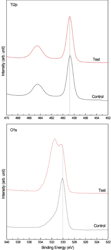
(458.9 ± 0.1 eV), O 1 s (531 ± 0.5 eV) (Figure 2A), and Mg 2p (50.4 ± 0.1 eV)
core-level energy regions of the electron orbitals before and after argon ion (Ar+) sputter cleaning (Figure 2B). Table 1 shows the quantitative differences between the chemical compositions of the samples. The test sample showed the major doublet peaks of the O 1 s at 530.8 eV and 531.7 eV, which may be attributed to the Mg titanate and –OH functional groups. The blasted implants consisted mainly of TiO2. Figure 3 shows the XRD patterns of the amorphous structure in the control group and a mixture of anatase and rutile phase in the test group (Figure 3).
Figure 1. Scanning electron microscopy image of the surface blasted with titanium particles (control), and Mg-incorporated anodized surface (test) (Scale bar: 5 µm).
Figure 2A. Ti 2p and O 1 s spectra of control and test surface. The dashed line indicates the binding energy of peak position at Ti 2p and O 1 s for the control surface.
Figure 2B. Mg cation incorporation during the MAO process, characterizing the binding energy at the Mg 2p of as-received and Ar + sputter-cleaned surfaces. The dashed line indicates the binding energy of peak position at Mg 2p of as-received surface.
Figure 3. X-ray diffraction patterns of control and test surface. Amorphous, anatase and rutile phase of TiO2 were detected on the
control and test groups. Ti = titanium; A = anatase; R = rutile.
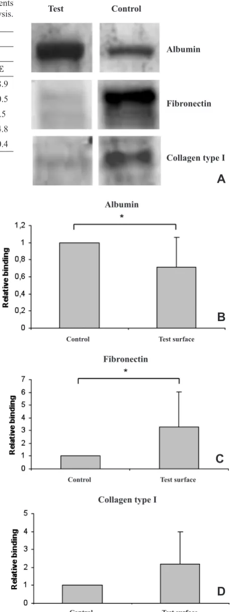
Figure 4. Attachment of extracellular matrix and blood plasma proteins to control and test surfaces (n = 3 in each group, performed
in triplicate). Purified albumin, fibronectin, or collagen type I were allowed to attach to surfaces for 16 h at 37 ˚C. Relative binding
was measured by electrophoresis followed by Coomassie blue
staining or immunoblotting of solubilized proteins (A). Significant
differences by densitometry (B - D) using paired Student’s t-test
(P ≤ 0.05) are indicated (*).
Control Test surface
Control Test surface
Control Test surface
Albumin
Fibronectin
Collagen type I
Albumin
Fibronectin
Collagen type I
Test Control
Adsorption of purified albumin, fibronectin, and
collagen type I to control and test surfaces
The intensity of the protein stainings for each surface is shown in Figure 4A. The results of this study showed
that significantly more albumin adhered to control surfaces than to test surfaces, and significantly less purified fibronectin adhered to control surfaces than to test surfaces (P ≤ 0.05) (Figure 4B and 4C). For collagen type I, no significant difference was detected,
although a binding tendency was found in favour of the test surface (P = 0.09) (Figure 4D).
DISCUSSION
This study focused on the effect of titanium surface property changes with particular attention to the initial protein behaviour. Surface characterization determined major differences of surface chemistry and crystal structure but minor differences of surface roughness between the control and test surfaces.
In the test group, the characteristic element of Mg, 7 - 9 at.%, was incorporated into the oxide layer through
the field-associated ion incorporation during the MAO
process [22,23]. The finding of the hydroxyl group in the test surface is consistent with the findings of previous
studies [22,24].
It has been reported that enhanced osteogenic cell responses in vitro, and bone apposition in vivo, have been observed in surfaces possessing an external layer of anatase and rutile phases [25]. The present results of surface characterization are congruent with those of the implants used for the previous in vivo studies. This was an important aspect of our study, since its aim was to validate in vivo initial protein interactions with in vitro
data, possibly correlating the latter to the enhanced bone
B
D
A
C
Table 1. Binding energiesa and atom concentration rateb of elements
at as-received and sputter cleaned surface in XPS analysis.
Atom
Control Test
Beforec Afterd Beforec Afterd
at.% BE at.% BE at.% BE at.% BE
Ti 12.5 458.8 19.3 458.8 10.0 458.8 17.9 458.9
O 55.8 530.2 67.9 530.3 53.1 531.5 60.7 530.5
Mg - - - - 6.9 50.4 9.2 50.5
C 31.3 284.8 12.8 284.8 27.9 284.8 9.8 284.8
N 0.4 400.2 - - 2.1 400.3 1.6 400.4
aBinding energy value in eV. bAtom concentration rate in at.%. cAs-received surface.
dSputter cleaned surface.
apposition seen in the animal studies [21,22,24,26]. The individual protein adsorption test showed that the amount of albumin adsorbed onto the test surface
was significantly lower than that adsorbed onto the control surface. Moreover, the amount of fibronectin adsorbed to the test surface was significantly higher
than that adsorbed to the control surface. The amount of collagen type I adsorbed onto the test surface was also higher, although the difference was not statistically
significant. The results of the study clearly showed the characteristics of each surface with regard to specific
protein binding. It has been reported that osteoblasts grown on Mg-incorporated surfaces show higher
expression of β1, and α5β1 integrin receptors than do
non-Mg-incorporated surfaces [27]. Since the β1, and α5β1 integrin receptors are known to be fibronectin
receptors, these results suggest that Mg attracts more
fibronectin to the surface than occurs with
non-Mg-incorporated surfaces.
In a preliminary experiment, we used human plasma obtained from healthy blood donors and incubated this on control and test samples for 16 h. The precipitated proteins were run on SDS-polyacrylamide gel-electrophoresis, and protein bands detected by Commassie blue staining were cut out of the gel and sent
for protein identification by mass spectrometry (Pick’n
Post Service, VWR International AB, Stockholm, Sweden). The results showed that at 16 h, the proteins above detection level were plasma albumin, and the amount of adsorption showed different results than that
from purified albumin. Similarly, analysis of fibronectin
by immunoblotting after incubation of human plasma on discs showed different results compared to
corresponding purified protein demonstrated in the
current study. It would be interesting to study more thoroughly the protein adsorption using human plasma.
However, this is a difficult task because the amount
and type of protein adsorption changes rapidly due to competitive protein adsorption [28] (Vroman effect). The Vroman effect is the competitive nature of protein adsorption onto the surface depending on the molecular weight of the protein [29]. In future studies, it will be interesting to observe different time points and clarify the mechanisms of this phenomenon.
Collagen type I is the major constituent of bone matrix protein [30], which is assembled in the presence of
plasma fibronectin [31]. It is an essential protein in osteogenesis [32], which occurs later in the biological process. The reason for observing collagen type I adsorption in the individual protein adsorption test was to investigate its reaction to the Mg surface, because of its central role as structural component in bone, and the lack or abnormality of both collagen type I and Mg causes osteogenesis imperfecta [33].
Although there was no significant difference, the test
surface tended to have higher amounts of collagen type I adsorption, which may be one of the factors for the enhanced bone apposition seen in animal studies. Albumin is a major protein included in plasma (approx. 60%, molecular weight 65 kD) which is also a well-known blocking protein used in laboratory experiments. It has been reported that albumin has characteristics that prevent other protein adsorption and cell adhesion on its coated surface [34]. The relationship between plasma
fibronectin and albumin has been investigated by
Grainger and colleagues [35], who stated that albumin
“masks” adsorbed plasma fibronectin and lowers the amount of cell attachment, and that on specific
hydrophobic surfaces, albumin out-competes with other
ECM proteins, including plasma fibronectin, even if the concentration of the plasma fibronectin is comparatively high. It is well known that plasma fibronectin binds more
to hydrophilic surfaces [36], whereas albumin binds more to hydrophobic surfaces [37]. Anodic oxidized Ti surfaces have been reported to present hydrophilicity [38,39]. It has also been reported that anodic oxidized Ti surfaces have high surface energy [40,41], which is essential for maintaining surface hydrophilicity [42]. This suggests that the enhanced adsorption of
fibronectin and reduction of albumin may be a result
of surface energy-related hydrophilicity as well as Mg incorporation. Since surface roughness showed
no significant differences, our study results strongly
suggest the involvement of theses abovementioned factors.
CONCLUSIONS
In this study, the effect of titanium property changes on
the amount of fibronectin, albumin, and collagen type I
adsorption was investigated. Mg-incorporated titanium oxide surfaces showed major differences of surface chemistry and crystal structure, albeit similar surface roughness values compared to the control TiO2 blasted surface. In the protein adsorption investigation, the test
surface significantly reduced the adsorption of albumin and significantly enhanced fibronectin adsorption
as compared to the control. The presence of Mg, the high surface energy, and hydrophilicity most likely
influenced the enhancement of protein adsorption.
REFERENCES
1. Park JY, Davies JE. Red blood cell and platelet interactions with titanium implant surfaces. Clin Oral Implants Res. 2000 Dec;11(6):530-9. [Medline: 11168246] [doi: 10.1034/j.1600-0501.2000.011006530.x]
2. Turbill P, Beugeling T, Poot AA. Proteins involved in the Vroman effect during exposure of human blood plasma to glass and polyethylene. Biomaterials. 1996 Jul;17(13):1279-87. [Medline: 8805975] [doi: 10.1016/0142-9612(96)88673-X] 3. Bennett JH, Moffatt S, Horton M. Cell adhesion molecules in human osteoblasts: structure and function. Histol Histopathol.
2001 Apr;16(2):603-11. Review. [Medline: 11332716]
4. Jimbo R, Sawase T, Shibata Y, Hirata K, Hishikawa Y, Tanaka Y, Bessho K, Ikeda T, Atsuta M. Enhanced osseointegration by
the chemotactic activity of plasma fibronectin for cellular fibronectin positive cells. Biomaterials. 2007
Aug;28(24):3469-77. Epub 2007 May 3. [Medline: 17512051] [doi: 10.1016/j.biomaterials.2007.04.029]
5. Kilpadi KL, Chang PL, Bellis SL. Hydroxylapatite binds more serum proteins, purified integrins, and osteoblast
precursor cells than titanium or steel. J Biomed Mater Res. 2001 Nov;57(2):258-67. [Medline: 11484189] [doi: 10.1002/1097-4636(200111)57:2<258::AID-JBM1166>3.0.CO;2-R]
6. Pankov R, Yamada KM. Fibronectin at a glance. J Cell Sci. 2002 Oct 15;115(Pt 20):3861-3. Review. [Medline: 12244123] [doi: 10.1242/jcs.00059] [FREE Full Text]
7. Bentmann A, Kawelke N, Moss D, Zentgraf H, Bala Y, Berger I, Gasser JA, Nakchbandi IA. Circulating fibronectin affects bone matrix, whereas osteoblast fibronectin modulates osteoblast function. J Bone Miner Res. 2010 Apr;25(4):706-15.
[Medline: 19821765] [doi: 10.1359/jbmr.091011]
8. García AJ, Reyes CD. Bio-adhesive surfaces to promote osteoblast differentiation and bone formation. J Dent Res. 2005 May;84(5):407-13. Review. [Medline: 15840774] [doi: 10.1177/154405910508400502] [FREE Full Text]
9. Globus RK, Doty SB, Lull JC, Holmuhamedov E, Humphries MJ, Damsky CH. Fibronectin is a survival factor for differentiated osteoblasts. J Cell Sci. 1998 May;111 (Pt 10):1385-93. [Medline: 9570756] [FREE Full Text]
10. Huang G, Zhang Y, Kim B, Ge G, Annis DS, Mosher DF, Greenspan DS. Fibronectin binds and enhances the activity of bone morphogenetic protein 1. J Biol Chem. 2009 Sep 18;284(38):25879-88. Epub 2009 Jul 18. [Medline: 19617627] [doi: 10.1074/jbc.M109.024125]
11. Wennerberg A, Albrektsson T, Andersson B, Krol JJ. A histomorphometric and removal torque study of screw-shaped titanium implants with three different surface topographies. Clin Oral Implants Res. 1995 Mar;6(1):24-30. [Medline: 7669864] [doi: 10.1034/j.1600-0501.1995.060103.x]
12. Valencia S, Gretzer C, Cooper LF. Surface nanofeature effects on titanium-adherent human mesenchymal stem cells. Int J Oral Maxillofac Implants. 2009 Jan-Feb;24(1):38-46. [Medline: 19344023]
13. Ellingsen JE, Johansson CB, Wennerberg A, Holmén A. Improved retention and bone-tolmplant contact with fluoride-modified titanium implants. Int J Oral Maxillofac Implants. 2004 Sep-Oct;19(5):659-66. [Medline: 15508981]
14. Buser D, Broggini N, Wieland M, Schenk RK, Denzer AJ, Cochran DL, Hoffmann B, Lussi A, Steinemann SG. Enhanced
bone apposition to a chemically modified SLA titanium surface. J Dent Res. 2004 Jul;83(7):529-33. [Medline: 15218041] [doi: 10.1177/154405910408300704] [FREE Full Text]
15. Sul YT, Byon ES, Jeong Y. Biomechanical measurements of calcium-incorporated oxidized implants in rabbit bone: effect of calcium surface chemistry of a novel implant. Clin Implant Dent Relat Res. 2004;6(2):101-10. [Medline: 15669710] [doi: 10.1111/j.1708-8208.2004.tb00032.x]
16. Sul YT, Johansson CB, Kang Y, Jeon DG, Albrektsson T. Bone reactions to oxidized titanium implants with electrochemical anion sulphuric acid and phosphoric acid incorporation. Clin Implant Dent Relat Res. 2002;4(2):78-87. [Medline: 12121607] [doi: 10.1111/j.1708-8208.2002.tb00156.x]
17. Sul YT, Johansson C, Albrektsson T. A novel in vivo method for quantifying the interfacial biochemical bond strength of bone implants. J R Soc Interface. 2010 Jan 6;7(42):81-90. Epub 2009 Apr 15. [Medline: 19369221] [doi: 10.1098/rsif.2009.0060] [FREE Full Text]
18. Sul YT, Johansson C, Albrektsson T. Which surface properties enhance bone response to implants? Comparison of oxidized magnesium, TiUnite, and Osseotite implant surfaces. Int J Prosthodont. 2006 Jul-Aug;19(4):319-28. [Medline: 16900812]
19. Marie PJ, Travers R, Delvin EE. Influence of magnesium supplementation on bone turnover in the normal young mouse.
Calcif Tissue Int. 1983 Sep;35(6):755-61. [Medline: 6652550] [doi: 10.1007/BF02405119]
20. Rude RK, Singer FR, Gruber HE. Skeletal and hormonal effects of magnesium deficiency. J Am Coll Nutr. 2009
Apr;28(2):131-41. Review. [Medline: 19828898]
ACKNOWLEDGMENTS AND DISCLOSURE STATEMENTS
This research was supported by the research grant
21. Sul YT, Johansson C, Byon E, Albrektsson T. The bone response of oxidized bioactive and non-bioactive titanium implants. Biomaterials. 2005 Nov;26(33):6720-30. [Medline: 15975649] [doi: 10.1016/j.biomaterials.2005.04.058] 22. Sul YT, Byon E, Wennerberg A. Surface characteristics of electrochemically oxidized implants and acid-etched implants:
surface chemistry, morphology, pore configurations, oxide thickness, crystal structure, and roughness. Int J Oral Maxillofac
Implants. 2008 Jul-Aug;23(4):631-40. [Medline: 18807558]
23. Kang BS, Sul YT, Oh SJ, Lee HJ, Albrektsson T. XPS, AES and SEM analysis of recent dental implants. Acta Biomater. 2009 Jul;5(6):2222-9. Epub 2009 Feb 7. [Medline: 19261554] [doi: 10.1016/j.actbio.2009.01.049]
24. Sul YT, Kang BS, Johansson C, Um HS, Park CJ, Albrektsson T. The roles of surface chemistry and topography in the strength and rate of osseointegration of titanium implants in bone. J Biomed Mater Res A. 2009 Jun 15;89(4):942-50. [Medline: 18470920] [doi: 10.1002/jbm.a.32041]
25. Uchida M, Kim HM, Kokubo T, Fujibayashi S, Nakamura T. Effect of water treatment on the apatite-forming ability of NaOH-treated titanium metal. J Biomed Mater Res. 2002;63(5):522-30. [Medline: 12209896] [doi: 10.1002/jbm.10304] 26. Takemoto M, Fujibayashi S, Neo M, Suzuki J, Matsushita T, Kokubo T, Nakamura T. Osteoinductive porous titanium
implants: effect of sodium removal by dilute HCl treatment. Biomaterials. 2006 May;27(13):2682-91. Epub 2006 Jan 18 . [Medline: 16413052] [doi: 10.1016/j.biomaterials.2005.12.014]
27. Sul YT, Jönsson J, Yoon GS, Johansson C. Resonance frequency measurements in vivo and related surface properties of magnesium-incorporated, micropatterned and magnesium-incorporated TiUnite, Osseotite, SLA and TiOblast implants. Clin Oral Implants Res. 2009 Oct;20(10):1146-55. Epub 2009 Aug 30. [Medline: 19719742] [doi: 10.1111/j.1600-0501.2009.01767.x]
28. Zreiqat H, Howlett CR, Zannettino A, Evans P, Schulze-Tanzil G, Knabe C, Shakibaei M. Mechanisms of magnesium-stimulated adhesion of osteoblastic cells to commonly used orthopaedic implants. J Biomed Mater Res. 2002 Nov;62(2):175-84. [Medline: 12209937] [doi: 10.1002/jbm.10270]
29. Vroman L, Adams AL. Identification of rapid changes at plasma-solid interfaces. J Biomed Mater Res. 1969 Mar;3(1):43-67.
[Medline: 5784967] [doi: 10.1002/jbm.820030106]
30. Robey PG, Fedarko NS, Hefferan TE, Bianco P, Vetter UK, Grzesik W, Friedenstein A, Van der Pluijm G, Mintz KP, Young MF, et al. Structure and molecular regulation of bone matrix proteins. J Bone Miner Res. 1993 Dec;8 Suppl 2:S483-7. Review. [Medline: 8122516] [doi: 10.1002/jbmr.5650081310]
31. Guarnieri D, Battista S, Borzacchiello A, Mayol L, De Rosa E, Keene DR, Muscariello L, Barbarisi A, Netti PA. Effects
of fibronectin and laminin on structural, mechanical and transport properties of 3D collageneous network. J Mater Sci
Mater Med. 2007 Feb;18(2):245-53. [Medline: 17323155] [doi: 10.1007/s10856-006-0686-5]
32. Sollazzo V, Palmieri A, Giradi A, Farinella F, Carinci F. Early effects of P-15 on human bone marrow stem cells. J Oral Maxillofac Res. 2010;1(1):e4. URL: http://www.ejomr.org/JOMR/archives/2010/1/e4/e4ab.htm
33. Solomons CC, Styner J. Osteogenesis imperfecta: effect of magnesium administration on pyrophosphate metabolism. Calcif Tissue Res. 1969;3(4):318-26. [Medline: 4310540] [doi: 10.1007/BF02058674]
34. Yamazoe H, Uemura T, Tanabe T. Facile cell patterning on an albumin-coated surface. Langmuir. 2008 Aug 19;24(16):8402-4. Epub 2008 Jul 16.[Medline: 18627191] [doi: 10.1021/la801221r]
35. Grainger DW, Pavon-Djavid G, Migonney V, Josefowicz M. Assessment of fibronectin conformation adsorbed to polytetrafluoroethylene surfaces from serum protein mixtures and correlation to support of cell attachment in culture. J
Biomater Sci Polym Ed. 2003;14(9):973-88 [Medline: 14661874] [doi: 10.1163/156856203322381456]
36. MacDonald DE, Deo N, Markovic B, Stranick M, Somasundaran P. Adsorption and dissolution behavior of human plasma
fibronectin on thermally and chemically modified titanium dioxide particles. Biomaterials. 2002 Feb;23(4):1269-79.
[Medline: 11791930] [doi: 10.1016/S0142-9612(01)00317-9]
37. Zunszain PA, Ghuman J, Komatsu T, Tsuchida E, Curry S. Crystal structural analysis of human serum albumin complexed with hemin and fatty acid. BMC Struct Biol. 2003 Jul 7;3:6. Epub 2003 Jul 7. [Medline: 12846933] [doi: 10.1186/1472-6807-3-6] [FREE Full Text]
38. Jimbo R, Sawase T, Baba K, Kurogi T, Shibata Y, Atsuta M. Enhanced initial cell responses to chemically
modified anodized titanium. Clin Implant Dent Relat Res. 2008 Mar;10(1):55-61. [Medline: 18254741] [doi: 10.1111/j.1708-8208.2007.00061.x]
39. Jimbo R, Ono D, Hirakawa Y, Odatsu T, Tanaka T, Sawase T. Accelerated Photo-Induced Hydrophilicity Promotes Osseointegration: An Animal Study. Clin Implant Dent Relat Res. 2009 Aug 3. [Epub ahead of print] [Medline: 19681935] 40. Yang X, Jiang B, Huang Y, Tian Y, Chen H, Chen J, Yang B. Collagen nanofilm immobilized on at surfaces by
electrodeposition method. J Biomed Mater Res B Appl Biomater. 2009 Aug;90(2):608-13. [Medline: 19165768] [doi: 10.1002/jbm.b.31323]
41. Sawase T, Jimbo R, Wennerberg A, Suketa N, Tanaka Y, Atsuta M. A novel characteristic of porous titanium oxide implants. Clin Oral Implants Res. 2007 Dec;18(6):680-5. Epub 2007 Sep 13. [Medline: 17868377] [doi: 10.1111/j.1600-0501.2007.01404.x]
To cite this article:
Jimbo R, Ivarsson M, Koskela A, Sul YT, Johansson CB. Protein Adsorption to Surface Chemistry and Crystal Structure
Modification of Titanium Surfaces.
J Oral Maxillofac Res 2010 (Jul-Sep);1(3):e3
URL: http://www.ejomr.org/JOMR/archives/2010/3/e3/e3ht.pdf
doi: 10.5037/jomr.2010.1303
Copyright © Jimbo R, Ivarsson M, Koskela A, Sul YT, Johansson CB. Accepted for publication in the JOURNAL OF ORAL & MAXILLOFACIAL RESEARCH (http://www.ejomr.org), 18 May 2010.
This is an open-access article, first published in the JOURNAL OF ORAL & MAXILLOFACIAL RESEARCH, distributed