_____________________________________________________________________________________________________ *Corresponding author: E-mail: [email protected];
Antimicrobial Activities of Different Soaps on
Selected Human Skin Pathogens
Olakunle D. Teniola
1, Veronica Temitayo Folounsho
2, Pelumi S. Ogunlusi
1*,
Ayomikun E. Aderounmu
1and Adebukunola M. Omemu
31Department of Biological Sciences, Ondo State University of Science and Technology, Okitipupa,
P.M.B. 353, Ondo State, Nigeria.
2
Department of Microbiology, Adekunle Ajasin University, Akungba – Akoko, Ondo State, Nigeria.
3Department of Food Services and Tourism, Federal University of Agriculture, Abeokuta, Ogun State,
Nigeria.
Authors’ contributions
This work was carried out in collaboration among all authors. Author ODT designed and supervised the study. Authors PSO and AEA performed the laboratory experiment, statistical analysis and wrote the first draft of the manuscript. Author VTF managed the analyses of the study. Author AMO managed the literature searches. All authors read and approved the final manuscript.
Article Information
DOI: 10.9734/JAMB/2019/v17i230141 Editor(s): (1) Dr. Muhsin Jamal, Assistant Professor, Department of Microbiology, Abdul Wali Khan University, Garden Campus, Pakistan. Reviewers: (1) Syed Umer Jan, University of Balochistan, Pakistan. (2) Morebise Olugbenga, All Saints University School of Medicine, Dominica. Complete Peer review History:http://www.sdiarticle3.com/review-history/50125
Received 02 May 2019 Accepted 10 July 2019 Published 24 July 2019
ABSTRACT
Aims: To evaluate the antimicrobial activities of various branded soaps against selected microbes present on the normal and infected skin.
Study Design: Experimental design.
Place and Duration of Study: Department of Biological sciences (Ondo State University of Science and Technology) between April 2017 and October 2017.
Methodology: The identities of the reference organisms used were confirmed using standard microbiological techniques. A total of 10 soaps were assayed for their antimicrobial properties. Of these, three were medicated soaps, two laundry soaps, two beauty soaps, two toilet soaps and one black soap (traditionally made). Statistical analysis for zones of inhibition revealed variability of antimicrobial activity among different categories of the soaps.
Results: Soaps within the same categories showed positive correlation. Staphylococcus aureus was the most susceptible microbe with a zone of inhibition of 26.0±0.88 mm while Candida albicans was the least susceptible with a zone of inhibition of 9.0±0.67 mm. Averagely, Sample A3 exhibited the least zone of inhibition (13.0±1.70) mm. The results showed that majority of antimicrobial soaps have antibacterial activity though lack antifungal effect. Sample X1 (traditional black soap) and A3 are the only effective antifungal agents used. The study also revealed physical changes occur in the microbial structure of the test microorganisms.
Conclusion: This study has revealed that black soaps and medicated soaps are better antimicrobial agents than beauty soaps. Hence, it justifies the use of medicated soaps for control of skin related infections. However, better promotion is required for traditional black soap in order to maximize its antimicrobial potentials.
Keywords: Antimicrobial activity; pathogenic organisms; skin pathogens; soaps; zone of inhibition.
1. INTRODUCTION
The human skin is the largest organ in the body, forming the outer surface of the entire body and acts to keep the internal tissues free from infection. It does this by forming a physically protective water proof layer that blocks the entry of bacteria, viruses, fungi and parasites [1]. Every person has a different complement of friendly bacteria on their skin surface and there can be as many as 180 different species growing there. These include: Staphylococcus epidermidis, Staph. hominis, Staph. aureus, Micrococcus luteus, Arcanobacterium haemolyticum and Propionibacterium acnes.
Other commensals are part of the
Corynebacterium group, the Brevibacterium species and the Dermabacter group [2]. Transient bacteria may be deposited on the skin surface from environmental sources and cause skin infections. Examples of such bacteria are Pseudomonas aeruginosa [3] and Staphylococcus aureus [4]. Some friendly bacteria species are known to normally cover the human skin and are known as the normal flora of the skin. This normal flora protects the skin by covering all the spaces thereby preventing other harmful bacteria species from growing on the body. Wound is defined as a break in integrity of the skin or discontinuity of the skin as a result of breakage [5]. Wound healing or restoration of skin continuity, a biological process can be accomplished by regeneration, cell proliferation and collage production which can be encouraged by washing the wound surface and other infected skin lesions like atopic dermatitis especially with antiseptic soap which due to its content of phenolic compounds help in keeping off organisms like Staphylococcus aureus, Escherichia coli and Pseudomonas aeruginosa away from the sites .Antimicrobial activity is the ability to either destroy or inhibit the growth of microorganisms. This can be referred to as either
cidal or static effects respectively. This is significant with respects to the human body in preventing sepsis and skin infections. This research is aimed at evaluating the antimicrobial effects of some readily available toilet, medicated, beauty and traditional soaps on common skin microflora.
2. MATERIALS AND METHODS
2.1 Collection of Soap Samples and Test Microorganisms
Ten different soaps were collected from Synako pharmaceutical store, Okitipupa, Ondo State. The soaps are categorized as follows: Antimicrobial soaps comprising Sample A1, Sample A2, and Sample A3; Beauty soaps comprising of Sample B1 and Sample B2; Laundry soaps comprising of Sample L1 and Sample L2; Toilet soap comprising of Sample T1 and Sample T2; and Traditionally made Black soap (X1). The test microorganisms used in this
study are: Staphylococcus aureus,
Pseudomonas aeruginosa, Klebsiella pneumoniae, Escherichia coli, and Candida albicans. These were collected on agar slants from the medical microbiology department, Ondo State Specialist Hospital, Okitipupa, Ondo state. The test microorganisms were screened and their morphological and biochemical characteristics were confirmed [6]. The isolates were subcultured onto Nutrient agar (oxoid CM0003) and Saboraud dextrose agar (Oxoid CM0041) slant and were maintained at 4oC [7].
2.2 Sterilization of Materials and Preparation of Media and Reagent
sterilized by autoclaving at 121oC at 15psi for 15 minutes. The autoclaved media were then allowed to cool to 45oC before dispensing into petri dishes and allowed to solidify aseptically.
2.3 Preparation of Stock Solutions of Soap Suspension
A sterile blade was used to scrape one gram (1 g) each of the soaps and each quantity was dissolved in 9ml of sterile distilled water to give a stock solution of 10-1. (dilution factor of the solution which entails combining 1 unit gram of the solute to 9unit volume of solvent to give 10 units of total volume). These stock solutions were then stored in a refrigerator in well-sealed containers.
2.4 Preparation of Disks and
Impregnation with Soap Samples
Sterilized Whatman filter paper disks of 6mm were soaked in the different soap solutions for a period of 1 hour to ensure that the disks were fully saturated. The disks were then aseptically transferred directly into the sensitivity plates using sterile forceps.
2.5 Susceptibility of Test Organisms to Soap Suspensions
2.5.1 Disk agar diffusion methods
The antimicrobial susceptibility test used was the Kirby-Bauer NICCS modified disk diffusion method as described by Bauer et al. [8]. The test organisms from an overnight culture plate, incubated at 37°C were suspended in saline solution (0.85% NaCl) and adjusted to match a turbidity of 0.5 McFarland Standard. The standardized suspension was used to inoculate the surfaces of Mueller Hinton agar (Oxoid CM0337) plates and SDA plates using sterile cotton swab. The plates were left for about 30 minutes; the impregnated disks were aseptically transferred into the sensitivity plates with the aid of a sterile forceps. Subsequently, the plates were inverted, incubated at 37°C for 24 hours for bacteria and at 30oC for 24-48 hours for fungi and then examined for zone of inhibition around the disk [9].
2.6 Statistical Analysis
Data obtained from this study were analyzed using ANOVA and descriptive statistics in form of
means and standard deviation and correlation were also used to assess the data.
3. RESULTS
Table 1 shows the result of the confirmed test microorganisms collected from the medical microbiology department of Ondo State specialist hospital, Okitipupa. Table 2 shows the antimicrobial zones of inhibition (in millimeters) of the isolated pathogenic microorganisms using different types of soaps. Results from this study revealed that most of the assayed soaps have antimicrobial effects against the test organisms. The test organisms however showed different levels of sensitivity to different soap samples. sample X1 was seen to have the highest zone of inhibition against Staphylococcus aureus (26 mm±0.88), while sample A3 had the least zone of inhibition against Candida albicans (9 mm±0.67). When the efficacy of the soaps was compared using disc agar diffusion method, sample X1 was found to be the most effective with large zones of inhibition against all tested isolates while sample B1 exhibited the least antimicrobial activity. Sample A1, an antibacterial soap was seen to be effective against all the test bacteria isolates with its maximum zone of inhibition recorded against P. aeruginosa (23.0 mm±0.58), followed by S. aureus (20 mm±0.88), E. coli (17.0 mm±0.58), and the least against K. pneumoniae (12.0 mm±0.67). Sample A2, which is also a medicated soap was effective against all test isolates except C. albicans with its highest zone of inhibition recorded against S. aureus (20.0 mm±0.58) and least recorded against Klebsiella pneumoniae (14.0 mm±0.58). Sample B2, a beauty soap also showed antibacterial activity but not as high as compared to medicated soaps. Table 3 shows the active ingredients present in the assayed soaps.
The laundry soaps (L1 and L2) also showed zones of inhibition against a few test microorganisms but with rather low zones of inhibition and were not as effective when compared to the medicated soaps used. For the average zones of inhibition of the test microbial isolates against different soap samples, S. aureus had the highest average (21.0±0.52 mm) followed by P. aeruginosa (18.0±0.58 mm), E.
coli (14±0.51 mm), K. pneumoniae
Table 1. Confirmation of identities of test microorganisms Isolate C ol oni al mor p hology Mi c ro sc opy Gram st ain In dol e Coa gul as e C atal a s e Me thyl R ed Ox ida se Lacto s e fer m entati on Possi bl e isola te Bacteria 1 Small smooth
yellow colonies with glistering surface Cocci in grape like clusters
+ + + + + - + Staphylococcu s aureus
2 Large circular smooth white colonies with moist surface
Short rods - +
ND
+ + - + Escherichia coli3 Metallic green colonies
Rod shaped - - - + - + - Pseudomonas aeruginosa
4 Slimy grayish white colonies
Coccobacilli - -
ND
+ - - + Klebsiella pneumoniaeFungi
5 Cream colored pasty colonies
Budding
yeast cells
+
ND ND
ND ND ND ND
Candida albicans
+ positive, - negative, ND not detected
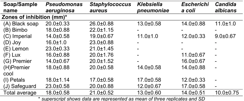
Table 2. Summary of antimicrobial susceptibility profiles of the soaps used against the test microbes Soap/Sample name Pseudomonas aeruginosa Staphylococcus aureus Klebsiella pneumoniae Escherichi a coli Candida albicans Zones of inhibition (mm)*
(A) Black soap 20.0±0.33 26.0±0.88 13.0±0.58 14.0±0.88 11.0±1.0
(B) Bimbo 18.0±0.88 22.0±1.15 - - -
(C) Imperial 14.0±0.58 19.0±0.67 11.0±1.0 12.0±0.33 9.0±0.67
(D) Joy 16.0±1.0 23.0±0.88 - - -
(E) Lemon 23.0±0.33 21.0±1.45 - - -
(F) Lux 16.0±0.88 20.0±1.76 - 11.0±0.67 -
(G) Premier 14.0±0.67 20.0±1.52 - 16.0±0.67 -
(H)Premier cool
18.0±0.88 20.0±0.58 14.0±0.58 14.0±0.88 -
(I) Petals 18.0±1.14 17.0±0.58 17.0±0.58 12.0±0.33 -
(J) Safeguard 23.0±0.58 20.0±0.88 12.0±0.67 17.0±0.58 -
Total average 18.0±0.58 21.0±0.52 13.0±0.60 14.0±0.51 10.0±0.75
* superscript shows data are represented as mean of three replicates and SD
bacteria showing resistance to as much as five soap samples. C. albicans was barely susceptible to the soaps showing very small zones of inhibition against Sample X1 and soap A3 only. E. coli was resistant to a few soaps namely Sample L2, B1, and T2.
The medicated soaps used (A1, A2, A3) were all effective against the test microorganisms with the
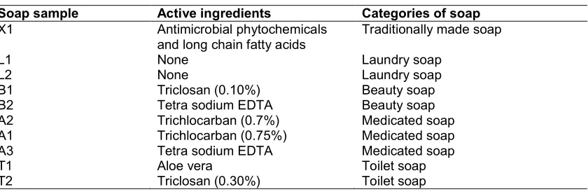
Table 3. Assayed soaps and their active ingredients as per label disclosure
Soap sample Active ingredients Categories of soap
X1 Antimicrobial phytochemicals
and long chain fatty acids
Traditionally made soap
L1 None Laundry soap
L2 None Laundry soap
B1 Triclosan (0.10%) Beauty soap
B2 Tetra sodium EDTA Beauty soap
A2 Trichlocarban (0.7%) Medicated soap
A1 Trichlocarban (0.75%) Medicated soap
A3 Tetra sodium EDTA Medicated soap
T1 Aloe vera Toilet soap
T2 Triclosan (0.30%) Toilet soap
positive correlation (p<0.05) between soaps with similar ingredients or similar antimicrobial activities. (Pearson’s correlation coefficient was used as the statistical measure). This study shows a positive correlation between soap X1 and A3 (r=0.98; p=0.02); the two soaps being the only active antifungal agents used. The result of the study also shows an evaluation of the correlation between the beauty soaps; soap B1 and B2 with regard to the zone of inhibition of the test microorganisms affected. There is a statistically significant positive correlation between the two (r=0.85; p=0.03). Laundry soaps, L1 and L2 containing no active antimicrobial agent also showed a significant positive correlation (r=0.95, p=0.004) with regard to the zone of inhibition of the test microorganisms affected. It should be noted that laundry soaps show a greater correlation at an even lower significance level (p<0.01). This indicates that fewer added ingredients to soap reduces the degree of variability of antimicrobial properties of the soaps.
When the efficacy of Sample A1 and A2 were compared, it was estimated that their antibacterial activity was almost the same against S. aureus and K. pneumoniae. This is evidenced as a significant positive relationship between both soaps. (r=0.85; p=0.001). The Figure below (Figs. 1-5) shows a graphical representation of the relationship between the different soaps and their inhibitory activities against different microorganisms. From Fig. 1, it is clearly seen that S. aureus had the highest zone of inhibition with sample X1 followed by inhibition with sample B1 with the least inhibitory soap being Sample T1. This is in contrast to the zone of inhibition seen around P. aeruginosa as shown in Fig. 2. The highest zone of inhibition is seen in Sample A1 and T2, both of which contain antimicrobial agents followed by black soap with
the least seen in Sample L1 and A3. Fig. 3 reveals the susceptibility profiles of different soaps against K. pneumonia with the least active soap against it being Sample A3. Fig. 4 shows a similar antimicrobial susceptibility profile to that of P. aeruginosa with the highest zone of inhibition seen with Sample A1. The least inhibitory soaps were Sample L2, B1, and T2 with each only active against 2 tested pathogens. However, Sample B1 being a beauty soap, had the least zone of inhibition and as a result is the mildest soap to be used against skin diseases or for skin cleaning. Plate 1 shows the effects of Sample X1 on the microbial cell structure and Plate 2 reveals the zones of inhibition of the different soap samples on S. aureus. It was discovered that for S. aureus, the cells appeared visibly thinner after exposure and their arrangement was more dispersed. For E. coli, the cells which were previously seen as short rods were later seen to have shrunk, appearing even smaller and in cluster. This could be a response to the inhibitory effect of the soap as seen in plate 1. For K. pneumoniae, the cells majorly appeared dispersed, thinner and were hardly visible under the microscope while a change in the arrangement of cells was observed in P. aeruginosa.
4. DISCUSSION
soaps have demonstrated satisfactory effect, particularly the antibacterial activity, hence buttressing the information written on the soap labels that they possess antibacterial activity.
The best in antimicrobial activity of all the soaps used is Sample X1 (black soap). This could be attributed to the presence of some antimicrobial phytochemicals such as alkaloids, tannins, flavonoids, cyanogenic glycosides and saponins, which may in turn account for the antifungal
efficacy against C. albicans. [12]. This results as obtained from this investigation are in agreement with the report of [13] on the efficacy of Cassia senna formulated black soap against some pathogenic microorganisms on human skin. S. aureus and C. albicans have been incriminated in causing skin infections including boils, thrush, impetigo etc. The susceptibilities of these organisms to black soap indicate the therapeutic potentials of black soap in treatment of such diseases.
Fig. 1. Effect of different soap samples on microbial inhibition of Staphylococcus aureus
Fig. 3. Effect of different soap samples on microbial inhibition of Klebsiella pneumoniae
Fig. 4. Effect of different soap samples on microbial inhibition of Escherichia coli
Also, one of the major components of black soap is palm kernel oil which is found to cause distortion in the peptidoglycan layer of Gram positive organisms (S. aureus) due to its long chain of fatty acids. This explains why S. aureus had the highest zone of inhibition in comparison to the rest of the microorganisms used. The major fatty acids in palm kernel oil used for the production of black soap are lauric acid, myristic acid, and oleic acid. The effect of long chain fatty
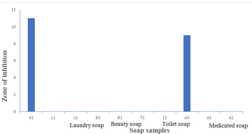
Fig. 5. Effect of different soap samples on microbial inhibition of Candida albicans
and shea butter (used in the preparation of the soap) may have significant effect on the properties and quality of the soap used. The resistance of E. coli to antimicrobial agents is usually due to chromosomal mutation which lowers the permeability of the bacteria to the agents or acquisition of resistance (R) plasmids and tranponsoms.
Beauty soaps (B1 and B2) contain some natural and plant extracted ingredients in their composition which have the ability to inhibit the growth of some bacteria. The inhibition of the growth pattern of the isolates indicates the varying abilities of the organism to resist the antimicrobial effect of the soaps. However, these variations could be due to differences in the nature and structures of the bacterial cell wall since it is the ultimate target of any antimicrobial agent or disinfectant which agrees with the research by Obi [15]. The active ingredient in the soap is what distinguishes each soap from another. The medicated soaps in this study were found to contain trichlocarban and triclosan as active antimicrobial agents (Table 3). A statistically significant positive correlation was observed between medicated soaps that had trichlocarban as the main ingredient with Pearson’s coefficient ranging from 0.85 to 0.98. However, some other medicated soaps with different active agent did not show good correlation. As a result, it can be concluded that the active agents alone may not be sufficient to judge the antimicrobial efficacy of a soap, as
other factors such as concentration of active ingredient and other additives might influence the antimicrobial properties. This is corroborated by the work of Geraldo et al. [16]
which shows that combination of
benzenethonium chloride, polyhexamethylene biguanide, and farnesol is superior to the use of triclosan alone. Changes seen in the microbial cell structure under the microscope revealed that the activity of the soaps majorly affects the disruption of cell wall and cell membrane. In general, traditional black soap was most effective followed by medicated soaps which are more effective than other categories of soap used.
5. CONCLUSION
The antimicrobial activity exhibited by the black soap against the test organisms (S. aureus and C. albicans) that are associated with various skin infections, have provided scientific justification for the ethno medical uses of the soap by Hausa, Yoruba, and Nupe tribes in Nigeria. This study has justified the use of medicated and black soaps for the control of skin related infections.
From the results of this study, it is recommended that prolonged usage of medicated soaps should be discouraged. It is also recommended that further studies should be conducted on the traditional black soap and its efficacy and other studies to determine the bactericidal or bacteriostatic properties of soaps.
ACKNOWLEDGEMENTS
The authors appreciate the Department of Biological sciences, Ondo State University of Science and Technology, Okitipupa for their efforts in providing a conducive laboratory suitable for the completion of this work.
COMPETING INTERESTS
Authors have declared that no competing interests exist. The products used for this research are commonly and predominantly use products in our area of research and country. There is absolutely no conflict of interest between the authors and producers of the products because we do not intend to use these products as an avenue for any litigation but for the advancement of knowledge. Also, the research was not funded by the producing company rather it was funded by personal efforts of the authors.
REFERENCES
1. Grice EA, Kong HH, Renaud G, Young AC, Bouffard GG, Blakesley RW, Wolfsberg TG, Turner ML, Segre JA. A
diversity profile of the human skin microbiota. Genome Resource. 2008;18:
1043-50.
2. Lambers H, Piessens S, Bloem A. Natural skin surface pH is on average below which is beneficial for its resident flora. International Journal of Cosmetic Science. 2009;28:359-370.
3. Fluit AC, Schmits FJ, Verhoef J. Frequency and isolation of pathogens from
blood stream nosocomial pneumonia, skin and soft tissue. European Journal of Microbiology Infection. 2001;20:188-191.
4. Higaki S, Kitagawa T, Kagoura M, Morohashi M, Yamagishi T. Predominant Staphylococcus aureus isolated from various skin disease. Journal of International Medical Research. 2000;28: 87-119
5. Al-saimary I, Bakr S, Al-Hamdi K. Staphylococcus aureus as a causative
agent of
atopic dermatitis/eczema syndrome (ADES) and its therapeutic implications.
Advances in
Bioresearch. 2013;4:116-120.
6. Cheesbrough M. District laboratory practice in tropical countries, part 2. Cambridge University Press, Cambridge. 2005;159-162.
7. Raygada JL, Levine DP. Managing CAMRSA infections: Current and emerging options. Infections in Medicine. 2009;26(2):67.78.
8. Bauer AW, Kirby WM, Sherris JC, Jurck M. Antibiotic susceptibility testing by standard single disc method. American Journal Clinical Pathology. 1966;451:493-496.
9. Selvamohan V, Sandhya T. Studies on the bactericidal activity of different soaps against bacterial strains. Journal of Microbiology and Biotechnology Research. 2012;5:646- 650.
10. Rama Bhat P, Prajna PS, Vinita Preethi Menezez, Pavithra Shettuy. Antimicrobial activities of soaps and detergent. Advances in Biotechnology Research; 2011.
11. Bhargava HN, Leonard PA. Triclosan, application and safety. American Journal of Infection Control. 1996;24:209-218.
12. Oluranti OO, Olawuyi OJ, Jonathan SG. Therapeutic properties of some Nigerian higher fungi. Nature and Science. 2012;10(10):135-143.
14. Arora I. Textbook of microbiology. Satish Kumar publishers, India. 2004;56-68.
15. Obi CN. Antibacterial activities of some medicated soaps on selected human pathogens. Annual Journal of Microbiology Res. 2014;2(6):178-181.
16. Geraldo IM, Gilman A, Shintre MS, Modak SM. Rapid antibacterial activity of 2 novel hand soaps: Evaluation of the risk of development of bacterial resistance to the antibacterial agents. Infect
Control Hospital Epidemiology.
2008;29:736-41.
_________________________________________________________________________________ © 2019 Teniola et al.; This is an Open Access article distributed under the terms of the Creative Commons Attribution License
(http://creativecommons.org/licenses/by/4.0), which permits unrestricted use, distribution, and reproduction in any medium,
provided the original work is properly cited.
Peer-review history: