ISSN 0015–5659 journals.viamedica.pl
Address for correspondence: Dr. A. Kobus, Department of Dentistry Propaedeutics, Medical University of Bialystok, ul. Szpitalna 30, 15–295 Białystok, Poland, tel: +48 660464630, e-mail: [email protected]
Dentin dysplasia type I
A. Kobus
1, M. Świsłocka
2, A. Kierklo
1, J. Borys
3, E. Domel
3, J. Różycki
4, S. Ławicki
5, J. Bagińska
11Department of Dentistry Propaedeutics, Medical University of Bialystok, Poland
2Resident in the Course of Specialisation in Conservative Dentistry with Endodontics, Karedent Sp. J., Bialystok, Poland 3Maxillofacial Surgery Clinic, Medical University of Bialystok, Poland
4Department of Radiology, Medical University of Bialystok, Poland
5Department of Population Medicine and Civilisation Disease Prevention, Medical University of Bialystok, Poland
[Received: 13 November 2018; Accepted: 10 January 2019]
This paper describes a rare case of genetically determined dentin dysplasia type I in 26-year-old male patient. The paper highlights anatomical and radiological aspects of dental abnormalities and emphasizes the significance of the education of both general practitioners and paediatricians as regards referring patients with diagnosed dentin dysplasia for a multi-specialty therapy. (Folia Morphol 2019; 78, 3: 637–642)
Key words: dental abnormalities, dentin dysplasia, pulp obliteration, computed tomography
INTRODUCTION
Dentin dysplasia is a congenital disease entity
which may occur separately or as one of the
symp-toms of an underlying disease [7]. It is one of dental hard tissue disorders of genetic origin apart from such entities as amelogenesis imperfecta and dentinogen
-esis imperfecta [7, 11, 22].
The incidence of dysplasia is estimated according to Witkop at 1:100000 cases [2, 7]. It is a geneti -cally determined pathology which occurs with the
same frequency in both genders [13, 14, 23]. It is inherited according to the autosomal dominant in
-heritance pattern. The mutation probably occurs on
chromosome 4q 13-21 [6]. The disorder formation
mechanism has not been fully explained. Probably as
a result of the mutation, abnormal Hertwig’s epithelial root sheath cells migrate to the dental papilla which
induces an abnormal differentiation of odontoblasts. Pathological dentin is formed as a consequence [15].
However, more recent research suggests that during the incorporation of Hertwig’s epithelial root sheath
into dental papilla the mutations of the dentin matrix
acidic phosphoprotein gene and the dentin sialophos
-phoprotein gene may occur [15, 17].
Dentin dysplasia is divided into dysplasia type I, also referred to as root dysplasia, and dysplasia type II,
i.e. crown dentin dysplasia. Lesions with symptoms
of both type I and type II are referred to as type III,
i.e. focal or fibrous dysplasia [24]. Type III occurs
least frequently and affects the permanent dentition only [3]. The dysplasia types are diagnosed and dif
-ferentiated on the basis of the medical history and
physical examination as well as a very characteristic X-ray image.
In the case of crown dentin dysplasia (type II according to Shields classification) the symptoms
occur in both permanent and deciduous dentition.
In primary teeth, normal crown shapes with the
col-our changed to amber or blue may be observed [7,
20]. The crowns are subject to a quick abrasion, and
the roots have normal length and shape [1, 7, 20].
However, permanent teeth may have an only slightly changed colour without an increased susceptibility
prone to caries. The teeth are not prematurely lost because their mobility is not increased. There are also no changes in marginal periodontium. In an
X-ray image, permanent teeth have the appearance of so-called shell teeth, and pulp chambers show an
abnormal shape depending on the degree of pulp cavity obliteration [14, 23]. The pulp chamber oblite
-ration begins to develop just after the tooth eruption. Incisors, canine and premolar teeth have chambers
in the shape of a tube bulging towards the roots, and in the molars they take the shape of a flame
or a thistle. Moreover, the occurrence of multiple
intra-pulpal calcifications (denticles, pulp stones)
may be observed [11, 12]. The occurrence of bone structure density reduction around the root apexes
is characteristic for this disease entity; however, such
changes are not related to caries. The pathomecha
-nism of formation of periapical lesions has not yet been fully explained. According to one of the hypoth -eses, probably due to a weaker and abnormal bone development, the inflammatory condition penetrates through the weaker mineralisation line in the region
of tooth neck or through marginal periodontium. It
is also likely that the Hertwig’s epithelial root sheath, by not reaching the normal root length, modifies its
activity and forms a cyst as a result. A second theory says that the “autointoxication” of the pulp occurs during its obliteration.
In the case of root dentin dysplasia (type I accord-ing to Shields classification) the pathological changes
may be observed in both deciduous and permanent
dentition. Initially, pathologically changed teeth were
referred to as rootless teeth. In the clinical examina -tion, no abnormalities in the tooth crown structure
are observed. The shape is normal, and the enamel is
shiny, hard and does not show any colour changes. In
the clinical examination, also a transverse line in the
region of dental neck separating the crown portion from the root portion can be seen. Due to a weaker mineralisation at this place, a tooth crown fracture
may occur [4, 16]. In the course of further assessment,
increased tooth mobility is found, whereas its degree
depends on the disease progression. It is associated
with shortened or even missing roots [14, 23], which
is a reason for early tooth loss in both deciduous and permanent dentition. Also teeth in dysplasia type
I are frequently extracted in connection with pain complaints which are not caused by complicated
dental caries, but by granulomas or cysts similar to
lesions occurring in crown dysplasia [10]. The
en-dodontic treatment is practically impossible due to obliterated pulp chambers and underdeveloped roots.
The X-ray image of teeth with root dentin dysplasia
is very characteristic. The roots are shortened, have a pointed shape and changed proportions in relation to the pulp chamber. Depending on the progression of underdevelopment of the root and the pulp cham
-bers and canals, Carroll et al. [15] divided the disease
into subtypes Ia, Ib, Ic and Id, where the subtype Ia
means the mildest and Id the severest form of the disorder. In the case of deciduous teeth, the pulp chamber is subject to a complete obliteration. On the other hand, permanent teeth have a reduced dentin contrast, and the pulp chambers are completely or partially obliterated. In the molars, the pulp chamber frequently takes the form of a crescent. In contrast
to crown dysplasia, the obliteration develops yet before the eruption of teeth. In dysplasia type I we may also find denticles which make the treatment even more difficult.
CASE REPORT
A 26-year-old male patient came to a dental office
due to sensitivity to hot and cold and the episodes of
idiopathic pain in right lower second molar (tooth 47).
Based on reported symptoms and decreased response to pulp vitality tests, the decision of endodontic treat -ment was made. However, the trepanation of pulp chamber failed. An intraoral X-ray was taken and
a completely obliterated pulp chamber and consider -ably shortened roots without a visible lumen of root canals were found. Then the patient was referred for an X-ray scan (a panoramic radiograph) and for the
specialist endodontic treatment.
At the first visit, following detailed medical and
previous treatment history taking, no systemic diseas -es were found. Patient previously suffered from pain complaints which were initially located mainly within
the gums, and then the responses of the teeth to hot
and cold appeared. In such cases he was treated only by the oral antibiotic therapy because he was told to have “difficult canals”. He also reported that similar symptoms have occurred in the patient’s siblings as well as in his father’s twin brother and his children.
No abnormalities were found in the extraoral ex-amination. The face was symmetrical, the ostia of
the trigeminal nerve painless, the submandibular and submental lymph nodes not enlarged. The intraoral
abnor-malities within the mandibular alveolar region were found. In the maxilla, however, the alveolar process distension (deformation) located between the left maxillary first premolar and left maxillary first molar and painless on pressure was observed (Fig. 1). The midline of maxillary teeth was shifted to the right. The teeth were abnormally positioned in the arch and the mandibular second premolar and first molar teeth were missing. No pathological tooth mobility was found. Clinically, the teeth had a milk-yellow colour, normal crown structure and proportion (Fig. 2). The subject had fillings in numerous teeth and a carious defect in the first right mandibular premolar as well as a temporary filling in the second right mandibular molar. The vitality test showed a lack of response to cold of the left upper lateral teeth and few right lower lateral teeth (teeth no. 24, 25, 26, 44 and 47).
Based on an analysis of the panoramic radiograph, an abnormal structure of pulp cavities and roots as
well as the presence of translucent lesion located within the bones at the roots of multiple teeth were
noticed. In the maxilla, in the region of left premolars
and molars, a shadowing with the diameter of about 3 cm, situated in the left maxillary sinus was found (Fig. 3). The decision to perform a cone-beam comput-ed tomography (CBCT) scan was taken. The scan was performed using a NewTom 5G device in a resolution with the imaging field of 12 × 8 cm. An analysis of the scans showed that the entire dentition except for the canines was characterised by a shortening of the root length. The changes were mostly visible in the root structure of molar teeth (except for third molars) which were characterised by a pointed shape. In addition, they practically did not show any division
into separate roots or such division started in 1/3 of
the periapical region (Fig. 4). An almost complete obliteration of pulp cavities of lateral teeth was also found. The canal lumen was visible only within
max-illary incisors and canines; a small outline of the pulp cavity could also be seen in the mandibular premolars
(35 and 44). In the lumen of pulp cavities of ante-rior teeth, the presence of numerous spherical well Figure 3. Panoramic radiograph showing the characteristic appearance of teeth with thick and short roots and pulp nodules in dysplasia type I.
Figure 2. Clinical dental status.
mineralised formations tightly filling the space, sug-gesting the presence of denticles, was found (Fig. 5).
In the projection of root apexes of the right upper lateral teeth and in the mandible around the roots of the teeth 37, 35 and 44 the bone structure density
reductions with a considerable degree of progression were present. At the level of the left maxillary lateral teeth (24, 25 and 26) a shadowing with the diameter
of 3 cm indenting into the lumen of the left sinus
was found.
Based on clinical and radiological examination, the
patient was diagnosed with the dentin dysplasia type I with a considerable degree of progression.
The interdisciplinary treatment was started with
oral hygiene instructions. Oral hygiene procedures
as well as a closed curettage within the right lower second molar (47) were performed in order to reduce the inflammation around the root apex. The endodon-tic treatment was not possible due to the degree of
pulp cavity obliteration. After the pain subsided, the
defect in the tooth 47 was filled with the composite material. Furthermore, the patient was referred to the
Maxillofacial and Plastic Surgery Clinic to have the cyst
within the left maxillary sinus enucleated. Based on
the maxillary computed tomography scan the range
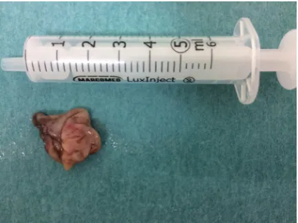
of the lesion was determined and its enucleation to-gether with the extraction of the teeth 24, 25 and 26 was scheduled. The cystectomy (Fig. 6) and the root resection of the teeth 24 and 25 with the retrograde filling of root canals of these teeth were performed. The tooth 26 was not affected by the lesion. A
patho-morphological examination of the specimen revealed an odontogenic epithelial root cyst. Due to the pa -tient’s young age, during the procedure the decision to leave the teeth 24 and 25 in the oral cavity was taken. Half year observation showed good oral tissues
healing. At a later stage, an orthodontic consultation and a prosthetic reconstruction of existing dental de -fects were scheduled. The patient still remains under
the care of a specialist team.
DISCUSSION
The dentin dysplasia still remains a disease entity
with an unexplained aetiology. It may also cause difficulties at both diagnostics and treatment stages. Due to the lack of changes in crown shape and col-our, the root dentin dysplasia (type I) is still in most
cases discovered by accident. Patients most frequently decide to see a dentist because of pain complaints,
and the difficulties in locating the canal ostia force dentists to consult specialists who diagnose a dyspla-sia based on a characteristic X-ray image (panoramic Figure 4. Sagittal section, left side. Visible characteristic structure
of teeth with dysplasia type I and root cyst (an arrow) indenting into the lumen of the left sinus (visible above the first left upper molar tooth).
Figure 5. Sagittal sections through the right lower canine tooth. Visible denticles within the pulp cavity (arrows).
radiograph). Previously, such extraoral images were
the main source of information about the status and
the anatomy of patient’s teeth; however, at present,
in each case of suspected complex structure of pulp cavities it is recommended to perform a CBCT scan [8]. The CBCT scan makes it possible to precisely deter
-mine the degree of dental canal obliteration and
the place where denticles occur, which increases the
chance of proper treatment. In spite of clear indica
-tions for a CBCT scan, no case study of root dentin
dysplasia with the use of such imaging was found in
the available literature.
The significance of multi-specialty therapy to which each patient with diagnosed dentin dysplasia
should be subjected already at the stage of deciduous dentition is to be emphasised. Unfortunately, due to
both parents’ and dentists’ insufficient knowledge and awareness of hereditary origin of this disease,
a premature loss of teeth rarely induces them to perform an extended diagnostics in family members.
This is why the education of both general practitioners and paediatricians who frequently are primary care physicians is so significant.
The aim of the therapy is to make sure that the teeth remain in the oral cavity for as long as possi
-ble. It comprises complex restorative, orthodontic,
periodontal, surgical as well as prosthetic treatment.
It is necessary to put emphasis on the prevention
which consists not only in removing dental plaque at a dental office with the use of fluoride preparations,
but also in providing appropriate patient education. Professional oral hygiene instructions including also
proper dietary habits and frequent follow-up visits allow reducing the risk of occurrence of dental caries and periodontal diseases which may further weaken
the periodontal apparatus. In many cases, like in the case described above, the endodontic treatment is
not possible, which is why it is so important to
pre-vent pulp complications [5]. It is frequently possible
to suppress the inflammation around root apexes by curettage procedures with the application of
medi-cines into gingival pockets [9, 19, 21].
The orthodontic treatment in dentin dysplasia is quite controversial because of the action of excessive
forces which may lead to a faster bone resorption,
and consequently to an increased tooth mobility.
However, it is important to eliminate excessive
oc-clusal overloads or ococ-clusal disease symptoms and to restore normal occlusion. The surgical treatment consists not only in dental extractions, but also, due
to resection or hemisection procedures, enables the diseased teeth to survive longer despite the lack of possibility to carry out proper endodontic treatment. The described case indicates the possibility of tooth retention even in the event of occurrence of an ex
-tensive root cyst. Unfortunately, in spite of the use of different treatment methods, a tooth loss frequently occurs in root dentin dysplasia patients. Then it is necessary to provide a prosthetic rehabilitation in
order to restore the chewing function and improve the aesthetics of the patient’s dentition.
CONCLUSIONS
In spite of an increasing number of reports of
diagnosed new dentin dysplasia cases, the treatment of this disease entity is difficult and unpredictable.
Therefore, it is important to keep records including not only case studies or photographs, but also
a long-term patient observation, in order to choose the best possible therapy which should be individual
for each patient.
REfERENCES
1. Cabay RJ. An overview of molecular and genetic alterations in selected benign odontogenic disorders. Arch Pathol Lab Med. 2014; 138(6): 754–758, doi: 10.5858/arpa.2013-0057-SA, indexed in Pubmed: 24878015.
2. Cherkaoui Jaouad I, El Alloussi M, Laarabi FZ, et al.
Inhab-itual autosomal recessive form of dentin dysplasia type I in
a large consanguineous Moroccan family. Eur J Med Genet. 2013; 56(8): 442–444, doi: 10.1016/j.ejmg.2013.05.003,
indexed in Pubmed: 23712319.
3. Ciola B, Bahn SL, Goviea GL. Radiographic manifestations
of an unusual combination Types I and Type II dentin
dysplasia. Oral Surg Oral Med Oral Pathol. 1978; 45(2): 317–322, indexed in Pubmed: 272613.
4. Comer TL, Gound TG. Hereditary pattern for dentinal
dysplasia type Id: a case report. Oral Surg Oral Med Oral
Pathol Oral Radiol Endod. 2002; 94(1): 51–53, indexed in Pubmed: 12193893.
5. Council O. Guideline on dental management of heritable
dental developmental anomalies. Pediatr Dent. 2013;
35(5): 179–184.
6. Dean JA, Hartsfield JK, Wright JT, et al. Dentin dysplasia, type II linkage to chromosome 4q. J Craniofac Genet Dev Biol. 1997; 17(4): 172–177, indexed in Pubmed: 9493074. 7. de La Dure-Molla M, Philippe Fournier B, Berdal A.
Iso-lated dentinogenesis imperfecta and dentin dysplasia:
revision of the classification. Eur J Hum Genet. 2015; 23(4): 445–451, doi: 10.1038/ejhg.2014.159, indexed in
Pubmed: 25118030.
8. Fayad M, Nair M, Levin M, et al. AAE and AAOMR Joint
Position Statement. Use of Cone Beam Computed To
-mography in Endodontics 2015 Update. Oral Surg Oral
9. Fulari SG, Tambake DP. Rootless teeth: Dentin dyspla-sia type I. Contemp Clin Dent. 2013; 4(4): 520–522,
doi: 10.4103/0976-237X.123063, indexed in Pubmed:
24403801.
10. Da Rós Gonçalves L, Oliveira CA, Holanda R, et al. Periodon-tal status of patients with dentin dysplasia type I: report of three cases within a family. J Periodontol. 2008; 79(7): 1304–1311, doi: 10.1902/jop.2008.070426, indexed in
Pubmed: 18597615.
11. Hart PS, Hart TC. Disorders of human dentin. Cells Tissues
Organs. 2007; 186(1): 70–77, doi: 10.1159/000102682,
indexed in Pubmed: 17627120.
12. Kim JW, Simmer JP. Hereditary dentin defects. J Dent Res. 2007;
86(5): 392–399, doi: 10.1177/154405910708600502,
indexed in Pubmed: 17452557.
13. Li F, Liu Y, Liu H, et al. Phenotype and genotype analyses in seven families with dentinogenesis imperfecta or dentin dysplasia. Oral Dis. 2017; 23(3): 360–366, doi: 10.1111/ odi.12621, indexed in Pubmed: 27973701.
14. Neville BW, Damm DD, Allen CM. Abnormalities of Teeth. In: Oral & Maxillofacial Pathology. 3rd Ed. Philadelphia, Pa: WB Saunders Company 2009: 99–112.
15. O Carroll MK, Duncan WK, Perkins TM. Dentin dysplasia:
review of the literature and a proposed subclassification based on radiographic findings. Oral Surg Oral Med Oral Pathol. 1991; 72(1): 119–125, indexed in Pubmed: 1891231. 16. Perzanowska E, Chałas R. Dentin dysplasia type I- a
diag-nostic challenge both for the dentist and paleodontologist.
Bull Int Assoc Paleodont. 2016; 10(2): 46–51.
17. Qari H, Kessler H, Narayana N, et al. Symmetric multi -quadrant isolated dentin dysplasia (SMIDD), a unique
presentation mimicking dentin dysplasia type 1b. Oral
Surg Oral Med Oral Pathol Oral Radiol. 2017; 123(5): e164–e169, doi: 10.1016/j.oooo.2016.11.024, indexed
in Pubmed: 28215628.
18. Ranta H, Lukinmaa PL, Knif J. Dentin dysplasia type II:
absence of type III collagen in dentin. J Oral Pathol Med.
1990; 19(4): 160–165, indexed in Pubmed: 2195160. 19. Ravanshad S, Khayat A. Endodontic therapy on a dentition
exhibiting multiple periapical radiolucencies associated
with dentinal dysplasia Type 1. Aust Endod J. 2006; 32(1): 40–42, doi: 10.1111/j.1747-4477.2006.00008.x, indexed
in Pubmed: 16603045.
20. Shields ED, Bixler D, el-Kafrawy AM. A proposed classifica-tion for heritable human dentine defects with a descripclassifica-tion of a new entity. Arch Oral Biol. 1973; 18(4): 543–553, indexed in Pubmed: 4516067.
21. Steidler NE, Radden BG, Reade PC. Dentinal dysplasia: a clinicopathological study of eight cases and review of the literature. Br J Oral Maxillofac Surg. 1984; 22(4): 274–286, indexed in Pubmed: 6235842.
22. Toomarian L, Mashhadiabbas F, Mirkarimi M, et al. Dentin dysplasia type I: a case report and review of the literature. J Med Case Rep. 2010; 4: 1–6, doi: 10.1186/1752-1947-4-1, indexed in Pubmed: 20205797.
23. White SC, Pharoah MJ. Dental anomalies. In: Oral Radiolo -gy: Principles and Interpretation. 5th Ed. St. Louis, Mosby/
Elsevier 2019: 307–311.
24. Witkop CJ. Amelogenesis imperfecta, dentinogenesis imperfecta and dentin dysplasia revisited: problems in