Slow Blood Sampling From an Umbilical Artery Catheter Prevents a
Decrease in Cerebral Oxygenation in the Preterm Newborn
Gabriele Schulz, MD; Esther Keller; Daniel Haensse, MSc; Romaine Arlettaz, MD; Hans Ulrich Bucher, MD; and Jean-Claude Fauche`re, MD
ABSTRACT. Objective. Blood sampling from an um-bilical artery catheter (UAC) placed in a high position (thoracal 6 –9) has the potential to produce clinically sig-nificant changes in cerebral blood flow and, thereby, in cerebral oxygenation. This may contribute to cerebral impairment in preterm newborn infants. Therefore, we set up a study to determine the effects of different sam-pling speeds through a UAC on cerebral oxygenation in preterm infants.
Methods. Thirty pairs of measurements were con-ducted on 20 preterm infants (median gestational age: 30.14 weeks; median birth weight: 1170 g). For each in-fant, 2 blood samplings (both 2.3 mL, including flush volume) through the UAC in high position were taken at 2 different speeds (20 and 40 seconds) in alternating sequence. Cerebral oxygenation was measured noninva-sively by near-infrared spectroscopy. Concentration changes in cerebral oxygenated hemoglobin (O2Hb) and
deoxygenated hemoglobin (HHb), along with the tissue oxygenation index (TOI; O2Hb/[O2Hb ⴙ HHb] ⴛ 100),
were recorded while blood was withdrawn and subse-quently reinfused.
Results. A significant decrease in O2Hb and TOI
oc-curred during blood sampling within 20 seconds (median
⌬O2Hb: ⴚ1.5 mol/L; range: ⴚ4.1–2.3; median ⌬TOI:
ⴚ0.6%; range:ⴚ6.3–2.3), whereas HHb increased (median
⌬HHb: 0.4 mol/L, range: ⴚ1.1–3.9). No significant change was found in O2Hb, HHb, and TOI when
sam-pling time was extended to 40 seconds.
Conclusion. Our results show that blood withdrawal over 20 seconds from a UAC in high position signifi-cantly decreases cerebral O2Hb and TOI in preterm
in-fants. Prolonging sampling time to 40 seconds can pre-vent this phenomenon.Pediatrics2003;111:e73–e76. URL: http://www.pediatrics.org/cgi/content/full/111/1/e73;
near-infrared spectroscopy, cerebral tissue oxygenation, neonate, tissue oxygenation index.
ABBREVIATIONS. UAC, umbilical artery catheter; NIRS, near-infrared spectroscopy; TOI, tissue oxygenation index; O2Hb, oxy-genated hemoglobin concentration; HHb, deoxyoxy-genated hemoglo-bin concentration.
N
eonatal mortality has markedly decreased in the past decades because of better prenatal fetal care and, postnatally, because of the ever-expanding diagnostic and therapeutic means available to neonatologists. The limit of viability, which was approximately 28 weeks’ gestation 10 years ago, has been constantly lowered to below 26 weeks, with a current 52% survival rate for liveborn infants who are born at 25 weeks.1Unfortunately, the decrease in mortality has not been accompanied by a decrease in mid- and long-term morbidity affecting especially the neurodevelopmental outcome. If pre-natal damage to the central nervous system can fre-quently not be prevented, then everything needs to be done to decrease further all possible risks of post-natal cerebral lesions in infants who undergo inten-sive care, the most vulnerable among these being preterm infants.The vast majority of very preterm and severely ill newborn infants are monitored using an umbilical artery catheter (UAC). This allows for continuous blood pressure monitoring, convenient arterial blood gas measurements, and painless blood sampling, with only little disturbance to the infant. However, UACs have been associated with complications such as local vascular (blanching or cyanosis of feet or toes)2– 4 or more extensive ischemic compromise,4,5 aortic thrombi,2,5–7necrotizing enterocolitis,5arterial hypertension,2 hematuria,5 and hyperglycemia.8 –13 As high-placed UACs have been shown to be safer with regard to clinical vascular complications, many institutions prefer the high-positioned (thoracal 6 –9) UAC. A recent Cochrane Review concluded that there seems to be no evidence to support the use of low-placed UACs, and, therefore, high catheters should be used exclusively.14 In our institution, UACs are pulled back to a low position only in the presence of persistent hyperglycemia with a high-placed UAC.
Recently, it has been shown that sampling blood from a high-positioned UAC may potentially de-crease cerebral blood volume and cerebral oxygen-ation in very low birth weight infants.15 It is un-known whether this is caused by the volume of withdrawn blood or by the blood sampling rate. To determine whether different speeds of blood sam-pling via a UAC might affect or influence changes in cerebral oxygenation in sick preterm newborns who undergo intensive care, we set up this study to de-termine whether the sampling speed per se could From the Clinic of Neonatology, University Hospital, Zurich, Switzerland.
Received for publication Jan 29, 2002; accepted Sep 10, 2002.
Reprint requests to (J-C.F.) Clinic of Neonatology, University Hospital, Frauenklinikstrasse 10, CH-8091 Zurich, Switzerland. E-mail: jean-claude. [email protected]
influence cerebral tissue oxygenation. We hypothe-sized that by extending the sampling time, it would be possible to avoid significant alterations in cerebral oxygenation. We explored this issue by using near-infrared spectroscopy (NIRS), a noninvasive method to evaluate changes in cerebral tissue oxygenation, while a fixed amount of blood was withdrawn from a high-positioned UAC.
METHODS
After obtaining approval from the local ethical committee and written parental consent, we enrolled 20 neonates who were ad-mitted to the neonatal intensive care unit of the University Hos-pital (Zurich, Switzerland). Only preterm infants (⬍37 completed weeks’ gestation) with a UAC (3.5 Ch, filling volume 0.15 mL; Sherwood Medical, Tullamore, Ireland) in high position were included in this study. The position (thoracal 6 –9) was confirmed by radiograph. To exclude measurement effects as a result of hemodynamic instability or of changes in vasoactive dosing, we excluded infants who were in an unstable hemodynamic situation or needed vasoactive drugs. Hemodynamic stability was defined by a stable arterial blood pressure as continuously measured and displayed on screen, by a normal heart rate without any arrhyth-mia, by normal capillary refilling, and by normal urine produc-tion.
A NIRS optode (Hamamatsu Photonics, Hamamatsu City, Ja-pan), consisting of an emitter and a receiver of near-infrared light with an interoptode distance of 50 mm, was placed on the skin overlaying the temple of the newborn, taking care to avoid the region over the sagittal sinus. Interference from light was pre-vented by shielding the optode. The light emitter optode carried 4 laser diodes operating at 775, 810, 847, and 919 nm, and the detector optode had 3 photodiodes, mounted in parallel with increasing distances, which permitted the calculation of an abso-lute tissue oxygenation index (TOI) value. The underlying techni-cal principles of spatially resolved spectroscopy has been previ-ously described in detail by Matcher and Suzuki.16,17 The NIRS probe was connected to a precalibrated measuring unit (NIRO-300, Hamamatsu Photonics).
NIRS measurements were performed without interruption at a sampling rate of 0.5 Hz during the whole procedure of blood withdrawing and reinfusing, the same being true for transcutane-ous arterial oxygen saturation (Nellcor 395, Nellcor Inc, Hayward, CA). Because a single lumen UAC was used, mean arterial pres-sure was recorded immediately before and after the blood sam-pling and catheter flushing procedure. After steady-state condi-tions were obtained for approximately 10 minutes, blood samples were withdrawn from the UAC at 2 different speeds, with the sampling speed order following an alternating sequence. For the 20-second sampling, 2 mL was aspirated in 15 seconds to fill the catheter with blood, and an additional 0.3 mL was taken in 5 seconds for the laboratory test. The surplus blood volume (2 mL) was then reinfused into the infant before the system was flushed with 2 mL of saline. For the 40-second sample, a similar procedure was undertaken, except that the first 2 mL was withdrawn over 30 seconds, and the blood sample itself (0.3 mL) was withdrawn over another 10 seconds. Both blood samples of the second sampling procedure were then returned to the infant, and the catheter afterward was flushed as usual. Between each sampling proce-dure, at least 10 minutes was allowed to elapse to permit all parameters to return to baseline conditions. Changes in the ther-apeutic and ventilator settings were avoided during the whole measurement, including the baseline and the 2 withdrawal record-ings. Demographic and physiologic data were also recorded.
Data Analysis
All continuously measured data were simultaneously shown on the optical display and recorded onto a computer and subse-quently analyzed off-line. For each analysis, a baseline sequence of 2 minutes was used. The time of blood withdrawal used for calculation was defined as the time between the 2 digital markers set at the beginning and at the end of the sampling procedure. The 10th and 90th percentile values of oxygenated hemoglobin con-centration (O2Hb), deoxygenated hemoglobin concentration (HHb), TOI, arterial oxygen saturation, and mean arterial pressure
and their median values were calculated for the baseline condi-tion. During withdrawal, the 90th percentile of the data set in case of an increase and the 10th percentile of the measurements in case of a decrease and their median values were used for calculation. This method was chosen to obtain more robust data by avoiding the determination of only 1 single incidentally extreme measured value as the lowest or highest point of the whole procedure. The Wilcoxon signed rank test was used to detect significant changes in O2Hb, HHb, or TOI during the rapid or during the slow sampling procedure. For decreasing parameters such as O2Hb and TOI, the 10th percentile values of the baseline and of the maximal decrease were compared. For the observed increase in HHb, the difference between the 90th percentile values of the baseline and of the maximum increase were compared. For the reasons already mentioned, the data beyond the 10th and 90th percentiles were not included in the analysis.P⬍.05 was considered as statistically significant. Calculations were performed using StatView (version 5.01 for Windows, SAS Inc, Cary, NC).
RESULTS
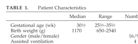
A total of 30 pairs of measurements were per-formed on 20 preterm infants. Five pairs of measure-ments had to be omitted because of artifacts caused by the movements of the infant, leaving 25 pairs of measurements for analysis in 20 preterm infants. Five newborns contributed to a second set of data. All preterm infants included were under stable clin-ical conditions during the measurements. The clini-cal data are shown in Table 1.
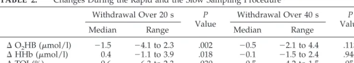
Figure 1 depicts the changes in O2Hb, HHb, and TOI during a 20-second withdrawal and subsequent reinfusion and the changes when withdrawal time was extended to 40 seconds in 1 infant studied. Table 2 summarizes these results by reporting the median and range values of the changes in O2Hb, HHb, and TOI during the rapid and during the slow blood sampling procedure. O2Hb and TOI decreased sig-nificantly when blood was sampled over 20 seconds while, simultaneously, HHb increased. No such sig-nificant differences were found for O2Hb, HHb, or TOI at the 40-second withdrawal time (Table 2). Moreover, there were no significant changes in cere-bral O2Hb, HHb, and TOI during reinfusion of the withdrawn volume and the flushing volume.
DISCUSSION
The present study shows that in preterm infants, withdrawing blood over 20 seconds from a high-positioned UAC significantly decreases cerebral O2Hb and TOI, with a simultaneous increase in HHb. These phenomenon could be prevented when sam-pling time was prolonged to 40 seconds.
These results suggest that rapidly withdrawing blood from a catheter in a high aortic position in-duces a significant fall in oxygenated blood supply to the central nervous system, the potential mechanism acting via a rapid steal of blood that cannot be
com-TABLE 1. Patient Characteristics
Median Range Number
Gestational age (wk) 301⁄7 253⁄7–352⁄7 Birth weight (g) 1170 650–2540
Gender (male/female) 16/9
Assisted ventilation 4
Nasal CPAP 17
Spontaneous breathing 4
CPAP indicates continuous positive airway pressure.
pensated for in time. The observed effect may well be age dependent, as the relationship between the with-drawn blood volume and the circulating blood vol-ume will certainly change with gestational and post-natal age. Although our study did not include term infants, it may well be that the observed decrease in cerebral O2Hb would not occur in term infants with a higher circulating blood volume, as the withdrawn 2.3 mL of blood would represent only a small frac-tion of their total circulating blood volume. As de-picted in Fig 1, the rapid withdrawal over 20 seconds was followed by a fall in O2Hb that lasted for 2.5 minutes. Although this was not consistently ob-served throughout our study, this nevertheless dem-onstrates that even short alterations in cerebral blood supply may be followed by lasting effects in cerebral oxygenation.
Alterations in cerebral hemodynamics during sam-pling blood from UACs have been shown by some investigators, primarily with the intention to study the differences between sampling from umbilical catheters in high or in low positions.14,18None of the these studies, however, investigated the influence that the duration of blood sampling may have on cerebral oxygenation. Lott et al18 described the im-pact of blood sampling procedure over 20 seconds from high UAC on cerebral blood flow velocity, as measured by cerebral ultrasound. Using NIRS, Roll et al15also investigated the effect of blood sampling from high UAC and found a significant decrease in cerebral blood volume and cerebral oxygenation. Their sampling procedure, however, was not per-formed within a set time but rather done “as usual.” As a consequence, their blood sampling durations spread over a wide range, between 26 and 75 sec-onds, with a median of 40 seconds. This may explain why our results are not in keeping with their find-ings, as we did not observe a significant decrease in O2Hb when sampling time was extended to 40
sec-onds. This discrepancy may also be related to several other methodological differences. For example, the volume of blood withdrawn was not standardized in their study, showing a range between 0.4 and 3.0 mL. In our study, not only were the sampling times set at 2 fixed values, namely at 20 and 40 seconds, but also the sampling volumes were kept identical over the whole study, with each infant being its own control. Importantly, we did not observe any significant changes in cerebral O2Hb, HHb, and TOI during reinfusion of the withdrawn volume and of the sub-sequent flushing volume. The reinfusion time was not fixed, but our recordings show that reinfusion was not accompanied by an alteration in oxy- or deoxyhemoglobin. The explanation for this could be that the reinfusion of these small flush volumes en-counters a strong opposite aortic flow that, with perhaps the exception of a very quick bolus injection or a greater flush volume, tends to dampen the he-modynamic effects on cerebral perfusion.
To our knowledge, this is the first study performed in preterm infants to evaluate the impact of various durations of the blood sampling procedure from high UAC on cerebral oxygenation under controlled conditions of withdrawal time and volume. Al-though the clinical importance of a short decrease in cerebral oxygenation, as observed during fast blood sampling, is not quite clear, our results add an addi-tional means of further reducing potential risk fac-tors for cerebral impairment in tiny infants already at high risk of mid- and long-term neurologic disabili-ties. As our constant aim should be to reduce every potential harm that our various interventions may have on these very premature and sick infants, these findings may be of clinical importance considering that these sampling procedures are routinely and repetitively performed on preterm infants who un-dergo neonatal intensive care. Furthermore, prolong-ing samplprolong-ing time is an easy alteration to make to
Fig 1. Original NIRS tracing during blood sampling at 2 different speeds (20 and 40 seconds) in 1 infant studied.
TABLE 2. Changes During the Rapid and the Slow Sampling Procedure Withdrawal Over 20 s P
Value
Withdrawal Over 40 s P
Value
Median Range Median Range
⌬O2HB (mol/l) ⫺1.5 ⫺4.1 to 2.3 .002 ⫺0.5 ⫺2.1 to 4.4 .113
⌬HHb (mol/l) 0.4 ⫺1.1 to 3.9 .018 ⫺0.1 ⫺1.5 to 2.4 .946
routine practices, and because it has the potential to improve further the daily care of these fragile infants, it will certainly be well understood and taken up by the nursing staff.
ACKNOWLEDGMENTS
We thank the parents of the infants studied and the neonatal intensive care unit nurses for making this study possible. We also thank Dr Theo Gasser and Dr Valentin Rousson, Department of Biostatistics, University of Zurich, for statistical counseling. We also acknowledge the editorial help of Helen Keller-Kurzman, BSc (Hon, Deakin). Esther Keller participated in this research project as part of her medical curriculum (Medical Faculty, University of Utrecht, the Netherlands).
REFERENCES
1. Costeloe K, Hennessy E, Gibson AT, Marlow N, Wilkinson AR. The EPICure Study: outcomes to discharge from hospital for infants born at the threshold of viability.Pediatrics.2000;106:659 – 671
2. Mokrohisky ST, Levine RL, Blumhagen JD, Wesenberg RL, Simmons MA. Low positioning of umbilical-artery catheters increases associated complications in newborn infants.N Engl J Med.1978;299:561–564 3. Kempley ST, Gamsu HR. Randomised trial of umbilical arterial catheter
position: Doppler ultrasound findings.Arch Dis Child.1992;67:855– 859 4. Umbilical Artery Catheter Trial Study Group. Relationship of intraven-tricular hemorrhage or death with the level of umbilical artery catheter placement: a multicenter randomized clinical trial.Pediatrics.1992;90: 881– 887
5. Kempley ST, Bennett S, Loftus BG, Cooper D, Gamsu HR. Randomized trial of umbilical arterial catheter position: clinical outcome.Acta Pae-diatr.1993;82:173–176
6. Neal WA, Reynolds JW, Jarvis CW, Williams HJ. Umbilical artery
catheterization: demonstrations of arterial thrombosis by aortography.
Pediatrics.1972;50:6 –13
7. Wesstro¨m G, Finnstorm O, Stenport G. Umbilical artery catheterization in newborns. Thrombosis in relation to catheter type and position.Acta Paediatr Scand.1979;68:575–581
8. Nagel JW, Sims JS, Aplin CE 2nd, Westmark ER. Refractory hypogly-cemia associated with a malpositioned umbilical artery catheter. Pedi-atrics.1979;64:315–317
9. Cowett RM, Tenenbaum DG, Fatoba O, Oh W. The effects of arterial glucose infusion above the celiac axis in the neonatal lamb.Biol Neonate.
1985;47:179 –185
10. Urbach J, Kaplan M, Blondheim O, Hirsch HJ. Neonatal hypoglycemia related to umbilical artery catheter malposition.J Pediatr. 1985;106: 825– 826
11. Malik M, Wilson DP. Umbilical artery catheterization: a potential cause of refractory hypoglycemia.Clin Pediatr.1987;26:181–182
12. Puri AR, Alkalay AL, Pomerance JJ, Neufeld ND, Thangavel M. Neo-natal hypoglycemia associated with umbilical artery catheter positioned at the eighth to ninth thoracic vertebrae.Am J Perinatol.1987;4:195–197 13. Carey BE, Zeilinger TC. Hypoglycemia due to high positioning of
umbilical artery catheters.J Perinatol.1989;9:407– 410
14. Barrington KJ. Umbilical artery catheters in the newborn: effects of position of the catheter tip.Cochrane Database Syst Rev. 2002 15. Roll C, Hu¨ning B, Ka¨unicke M, Krug J, Horsch S. Umbilical artery
catheter blood sampling decreases cerebral blood volume and oxygen-ation in very low birthweight infants.Acta Paediatr.2000;89:862– 866 16. Matcher SJ, Kirkpatrick P, Nahid K, Cope M, Delpy DT. Absolute
quantification methods in tissue near infrared spectroscopy.SPIE.1995; 2389:486 – 495
17. Suzuki S, Takasaki S, Ozaki T, Kobayashi Y. A tissue oxygenation monitor using NIR spatially resolved spectroscopy.SPIE.1997;3597: 582–592
18. Lott JW, Conner GK, Phillips JB. Umbilical artery catheter blood sam-pling alters cerebral blood flow velocity in preterm infants.J Perinatol.
1996;16:341–345
DOI: 10.1542/peds.111.1.e73
2003;111;e73
Pediatrics
Bucher and Jean-Claude Fauchère
Gabriele Schulz, Esther Keller, Daniel Haensse, Romaine Arlettaz, Hans Ulrich
Cerebral Oxygenation in the Preterm Newborn
Slow Blood Sampling From an Umbilical Artery Catheter Prevents a Decrease in
Services
Updated Information &
http://pediatrics.aappublications.org/content/111/1/e73 including high resolution figures, can be found at:
References
http://pediatrics.aappublications.org/content/111/1/e73#BIBL This article cites 17 articles, 5 of which you can access for free at:
Subspecialty Collections
sub
http://www.aappublications.org/cgi/collection/fetus:newborn_infant_ Fetus/Newborn Infant
following collection(s):
This article, along with others on similar topics, appears in the
Permissions & Licensing
http://www.aappublications.org/site/misc/Permissions.xhtml in its entirety can be found online at:
Information about reproducing this article in parts (figures, tables) or
Reprints
DOI: 10.1542/peds.111.1.e73
2003;111;e73
Pediatrics
Bucher and Jean-Claude Fauchère
Gabriele Schulz, Esther Keller, Daniel Haensse, Romaine Arlettaz, Hans Ulrich
Cerebral Oxygenation in the Preterm Newborn
Slow Blood Sampling From an Umbilical Artery Catheter Prevents a Decrease in
http://pediatrics.aappublications.org/content/111/1/e73
located on the World Wide Web at:
The online version of this article, along with updated information and services, is
by the American Academy of Pediatrics. All rights reserved. Print ISSN: 1073-0397.
the American Academy of Pediatrics, 345 Park Avenue, Itasca, Illinois, 60143. Copyright © 2003 has been published continuously since 1948. Pediatrics is owned, published, and trademarked by Pediatrics is the official journal of the American Academy of Pediatrics. A monthly publication, it
at Viet Nam:AAP Sponsored on August 30, 2020
www.aappublications.org/news