Iran University of Medical Sciences
____________________________________________________________________________________________________________________
1. Associate Professor, Department of Ergonomics, School of Health, Iran University of Medical Sciences, Tehran, Iran. [email protected] 2. Professor, Occupational Medicine Research Center (OMRC), Iran University of Medical Sciences, Tehran, Iran. [email protected] 3. Associate Professor, Occupational Medicine Research Center (OMRC), Iran University of Medical Sciences, Tehran, Iran. [email protected] 4. (Corresponding author) Occupational Medicine Resident, Co-member of Occupational Medicine Research Center, Iran University of
Medi-cal Sciences, Tehran, Iran. [email protected]
The effect of foot hyperpronation on spine alignment in standing
position
Mohammad Sadegh Ghasemi1, Jalil Koohpayehzadeh2, Hamidreza Kadkhodaei3
Ali Asghar Ehsani*4
Received:10 October 2016 Accepted:8 December 2016 Published:28 December 2016
Abstract
Background: According to clinical observations, foot hyperpronation is very prevalent and may cause malalignment of the lower extremity, leading to structural and functional deficits in standing and walking. This study aimed at investigating the effect of foot hyperpronation on spine alignment in the standing position.
Methods:Thirty-five healthy males with an age range of 18-30 years participated in this cross-sectional study. Evaluation was performed with two examiners in four standing positions (on the floor, and on the wedges an-gled at 10, 15, and 20 degrees) using a motion analysis system (Zebris). Moreover, each of the measurement methods was repeated for three short times. Paired t- test and repeated measures ANOVA test were used for statistical analysis.
Results:Significant differences were observed between all modes in the sacral angle, pelvic inclination, lum-bar lordosis, and thoracic kyphosis variables (except between the first and second mode). Finally, a positive correlation was obtained for the examiners and all the variables with an increasing slope of the angle of wedge.
Conclusion: The results of the present study revealed sacral angle, pelvic inclination, lumbar lordosis, and thoracic kyphosis were increased with an increase in bilateral foot pronation. In fact, each one of them is a com-pensatory phenomenon.
Keywords: Biomechanics, Hyperpronation, Motion Analysis System, Spine Alignment, Wedges, Zebris Sys-tem.
Cite this article as:Ghasemi MS, Koohpayehzadeh J, Kadkhodaei H, Ehsani AA. The effect of foot hyperpronation on spine alignment in standing position.Med J Islam Repub Iran2016 (28 December). Vol. 30:466.
Introduction
Understanding the biomechanical struc-ture of each part of the body is important for preventing and treating the musculo-skeletal system (1). The normal biomechan-ics of the foot might be disrupted because of abnormal function of the subtalar joint. In a closed kinematic chain, pronation of the subtalar joint is characterized by adduc-tion and plantar flexion of the talus and eversion of the calcaneus (2). Excessive pronation of the foot may cause malalign-ment of the lower extremity, frequently leading to structural and functional deficits in both standing and walking. The bilateral presence of excessive calcaneal eversion
generates internal rotation of the hips, and consequently, may lead to increased pelvic anteversion (3) and to the presence of lum-bar hyperlordosis (4). Thus, the presence of excessive calcaneal eversion may be related to the occurrence of pathological conditions of the lumbar spine (5). However, several researchers suggest a relationship between hyperpronation and alignment of the pelvis and lumbar spine (4-8). Hyperpronation causes more proximal biomechanical dys-function. The current research suggests that the dysfunction of the musculature of the lumbopelvic-hip complex may lead to low-er extremity functional changes and devel-oping some pathology traditionally
ed to excessive foot pronation. Good pos-ture is often idealized as the perfect align-ment of the weight-bearing segalign-ments (9, 10). Pelvic position is an important key in appropriate postural alignment and acts as an intermediary between the lower extremi-ties; moreover, spine is responsible for the anatomic connection and transmission of forces between the lower limbs and the up-per body. Pelvic position is associated with lumbar vertebrae position (4,11). When the center of the body is deviated from the ide-al ide-alignment, the body may use compensa-tory postural strategies until the center of gravity (COG) returns within the base of support and obtains a stable position (12). According to clinical observations, hy-perpronation is highly prevalent. Several authors have suggested the possibility of excess pronation during a two-legged stance and gait, affecting the posture of the pelvis (3,13) and lumbar spine (5,14). Few studies have described the relationship be-tween bilateral hyperpronation and align-ment of the spine in the standing position. Thus, this study aimed at investigating the immediate effect of induced hyperpronation of the feet on the spine alignment in the standing position using an ultrasound-basis motion analysis system.
Methods
Participants
Thirty-five healthy males, aged 18-30 years participated in this cross-sectional study. All participants were evaluated and selected in School of Rehabilitation Clinic at Iran University of Medical Sciences (Tehran, Iran) according to the following inclusion criteria: 1) No history of lower limb or spine surgery, musculoskeletal inju-ries, and neurological diseases; 2) No pain or pathology in the ankle, hips, pelvis, and spine for at least 1 year. Furthermore, those participants with structural lower limb abnormalities (e.g., arthritis, leg length discrepancy (>5mm), known muscle atrophic diseases, acute or chronic back pain, and postural abnormalities) were also excluded. The institutional ethics
commit-tee at University of Iran approved this study, and all participants signed an in-formed consent form. Moreover, approval was obtained from the Research Ethics Committee of Iran University of Medical Sciences. Research Ethics Board approvals were kept current for the duration of the study. Furthermore, the present study was conducted according to the Declaration of Helsinki, the Australian NHMRC National Statement on Ethical Conduct in Human Research 2007 (15) the Notes for Guidance on Good Clinical Practice as adopted by the Australian Therapeutic Goods Administra-tion 2000 CPMP/ICH/135/95 (16) and the ICH GCP Guidelines. The study was con-ducted in a biomechanical laboratory.
Measures and Procedures
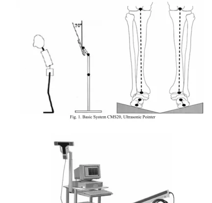
Three-dimensional motion analysis sys-tem (Zebris Medical GmbH, Germany), with ultrasonic pointer and basic system (CMS20) was applied for analyzing the spine (Fig. 1). To analyze with the pointer, we attached the triple reference marker to the participant’s body using a Velcro strip. It was used to eliminate fluctuations of the body’s position during the pointer meas-urement. First, the position of the measur-ing device was calibrated to define its posi-tion to the ground. Four points were indi-cated on the floor for calibration by the pointer. The points were fixed by pushing the button on the pointer and sensors of the pointer were facing the measuring unit. A new calibration was necessary only if the measuring unit or the reference marker had slipped. Initially, the barefoot participants were asked to stand in a relaxed position with their weight evenly distributed on both feet to obtain the same base of support con-sidering their pelvic width and the same natural foot alignment. Then, the partici-pants stood on the posture wedge angled at 10, 15, and 20 degrees, which was designed to induce hyperpronation (Fig. 2). Tracing was then made of the participant’s feet so that all measurements would be made with the participant in the same standing posi-tion. Each measurement was taken 3 times
during a normal relaxed standing position. Each participant remained in a standing po-sition with his back turned to the device, and the position of the examiner was pref-erably on the side opposite to the applied reference marker. While the participant was standing, the first examiner palpated the right and left ASIS, and PSIS, left and right acromions, and the spinous processes of C7 and S3, and marked them with a marker. The top of the pointer was put on the ana-tomical points and the key-button of the
pointer was pressed for approximately one second until the point appeared on the screen and a short acoustic signal was giv-en. After all pointers were entered, the spi-nal crest line was scanned with the tip of the pointer from C7 to S3. A line was drawn from C7 to S3 using an ultrasonic pointer. This measurement, like others, was taken 3 times by the same examiner with-out rest between the measurements. Each of the measurement methods was repeated 3 times. The second examiner took the
meas-Fig. 1. Basic System CMS20, Ultrasonic Pointer
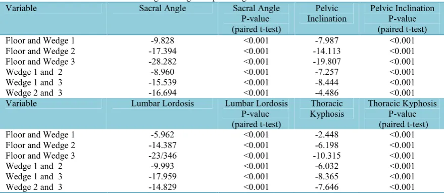
Fig. 2. (A) The Participant Standing on a Wedge, (B) Position of the Measuring Unit
urements 3 times on each participant to de-termine the average of these measurements. The first examiner was not aware of the original values, and thus, could not be in-fluenced by them, so reliability assessment was not compromised. The second examin-er recorded the measurement without al-lowing the first examiner to be informed about those measurements.
Data Reduction
Data were processed through the 3D Mo-tion Analysis Software (WinSpine Pointer Program for analyzing the posture, shape and mobility of the spine). Layouts of the program were database, real time meas-urement, signal viewer, and report. Sacral angle was defined by the angle between the tangent through S1 and the frontal plane. Pelvic inclination was described by the gle between the line drawn through the an-terior superior iliac spine and posan-terior su-perior iliac spine and the line drawn through the transverse plane in the sagittal plane. Thoracic kyphosis (the abnormally excessive convex kyphotic curvature of the spine) and lumbar lordosis (the normal in-ward lordosis curvature of the lumbar and cervical regions of the spine) angles were formed from all the thoracic vertebrae an-gles. The angles of the vertebrae were cal-culated using 3 points as described in the Annex. The means and standard deviations of the position of the spine were computed using the three trials for each participant in each study condition.
Statistical Analysis
Statistical analysis and graphic presenta-tion were prepared using software SPSS Version 17. Significance of the change in the segmental alignment between modes was determined using paired t-test.
Cumu-lative influence of the increasing wedge angle on the segmental alignment change was examined using repeated measure ANOVA. Significance level was adjusted by Bonferroni's equation for multiple com-parisons. Significance level was set at P < 0.0083.
Results
The mean (SD) age, height, and weight of the participants was 22.8 (28.9) years, 1.77 (4.98) m, and 78 (7.77) kg, respectively. The physical status of the participants is demonstrated in Table 1. Repeated measures were used for statistical analysis. No significant differences were found in the variables and examiners. A strong cor-relation was found for examiners and sacral angle between standing directly on the floor and three wedges angled at 0.929°, 0.931°, and 0.940°, respectively. The spaces added for examiners and pelvic inclination were as follow: Between standing directly on the floor and the first wedge (r=0.862), second wedge (r=0.871), and the third wedge (r=0.903). The spaces added for examiners and lumbar lordoses were as follow: Be-tween standing directly on the floor and the first wedge (r=0.817), second wedge (r=0.856), and the third wedge (r=0.871). The spaces added for examiners and thorac-ic kyphosis were as follow: Between stand-ing directly on the floor and the first wedge (r=0.807), second wedge (r=0.842), and the third wedge (r=0.844). The results demon-strated a positive correlation with the in-creased slope of the angle of the wedges, which was obtained for the examiners and variables. The means and standard devia-tions for each variable in the 4 standing po-sitions provided by the 2 examiners are demonstrated in Table 2.
No statistically significant difference was
Table 1. The Physical Status of the Participants
Variable Mean±SD Maximum Minimum
Age 22.8±2.89 18 28
Weight (kg) 78.0±7.77 61.8 93.5
Height (cm) 177.0±4.98 168 187
BMI* 24.8±2.73 19.8 31.2
* Body Mass Index (BMI) was calculated by the following formula: Weight (kg) / height (m). This index was used to define the nutritional status.
found between the examiners. An increase of 2.4, 1.8, and 2.2 in sacral angle demon-strated the transition from the floor to Wedge 1, Wedge 1 to Wedge 2, and Wedge 2 to Wedge 3.
Table 3 demonstrates a statistically signif-icant increase in sacral angle, pelvic incli-nation, lumbar lordosis, and thoracic ky-phosis in all modes (p<0.0083), except be-tween the first and second mode of the tho-racic kyphosis variable (p>0.0083).
Discussion
In terms of biomechanics, the human body has a multisegmental structure, initiat-ing major and powerful interactions tween adjacent segments. Interaction be-tween segments that are further apart may also hold a high significance for symptom-free musculoskeletal function. The pelvis bone is an important segment situated in the center of the body (4) and connects the movement of the lower limbs to the
seg-mental motion of the spine, and is a func-tional link through which loads are trans-ferred in a proximal and distal manner. Alt-hough suggested often, its position and movement as related to foot posture was mostly hypothesized (13). When postural alignment is optimal, little or no muscle activity is required to maintain medial-lateral stability. The gravitational torques acting on each body is opposed by equal torques, acting on the other side of the body (17). When the center of the gravity exits the ideal alignment, the body may use compensatory postural strategies until the COG returns within the base of support and obtains a stable position. Therefore, accord-ing to the body components such as inter-locking rings that each affects the other (18, 19), it is likely that changes of foot align-ment may lead to the presence of postural alterations and instability of the spine, bal-ance disorders, and structural abnormalities (20,21). Our results suggest that during the
Table 2. The Means and Standard Deviations of the Variables in the Four Standing Positions (on the Floor, on the Wedg-es Angled at 10, 15, and 20 DegreWedg-es) in the Sagittal Plane Determined by 2 Examiners (Mean±SD).
Variable Examiner Floor
Mean±SD First Wedge(10 Angle) Mean±SD
Second Wedge (15 Angle)
Mean±SD
Third Wedge (20 Angle)
Mean±SD Sacral angle Examiner 1
Examiner 2 30.2±5.2130.5±5.75 32.8±5.4532.7±5.31 34.5±5.5034.7±5.65 36.8±5.5636.9±5.78 Pelvic inclination Examiner 1
Examiner 2 34.6±5.2434.7±5.23 37.3±5.3337.3±5.36 39.0±4.9439.0±4.92 40.0±5.2340.2±5.40 Lumbar lordosis Examiner 1
Examiner 2 33.9±4.7533.9±4.71 35.7±4.1535.9±4.06 38.0±4.3038.2±4.25 40.5±4.6140.4±4.62 Thoracic kyphosis Examiner 1
Examiner 2 31.2±5.3231.0±5.00 31.9±5.1932.1±5.15 33.4±5.7833.4±5.77 34.8±5.5234.8±5.66
Table 3. Changes in Sagittal Spine Alignment between the Modes Variable Sacral Angle Sacral Angle
P-value (paired t-test)
Pelvic
Inclination Pelvic InclinationP-value (paired t-test) Floor and Wedge 1 -9.828 <0.001 -7.987 <0.001 Floor and Wedge 2 -17.394 <0.001 -14.113 <0.001 Floor and Wedge 3 -28.282 <0.001 -19.807 <0.001
Wedge 1 and 2 -8.960 <0.001 -7.257 <0.001
Wedge 1 and 3 -15.539 <0.001 -8.444 <0.001
Wedge 2 and 3 -16.694 <0.001 -4.486 <0.001
Variable Lumbar Lordosis Lumbar Lordosis P-value (paired t-test)
Thoracic
Kyphosis Thoracic KyphosisP-value (paired t-test) Floor and Wedge 1 -5.962 <0.001 -2.448 <0.001 Floor and Wedge 2 -14.387 <0.001 -6.198 <0.001 Floor and Wedge 3 -23/346 <0.001 -10.315 <0.001
Wedge 1 and 2 -9.993 <0.001 -6.032 <0.001
Wedge 1 and 3 -17.959 <0.001 -8.365 <0.001
Wedge 2 and 3 -14.829 <0.001 -7.646 <0.001
pronation of the subtalar joint, the calcane-us everts, cacalcane-using the talcalcane-us to slide medially and inferiorly. This medial downward movement of the talus induces an internal rotation of the tibia (14,22), and this may affect the knee joint function (23). Medial rotation of the femur causes the head of the femur to exert pressure on the posterior portion of the acetabulum (3,24). It has been hypothesized that internal rotation at the femur causes the head of the femur to exert pressure on the posterior portion of the acetabulum. This backward push on the posterior aspect of the pelvis would cause the pelvis to tilt anteriorly (15,25,26). Be-cause pelvis is tightly connected to the lumbar spine at the sacro-iliac joint by an extensive fibrous connection, an anterior tilt of the pelvis could increase the lumbar lordosis (27,28). Excessively tilted anterior-ly may lead to an increase in the lumbar anterior convexity. The line of gravity is at a greater distance from the lumbar joint ax-es than the optimal distance, and therefore, the extension moment in the lumbar spine is increased. The posterior convexity of the thoracic curve increases and becomes ky-photic to balance the lordotic lumbar curve and maintain the head over the sacrum (17). The change in thoracic kyphosis was small-er than the changes occurring in the seg-ments below, as the segment was located further away from the wedge. The most significant alteration in spine alignment occurred in the transition from standing di-rectly on the floor to standing on a 10 wedges. No significant difference was found between the examiners (p>0.0083), but there was a significant difference (p<0.0083) between the variables in all modes. The present study used the wedges to alter foot alignment in the standing posi-tion. Considering these findings, the transi-tion from standing directly on the floor to standing on 10, 15, and 20 wedges led to additional significant changes in spine alignment. Therefore, we hypothesize that hyperpronation may play an important role in an abnormal posture etiology, specifical-ly in babies. However, longitudinal studies
are necessary to find the long-term effects of excessive foot pronation and its possible influence on the body posture. Postural ab-normalities may lead to pain of the muscu-loskeletal system; however, correcting and training the posture is a method of reducing pain and has been used by physical thera-pists as a therapeutic approach (6,29,30). In the present study, the participants with ex-cessive foot pronation used short-term compensatory mechanisms to prevent im-balance and change their body posture.
Conclusion
The results of this study revealed that sa-cral angle, pelvic inclination, lumbar lordo-sis, and thoracic kyphosis increased with an increase in bilateral foot pronation. In other words, each one of them is a compensatory phenomenon.
References
1. Abboud RJ. Relevant foot biomechanics. Cur-rent Orthopaedics 2002;16(3):165-179.
2. Rockar JrPA. The subtalar joint: anatomy and joint motion. Journal of Orthopaedic and Sports Physical Therapy 1995;21(6):361-72.
3. Khamis S, Yizhar Z. Effect of feet hyperprona-tion on pelvic alignment in a standing posihyperprona-tion. Gait and Posture 2007;25(1):127-134.
4. Levine D, Whittle MW. The effects of pelvic movement on lumbar lordosis in the standing posi-tion. Journal of Orthopeadic and Sports Physical Therapy 1996;24(3):130-5.
5. Rothbart BA, Estabrook L. Excessive prona-tion: a major biomechanical determinant in the de-velopment of chondromalacia and pelvic lists. J Manipulative Physiol Ther 1988;11(5):373-9.
6. Donatelli R. The biomechanics of the foot and ankle. Philadelphia: F. A. Davis Company; 1996.
7. Tiberio D. Pathomechanics of structural foot deformities. Phys Ther 1988;68(12):1840-9.
8. Tiberio D. The effect of excessive subtalar joint pronation on patellofemoral mechanics: a the-oretical model. J Orthop Sports Phys Ther 1987; 9(4):160-165.
9. Genova JM, Gross MT. Effect of foot orthotics on calcaneal eversion during standing and treadmill walking for subjects with abnormal pronation. J Orthop Sports Phys Ther 2000;30(11):664-75.
10. Hellebrandt FA. Standing as a geotropic re-flex– the mechanism of the asynchronous rotation of motor units. Am J Physiol 1938;121:471-474.
11. Poussa MS, Heliovaara MM, Seitsamo JT, Kononen MH, Hurmerinta KA, Nissinen MJ.
velopment of spinal posture in a cohort of children from the age of 11 to 22 years. Eur Spine J 2005;14(8):738-742.
12. Shumway-Cook A, Woollacott MH. Motor control: theory and practical applications. London: Lippincott Williams & Wilkins; 2001:162-165.
13. Pinto RZA, Souza TR, Trede RG, Kirkwood RN, Figueiredo EM, Fonseca ST. Bilateral and uni-lateral increases in calcaneal eversion affect pelvic alignment in standing position. Manual Therapy 2008;13(6):513-519.
14. Tiberio D. Relationship between footprona-tion and rotafootprona-tion of the tibia and femur during walking. Foot Ankle Int 2000;21:1057-1060.
15. Health N, Council MR. National statement on ethical conduct in human research. Intern Med J. 2007.
16. OF CWPOE. Medicinal products. EEC note for guidance: good clinical practice for trials on medicinal products in the European Community. CPMP Working Party on Efficacy of Medicinal Products. Pharmacol Toxicol 1999;67:361-72.
17. Levangie PK, Norkin CC. Joint Structure & Function A Comprehensive Analysis. F. A. Davis Company; 2005:495-496.
18. Chaitow L, DeLany JW. Clinical Application of Neuromuscular Techniques. New york: Church-ill Livingstone; 2002.
19. Cutler WB, Friedmann E, Genovese-Stone E. Prevalence of kyphosis in a healthy sample of pre-and postmenopausal women. Am J Phys Med Re-habilitation 1993;72(4):219-25.
20.Sinaki M, Mikkelsen BA. Postmenopausal spinal osteoporosis: flexion versus extension exer-cises. Arch Phys Med Rehabil 1984;65(10):593-6.
21. Saunders D, Ryan RS. Evaluation, Treatment and Prevention of Musculoskeletal Disorders. Spine; 2004, 69.
22. Nawoczenski DA, Saltzman CL, Cook TM. The effect of foot structure on the three-dimensional kinematic coupling behavior of the leg and rear foot. Phys Ther 1998;78:404-416.
23. Legaye J, Duval-Beaupere G, Hecquet J, Marty C. Pelvic incidence: a fundamental pelvic parameter for three-dimensional regulation of spi-nal sagittal curves. European Spine Jourspi-nal 1998;7: 99-103.
24. Levens AS, Inman VT, Blosser JA. Trans-verse rotation of the segments of the lower extremi-ty in locomotion. J Bone Joint surg Am, 1948; 30:859-872
25. Day JW, Smidt GL, Lehmann T. Effect of pelvic tilt on standing posture. Physical Ther 1984; 64(4):510-6.
26. Duval K, Lam T, Sanderson D. The mechani-cal relationship between the rearfoot, pelvis and low-back. Gait Posture 2010; 32(4):637-640
27. Kisner C, Colby LA. Therapeutic Exercise: Foundations and Techniques. Philadelphia: F. A. Davis Company; 2002:844.
28. Kendall HO. Muscle Testing And Function. Baltimore: Williams & Wilkins; 1979:350.
29. Billis E, Katsakiori E, Kapodistrias C, Kapreli E. Assessment of foot posture: correlation between different clinical techniques. The foot 2007; 17(2):65-72.
30. Klingman RE, Liaos MS, Hardin KM. The ef-fect of subtalar joint posting on patellar glide posi-tion in subjects with excessive rearfoot pronaposi-tion. J Orthop Sports Physical Ther 1997;25(3):185-91.