Original Article
Comparison of Clinico-Pathological Changes in SPF Chickens
Infected with Different Iranian Genotypes of Infectious
Bronchitis Virus
Najafi H1, Ghalyanchi-Langeroudi A1, Hashemzadeh M2*, Madadgar O1, Karimi V2, Khaltabadi-Farahani R3, Maghsoudlo H4
1. Department of Microbiology and Immunology, Faculty of Veterinary Medicine, University of Tehran, Tehran, Iran.
2. Department of Research and Production of Poultry Viral Vaccine, Razi Vaccine and Serum Research Institute, Karaj, Iran.
3. Department of Clinical sciences, Faculty of Veterinary Medicine, University of Tehran, Tehran, Iran. 4. Iranian veterinary organization, Tehran, Iran.
Abstract
Background and Aims: Infectious bronchitis (IB), caused by infectious bronchitis virus (IBV), is an acute and highly contagious disease in chickens. IBV is mainly considered a respiratory infection, other clinical manifestations, including renal, enteric and reproductive signs can be observed. Since there has been no study on evaluation of changes in biochemical parameters during IB infection, this study was designed to assess the serum biochemical factors in experimentally infected chicks with two IBV isolates.
Materials and Methods: Two groups of 14-day-old SPF chickens were infected with two different isolates of IBV, Variant-2 like and IR-1 like genotypes respectively (35 chicks in each group). In addition, a group of 35 chickens remained non-infected as a control group. On days 1, 3, 5, 7, 14 and 21 and 28 post infection the sera of both infected groups and un-inoculated control group was collected to measure the biochemical factors, including uric acid, creatinine, alanine amino transferase (ALT) and aspartate amino transferase (AST) using a standard auto analyzer. We used multiple comparison ANOVA followed by a post-hoc test, (Level of significance <0.05).
Results: There were no significant differences between evaluated parameters of each infected group in comparison with the control group.
Conclusion: No changes were seen in serum factor levels between control and infected groups. In an experimental disease, there was no other infection to superimpose on IBV, so acute IBV infection was not strong enough to cause hepatic dysfunction or renal failure.
Keywords: Clinico - Pathological changes, Chickens, Infectious bronchitis virus
Introduction
*vian infectious bronchitis (IB) is one of the most serious diseases of chickens. It is of economic importance in the poultry industry worldwide and is associated with respiratory disease, reduction in weight gain, poor egg production and quality, and decreased feed conversion efficiency (1). Its etiologic agent is the avian infectious bronchitis coronavirus (IBV), which is a Gamma coronavirus of the coronavirus genus and replicates primarily in the upper respiratory tract, kidney, and oviduct of chickens. The etiologic agent, infectious bronchitis virus (IBV), belongs to the genus Gammacoronavirus within the Coronaviridae family. The IBV positive-sense single-stranded RNA genome (27.6 kb) encodes four structural proteins: the spike glycoprotein, the membrane glycoprotein, the envelope protein, and the phosphorylated nucleocapsid protein (2, 3). The primary tissue of avian IBV infection is respiratory tract, and the bird shows symptoms such as gasping, coughing, rales, and nasal discharge (2). The infection causes pathologic change in kidney, resulting in nephritis. In avian species, kidney function was evaluated by serum concentrations of uric acid, blood urea nitrogen, and creatinine(4). Blood creatinine is derived mainly from the catabolism of creatinine found in muscle tissue. Phosphocreatinine is used to store energy in muscle, and its catabolism to creatinine occurs at a steady rate. Excretion of creatinine is solely via the kidneys. It is freely filtered and reabsorbed in the tubules. In birds, creatinine is excreted in the urine before it has been converted to creatinine. In birds, uric acid is the major product of the catabolism of nitrogen. Synthesis occurs mainly in the liver and renal tubules. Approximately 90% of blood uric acid is eliminated by secretion into the lumen of the tubules. Only 50% of the healthy avian kidney is actually used for
*
Corresponding author: Masoud Hashemzadeh. Department of Research and Production of Poultry Viral Vaccine, Razi Vaccine and Serum Research Institute, Karaj, Iran. Tel: (+98) 26-34570038
Email: [email protected]
excreting protein waste, providing a large functional reserve. The evaluation of uric acid concentration in plasma or serum is widely used in birds for the detection of renal disease. IBV infection was also considered closely related with liver damage (4).ALT and AST belong to a group of enzymes that catalyze interconversion of amino acids and oxoacids by transfer of amino groups. While there are numerous enzymes involved in the conversion cascade, ALT and AST are the two enzymes of greatest clinical importance (5).As rare researches were done to evaluate the clinicopathological changes, as an indicator of liver and kidney dysfunction during IB infection. In Iran, several serotypes of IBV have been reported from different regions. The first isolation of IBV in Iranian chicken flocks was reported in1994 (6, 7). In spite of routine IBV vaccination, outbreaks of IB frequently occur in the field due to the presence of different serotypes as well as the emergence of multiple subtypes, generated by point mutations, insertions, deletions, or RNA recombination of the S1 genes. This study conducted to determine the serum level of ALT, AST, creatinine and uric acid in chickens that infected with two strains of IBV, one was classified as nephropathogenic and another with respiratory tropism. The isolates are among the most frequent IBV strains circulating in broiler farms in Iran and cause respiratory disease and nephritis.
Methods
Viruses
Two Iranian IBV field isolates were used in this study. Based on their S1 gene sequences the Isolate UTIVO-C (KR869776) belongs to IS/1494/06 (Variant-2), a nephropathogenic genotype and the isolate UTIVO-IR-1 like virus (KT210018) which characterized as an IBV strain showing respiratory disease in field. The isolates had been shown to be free from contamination by other avian pathogens using PCR assays detecting Newcastle disease virus, Avian influenza virus and Infectious larengtrachetis virus and Mycoplasma.
A
Experimental Design
SPF one-day-old chickens were divided randomly into three groups (thirty chickens in each two experimental group and twelve chickens in the control group). Each group was housed in different isolator. At age of 14 days, birds in the experimental group1 and group 2 were challenged via the intranasal route with viruses (Containing 104 EID50/0.1 ml of the virus) of UTIVO-C and UTIVO-IR-1 strains, respectively. UTIVO-C (Var-2 like; IS/1494/06 like)-virus is a nephropathogenic strain of IBV while IR-1 like virus an isolate with respiratory tract affinity.
Sample collection
On days 1, 3, 5, 7, 14 and 21 and 28 post infection, blood samples of both infected groups (5 blood sample from each group every time) and un-inoculated control group (2 blood sample every time) were collected. The sera was prepared by incubating blood samples in 37℃ for one hour, and then centrifuged at room temperature (1800g, 10 min). The sera were stored at -20℃ for biochemical measurement.
Laboratory analyses
Samples were analyzed at the clinical veterinary laboratory, faculty of veterinary medicine, University of Tehran. Uric acid, creatinine, alanine amino transferase activity and aspartate amino transferase were measured by a standard autoanalyzer with veterinary software (Selectra ProXS, 6003-200).
Data analysis
All data were analyzed with the SPSS 19.0, using multiple comparison ANOVA followed by a post-hoc test. Data are shown as mean+SE, and significant differences in frequencies were considered at P<0.05.
Results
There were no significant differences between evaluated parameters of each infected group in comparison with the control group. Comparison of creatinine, uric acid, AST and ALT levels of three different groups (UTIVO_C virus infected chickens, IR-1 like virus infected group and control group) are shown in tables 1 to 4, respectively. Despite no
biochemical changes being identified, both viruses caused respiratory disease and nephritis in chickens. In addition serologic, molecular and pathological findings demonstrated the pathologic effects caused by both viruses.
Discussion
Infectious bronchitis virus (IBV), a coronavirus of chickens, is one of the major causes of economic losses in the poultry industry. The name of the disease refers to its most frequent clinical manifestation, although it can infect many other epithelial cells, including the kidney, genital organs and many parts of the alimentary tract (1). Although vaccination is commonly adopted, outbreaks continue to occur worldwide with significant economic consequence. Different genotypes of IBV have been identified worldwide, and new variants continue to emerge (2). The first outbreak of IBV in Iranian chicken flocks was reported in 1994. Outbreaks of 793/B serotype were subsequently reported by several researchers in Iran, 793/B type, identified as the predominant circulating type of avian IBV in 1999 - 2004 in Iran (3, 4). Ghahremani et al showed existence of 2 serotype Massachusetts and 4/91 in commercial poultry flocks between 1998-2008 in Iran(5). IBV pathogenesis has been studied by various techniques (3). In Iran Mahdavi et al in an experimental histologic study suggested that 793/B may be a nephropathogenic virus (6) . In 2011 pathogenesis of 793/B like- virus has been studied by boroomand et al (7). Bijanzad et al (2013) characterize the Clinical signs and gross lesions of 793/B serotype of avian IBV in experimentally infected SPF chickens (3). Boroomand et al Assessed acute phase response following infectious bronchitis virus inoculation. Infected group were challenged intranasally with the IBV isolate IRFIBV32 and chickens in control group remained un infected. Blood samples were collected at prior and 1, 2, 3, 5, 7, 11, 13, 15, and 20 d postinoculation. In the serum the acute phase proteins (haptoglobin and serum amyloid A), pro-inflammatory cytokines (interferon-γ and tumor necrosis factor-α), and serum sialic acid
(total, TSA; lipid-bound, LBSA; and protein-bound, PBSA) concentrations were measured. All variables were significantly higher in the infected birds after virus inoculation compared with the healthy group (8). In Iran, there has been no study yet to determine the pathologic traits of new IBV strains. This is the first experimental study that investigates the blood biochemistry changes in Variant-2 like virus and IR-1 like virus-infected chickens. The kidneys are considered the second target organ damaged by toxins absorbed through the gut. In avian species, kidney function was
evaluated by serum concentrations of UA, BUN, and CR (9). The blood uric acid level is the primary indicator of renal function in birds. An elevated uric acid level is a reliable indicator that renal function is impaired (10). In all avian species that have been investigated, the reference interval for creatinine has been between 0.1-0.4 mg/dl. Severe kidney damage can lead to increased creatinine levels. Hyperuricemia can be expected if the glomerular filtration is decreased more than 70 to 80%. Decreased infiltration may occur from dehydration, intoxications or from some
Table 1: Creatinine levels (mg/dL) measured using an auto analyzer in serum samples of chicken in control and UTIVO-C and UTIVOIR-1 viruses infected groups on days 1, 3,5,7,14,21 and 28 post infection.
Control group UTIVO-C infected group UTIVOIR-1 infected group ANOVA
1 d.p.i 0.30+0.10 0.34+0.02 0.30+0.03 0 .679
3 d.p.i 0.35+0.05 0.36+0.04 0.38+0.02 0 .849
5 d.p.i 0.25+0.05 0.38+0.02 0.36+0.02 0 .044
7 d.p.i 0.30+0.00 0.34+0.02 0.34+0.02 0 .622
14 d.p.i 0.30+0.00 0.34+0.02 0.34+0.02 0 .622
21 d.p.i 0.35+0.05 0.40+0.00 0.38+0.02 0 .327
28 d.p.i 0.25+0.05 0.26+0.04 0.28+0.03 0 .892
Data are presented as mean+SEM. Comparisons among 3 groups were carried out by means of ANOVA.
Table 2: Uric acid levels (mg/dL) measured using an auto analyzer in serum samples of chicken in control and UTIVO-C and UTIVOIR-1 viruses infected groups on days 1, 3,5,7,14,21 and 28 post infection.
Control group UTIVO-C infected group UTIVOIR-1 infected group ANOVA
1 d.p.i 4.25+0.25 4.74+0.37 4.30+0.20 0.510
3 d.p.i 4.20+0.20 4.60+0.20 4.34+0.30 0.676
5 d.p.i 4.90+0.30 5.06+0.29 4.92+0.23 0.912
7 d.p.i 3.40+0.20 3.80+0.15 4.10+0.07 0.033
14 d.p.i 4.20+0.50 4.50+0.26 4.60+0.26 0.738
21 d.p.i 3.65+0.35 3.82+0.09 3.72+0.16 0.807
28 d.p.i 4.10+0.20 4.18+0.18 4.76+0.15 0.059
Data are presented as mean+SEM. Comparisons among 3 groups were carried out by means of ANOVA.
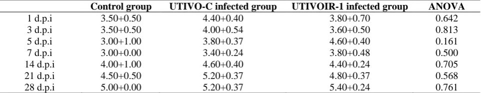
Table 3: Alanine amino transferase levels (IU/L) measured using an auto analyzer in serum samples of chicken in control and UTIVO-C and UTIVOIR-1 viruses infected groups on days 1, 3,5,7,14,21 and 28 post infection.
Control group UTIVO-C infected group UTIVOIR-1 infected group ANOVA
1 d.p.i 3.50+0.50 4.40+0.40 3.80+0.70 0.642
3 d.p.i 3.50+0.50 4.00+0.54 3.60+0.50 0.813
5 d.p.i 3.00+1.00 3.80+0.37 4.60+0.40 0.161
7 d.p.i 3.00+0.00 3.40+0.24 3.80+0.48 0.500
14 d.p.i 4.00+1.00 4.60+0.40 4.40+0.24 0.705
21 d.p.i 4.50+0.50 5.20+0.37 4.80+0.37 0.568
28 d.p.i 5.00+0.00 5.20+0.37 5.40+0.24 0.761
Data are presented as mean+SEM. Comparisons among 3 groups were carried out by means of ANOVA.
bacterial or viral infections. Ping liu et al evaluated the effect of nephrotrophic strains of infectious bronchitis virus on serum electrolytes, liver and renal function indexes in growing chickens of a layer strain. The chickens in the virus group and the control group were challenged by 0.2mL artificial nasal drip with virus and sterile saline solution per chicken, respectively. Blood parameters of chicken were evaluated on the 8th, 15th and 22nd day after challenge, respectively. The results showed that UA and CR values in the control group were significantly lower than in the virus group on the 15th day. Interestingly we found no differences in serum levels of creatinine and uric acid between infected and control groups even in group which was infected with nephtopathogenic strain. It can be concluded that the viruses could not cause kidney dysfunction. Liver is the primary organ collecting chemicals absorbed in the gut and also as the major detoxification organ; its function always harmed by the toxic properties of absorbed from agents, the leaking of cellular enzymes into the plasma is a noticeable indication of hepatocytes damage. IBV infection was considered closely related with liver damage. Abdel-Moneim et al. (2006) reported finding severe congestion of liver, spleen and lungs in birds died after IBV experimental infection (9). ALT activity occurs in many different tissues. Specific diagnostic value of these enzymes in birds is poor. In many cases, patients with severe liver damage have had normal ALT activities, reflecting a low level of enzyme activity in liver cells from certain species. It is coincided with our results. High AST activity has been described in liver,
skeletal muscle, heart, brain and kidney cells. The distribution of AST in avian tissues varies among the species. Elevated activities are usually indicative of liver or muscle damage. In general, AST activities in birds greater than 230 U/l are considered abnormal.
Ping liu et al studied on nephrotropic IBV strains and showed that AST values in the control group compared to virus group on the 8th, 15th high and 22nd day were low(9) . Our results did not show any abnormality in AST levels, maybe the IBV strains were not strong enough to injure hepatocytes membrane. In conclusion, abnormal renal or liver function did not observed in this study. Maybe the viruses did not injure the epithelial cells of target organs or cell damages were not severe enough to decrease the organ function. Therefore, liver and kidney histopathology studies are suggested to know whether the viruses could cause cell damage or not. It is of interest to note that chickens in an experimental study were just affected with IBV, whether in a field, IBV infection can also be further aggravated by the presence of viral or bacterial infections such as Escherichia coli, Mycoplasma gallisepticum, Mycoplasma synoviae and Ornithobacterium rhinotracheale which complicate the condition. Further studies mimicking field situation are needed to assess biochemical factors in chickens with mix viral and bacterial infections.
Acknowledgement
Research council, university of Tehran, under grant (No. 28692/6/1) and Iranian veterinary organization under grant (No. 22/39007),
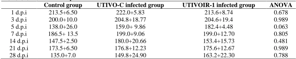
Table 4: Aspartate amino transferase levels (IU/L) measured using an auto analyzer in serum samples of chicken in control and UTIVO-C and UTIVOIR-1 viruses infected groups on days 1, 3,5,7,14,21 and 28 post infection.
Control group UTIVO-C infected group UTIVOIR-1 infected group ANOVA
1 d.p.i 213.5+6.50 222.0+5.83 213.6+8.74 0.678
3 d.p.i 200.0+10.0 204.8+18.77 204.6+19.4 0.989
5 d.p.i 138.0+26.0 159.0+ 9.86 182.4+4.48 0.063
7 d.p.i 186.5+ 13.5 199.0+9.06 199.0+12.70 0.805
14 d.p.i 147.5+2.50 180.0+20.66 153.4+15.73 0.481
21 d.p.i 173.5+6.50 176.8+12.23 175.6+12.67 0.989
28 d.p.i 135.0+7.0 149.8+24.90 163.2+22.30 0.788
Data are presented as mean+SEM. Comparisons among 3 groups were carried out by means of ANOVA.
financially supported this project. The authors gratefully acknowledge Dr. Abdolahi, Dr.Taha Zabihi and Mr.Javanmardi for their extensive technical support.
References
1. Jackwood MW. Review of infectious bronchitis virus around the world. Avian Dis, 2012. 56(4):634-41.
2. Sjaak de Wit JJ, Cook JK, Heijden HM. Infectious bronchitis virus variants: a review of the history, current situation and control measures. Avian Pathol. 2011;40(3):223-35.
3. Bijlenga G, et al. Development and use of the H strain of avian infectious bronchitis virus from the Netherlands as a vaccine: a review. Avian Pathol. 2004;33(6):550-7.
4. Liu P, et al. Clinicopathology of Gout in Growing Layers Induced by Avian Nephrotrophic Strains of Infectious Bronchitis Virus.
5. Pare J. Avian Medicine: Principles and Application. The Canadian Veterinary Journal. 1997;38(9):577.
6. Seyfi Abad Shapouri M, et al. A survey of the prevalence of infectious bronchitis virus type 4/91
in Iran. Acta Veterinaria Hungarica.
2004;52(2):163-6.
7. Bozorgmehri-Fard M, Charkhkar S, Hosseini, H Detection of the Chinese Genotype of Infectious Bronchitis Virus (QX-type) in Iran. Iranian Journal of virology. 2014;7(1&2):21-4.
8. Benyeda Z, et al. Comparison of the pathogenicity of QX-like, M41 and 793/B infectious bronchitis strains from different
pathological conditions. Avian Pathol.
2009;38(6):449-56.
9. Awad F, Baylis M, Ganapathy K. Detection of variant infectious bronchitis viruses in broiler flocks in Libya. International Journal of Veterinary Science and Medicine. 2014;2(1):78-82.
10. Bijanzad P, et al. Experimental study on histopathological changes and tissue tropism of Iranian infectious bronchitis serotype 793/B-like virus in SPF chickens. Journal of the South African Veterinary Association. 2013;84(1).
11. Shoushtari A, et al. 793/B type, the predominant circulating type of avian infectious bronchitis viruses 1999-2004 in Iran: a retrospective study. Archives of Razi Institute. 2008;63(1):1-5.
12. Ghahremani N, et al. Molecular Analysis of Infectious Bronchitis Viruses Isolated in Iran from 1998-2008. Journal of Animal and Veterinary Advances. 2011;10(22):2961-7.
13. Mahdavi S, et al. Experimental histopathologic study of the lesions induced by serotype 793/B (4/91) infectious bronchitis virus. Archives of Razi. 2007;62(1):15-22.
14. Boroomand Z, Asasi K, Mohammadi A. Pathogenesis and tissue distribution of avian infectious bronchitis virus isolate IRFIBV32 (793/B serotype) in experimentally infected broiler chickens. The Scientific World Journal. 2012. 15. Asasi K, et al. Changes of several acute phase factors in broiler chickens in response to infectious bronchitis virus infection. Poultry science. 2013;92(8):1989-96.
16. Tully TN, Dorrestein GM, Jones AK. Handbook of avian medicine. 2009;Elsevier/Saunders.