Bossenko K. V. Comparison of instrumental methods of hemostasis research. Journal of Education, Health and Sport. 2017;7(12):676-682. eISSN 2391-8306. DOI http://dx.doi.org/10.5281/zenodo.3252894
http://ojs.ukw.edu.pl/index.php/johs/article/view/7056
The journal has had 7 points in Ministry of Science and Higher Education parametric evaluation. Part B item 1223 (26.01.2017). 1223 Journal of Education, Health and Sport eISSN 2391-8306 7
© The Authors 2017;
This article is published with open access at Licensee Open Journal Systems of Kazimierz Wielki University in Bydgoszcz, Pola nd
Open Access. This article is distributed unde r the terms of the Creative Commons Attribution Noncommercial License which permits any noncommercial use, distribution, and reproduction in any medium, provided the original author(s) and source are credited. This is an open access article licensed under the terms of the Creative Commons Attribution Non Commercial License (http://creativecommons.org/licenses/by-nc/4.0/) which permits unrestricted, non commercial use, distribution and reproduction in any medium, provided the work is properly cited. This is an open access article licensed under the terms of the Creative Commons Attribution Non Commercial License (http://creativeco mmons.org/licenses/by-nc/4.0/) which permits unrestricted, non commercial
use, distribution and reproduction in any medium, provided the work is properly cited. The authors declare that there is no conflict of interests regarding the publication of this paper.
Received: 01.12.2017. Revised: 15.12.2017. Accepted: 25.12.2017.
COMPARISON OF INSTRUMENTAL METHODS OF HEMOSTASIS RESEARCH
K. V. Bossenko
Odessa National Medical University, Ukraine
Abstract
Urgency of the Problem. It is known that deep vein thrombosis of the lower extremities and pulmonary thromboembolism play an important role in the structure of postoperative morbidity and mortality, and these complications are characteristic of patients with different profiles. Considering the above, we consider it important to introduce into clinical practice new diagnostic methods that would be effective, reliable, and allow us to
The incidence of deep vein thrombosis (DVT) is 100 cases per 100,000 population, while thromboembolic complications taking the third place among cardiovascular diseases after coronary heart disease and stroke [1, 2]. According to various authors, in the overall mortality rate among hospitalized patients, pulmonary embolism (PE) ranges from 7,2 to 10,0%, and according to The Worcester DVT
Study, which was published in 1991, 170,000 new and 90, 000 repeat episodes of thrombosis and thromboembolism note that DVT and COPD cause 250,000 hospitalizations in the United States every year [1, 3]. However, it can not be ruled out that real rates of detection and mortality from thromboembolic diseases may be even higher, because DVT often runs asymptomatic. Not more than one in every five patients who died from pulmonary embolism had clinical signs of DVT, and only 10% of nonfatal venous thrombosis could have been diagnosed during the patient's life.
Thus, in the majority of cases, when pulmonary embolism is the direct cause of death, the pre-existing thrombosis is not diagnosed either clinically or laboratoryly, or by means of instrumental research methods, but is a discovery of autopsy. The same authors rightly point out that today there is no clinical, laboratory or instrumental evidence that would almost certainly indicate the presence of pulmonary embolism and DVT and that many clinical symptoms that were traditionally considered specific are found in 1-54% of cases (depending on symptom), but no more [4, 5].
Given the foregoing, it is important to introduce into clinical practice new diagnostic methods that would be effective, credible and allow for real -time research
Fig. 1. ARP-01М «Mednord»
The principle of the device is to record the viscous characteristics of blood or plasma in the process of its coagulation by measuring the energy of the extinction of the oscillation of the
mechanical resonance element (probe) located in the test sample placed in the thermostat cuvette. A wavelength piezoelectric converter results in flat sound oscillations of the probe with a given amplitude. The mechanical energy of the extinction of the oscillation of the probe, which depends on the change in the characteristics of the test medium, is transformed into a receiving piezoelectric converter in the electric potential and recorded by a potentiometer. In this case, the measurement of the investigated characteristics of the sample is continuous.
The device provides output to the personal computer a graph of change of the resistivity of the investigated medium by oscillation of the probe attached to the vibroelectric sensor, and the software (ICS hemo-3) provides the calculation of the corresponding amplitude and chronometric parameters:
Аі — current indicator of aggregate state of blood;
TI — current time, min.;
A0 — initial rate of aggregate state of blood, t0;
A1 — the amplitude of contacts of the phase of coagulation of the blood, RH; t1 — time of contact phase of coagulation, min;
ІCC — the intensity of the contact phase of coagulation; СТА — constant of thrombin activity;
TBC — time of blood coagulation;
ІRLC — the intensity of retraction and lysis of the clot.
In turn, the TEG 5000 Thrombelastograph has a slightly different operating principle, and therefore other measurement parameters. The TEG® analyzer measures the physical properties of a blood clot using a special cylindrical cup for which a blood sample is placed. The cup performs rotational movements relative to its axis at an angle of 4-45°. Each rotational cycle lasts 10 seconds. A pin immersed in a blood sample suspended on a torsion wire. The rotational moment of the cup rotation is transmitted to a dipped-like rod only after the bundle formed by the fibrin-platelet connection begins to connect the cup and rod together. The strength of these bonds determines the angle of rotation of the rod: if the blood does not curtail - does not transmit the rotation, the loose
clot only partly transmits the rotation, and a solid clot causes the rod to move synchronously with the cup. Thus, the angle of rotation of the rod directly depends on the strength of the formed clot.
As soon as the blister begins to crumble or collapse (lysis), the ligaments are torn, the interaction between the cup and the rod weakens, and the movement of the cup on the rod decreases. The rotation of the rod will be transformed from a mechanical to an electrical signal, which is fixed using a computer. As a result, we can measure the time when the beginning of the formation of the first strands of fibrin, the kinetics of clot formation, the clot strength (elasticity in din/cm2), and the process of dissolving the clot, whether fibrinolysis occurs or not (Figure 2). To interpret the graphical information displayed by the TEG® analyzer, five basic parameters of clot formation and its lysis are measured.
R — time from the moment when the sample was placed in the analyzer until the first fibrin threads were formed. It is a characteristic of the enzymatic part of the coagulation cascade.
K — The time from the beginning of the clot formation to achieve a fixed level of its strength reflects the kinetics of increasing the strength of the clot.
α — angle, built on the tangential to thromboelastogram from the point of formation
of a bunch. Displays the growth rate of fibrin nets and their structure formation (increasing the buccal strength). Characterizes the level of fibrinogen.
MA — maximum amplitude, characterizes the maximum dynamic properties of fibrin and platelet binding with GPIIb / IIIa and reflects the maximum strength of clot. At 80% of the MA is due to the number and properties (the ability to aggregate) platelets, by 20% - the
amount of fibrin formed.
LY30 —change in the area under the curve of the thromboleastogram during subsequent MA 30 mins in relation to the area under the curve of the thromboelastogram without signs of lysis (rectangle with altitude MA), expressed as a percentage. It is a characteristic of the dissolution process of the clot - lysis.
Materials and methods of research. The functional state of the hemostasis system in the group of 65 healthy volunteers, as well as 68 patients with post-thrombophlebitis syndrome (PTFS) was investigated. In the group of patients with PTFS, background studies of haemocoagulation status and daily dynamic monitoring of changes in the functional state of hemostasis after heparin administration, a comparative assessment of ARP-01M Mednord and TEG® data for 8 days before and after a single administration of Cardiomagnol (150 mg) were conducted. To evaluate the reproducibility of the technique, a series of measurements of hemostasis parameters were performed in each healthy volunteer.
Material for research (whole unstable blood) was collected in the surveyed according to the generally accepted method, siliconized needles with a wide lumen from the cubital vein.
Subsequently, a correlation analysis was performed with the results obtained using control hemostatic techniques.
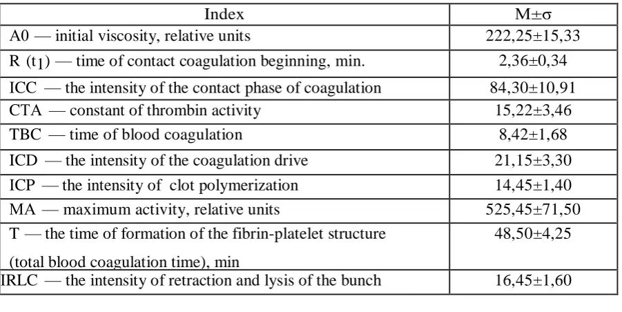
Table 1 Indicators ARP-01M "Mednord"
Index М±σ
А0 — initial viscosity, relative units 222,25±15,33
R (t1) — time of contact coagulation beginning, min. 2,36±0,34 ІCC — the intensity of the contact phase of coagulation 84,30±10,91
CТА — constant of thrombin activity 15,22±3,46
TBC — time of blood coagulation 8,42±1,68
ICD — the intensity of the coagulation drive 21,15±3,30
ІCP — the intensity of clot polymerization 14,45±1,40
МА — maximum activity, relative units 525,45±71,50
T — the time of formation of the fibrin-platelet structure (total blood coagulation time), min
48,50±4,25
ІRLC — the intensity of retraction and lysis of the bunch 16,45±1,60
Table 2
Indices of thromboelastogram TEG 500
Index М±σ
R — time of reaction, m i n 10,42±2,67
К — tine of clot formation, min 6,88±2,43
МА — maximum amplitude, mm 45,37±6,12
FА — fibrinolytic activity, % 12,41±3,58
Table 3 Correlation analysis of blood coagulation tests
АRP-01М «Mednord»TEG
5000
TEG 500 Correlation
CТА TBC ICD МА ІRLC К R R МА FА 0,95 0,66 0,75 0,96 0,86 Conclusions
[image:6.595.78.524.97.312.2] [image:6.595.97.502.539.680.2]to these qualities, it can be successfully used not only in the conditions of clinical laboratories, but also at the patient's bed, in the operating room, in the conditions of the ambulance.
2. APK APP-01M "Mednord" allows to determine the total assessment of all parts of the hemocoagulation and lysis, as well as their interaction. Its indicators are characterized by objectivity and informativity, which is confirmed by close correlations with the indicators of traditional coagulogical techniques.
3. The parameters IСD, СTA, ICC can be successfully used to control heparin therapy in patients, and indicators of TBC, t1 and A0 - to control disaggregation therapy.
4. The ability to display the process on paper using the printer and transfer research data to various computer databases allows to use the device not only for clinical nee ds, but also
for statistical and scientific analysis. References:
1. Futterman L. A silent killer – often preventable // L.Futterman and L. Lemberg /Am J Crit Care. – 2004. – Sep; 13 (5): 431-6.
2. Таrabrin OA., et al. Modern approaches to the control of peri-operational blood loss in hysterectomy patients // GO Mozhayev Ukr J Extremal Medicine. – 2012. – Vol. 13, № 2. – P. 66-71 (Rus).
3. Udut VV. Теchnology of lowfrequency piesothromoelastograaphy in the assessment of hemostatic potential/ herald for new medical technologies. E-edition. – 2016. – Vol. 10. – № 4. – P. 104 – 113 (Rus).
4. Tarabrin O. New method diagnostics coagulation disorders after surgery // O.Tarabrin, V. Suslov, V. Grubnik / Critical Care. – 2010. – March; 14 (Suppl. 1): 122.