Open Access
Research
Sepsis causes neuroinflammation and concomitant decrease of
cerebral metabolism
Alexander Semmler
1, Sven Hermann
2, Florian Mormann
3, Marc Weberpals
1,
Stephan A Paxian
1, Thorsten Okulla
1, Michael Schäfers
2, Markus P Kummer
1,
Thomas Klockgether
1and Michael T Heneka*
1Address: 1University Bonn, Department of Neurology, Bonn, Germany, 2University Münster, Department of Nuclear Medicine, Münster, Germany
and 3University Bonn, Department of Epileptology, Bonn, Germany
Email: Alexander Semmler - [email protected]; Sven Hermann - [email protected];
Florian Mormann - [email protected]; Marc Weberpals - [email protected]; Stephan A Paxian - [email protected]; Thorsten Okulla - [email protected]; Michael Schäfers - [email protected];
Markus P Kummer - [email protected]; Thomas Klockgether - [email protected]; Michael T Heneka* - [email protected]
* Corresponding author
Abstract
Background: Septic encephalopathy is a severe brain dysfunction caused by systemic inflammation in the absence of direct brain infection. Changes in cerebral blood flow, release of inflammatory molecules and metabolic alterations contribute to neuronal dysfunction and cell death.
Methods: To investigate the relation of electrophysiological, metabolic and morphological changes caused by SE, we simultaneously assessed systemic circulation, regional cerebral blood flow and cortical electroencephalography in rats exposed to bacterial lipopolysaccharide. Additionally, cerebral glucose uptake, astro- and microglial activation as well as changes of inflammatory gene transcription were examined by small animal PET using [18F]FDG, immunohistochemistry, and real time PCR.
Results: While the systemic hemodynamic did not change significantly, regional cerebral blood flow was decreased in the cortex paralleled by a decrease of alpha activity of the electroencephalography. Cerebral glucose uptake was reduced in all analyzed neocortical areas, but preserved in the caudate nucleus, the hippocampus and the thalamus. Sepsis enhanced the transcription of several pro- and anti-inflammatory cytokines and chemokines including tumor necrosis factor alpha, interleukin-1 beta, transforming growth factor beta, and monocot chemoattractant protein 1 in the cerebrum. Regional analysis of different brain regions revealed an increase in ED1-positive microglia in the cortex, while total and neuronal cell counts decreased in the cortex and the hippocampus.
Conclusion: Together, the present study highlights the complexity of sepsis induced early impairment of neuronal metabolism and activity. Since our model uses techniques that determine parameters relevant to the clinical setting, it might be a useful tool to develop brain specific therapeutic strategies for human septic encephalopathy.
Published: 15 September 2008
Journal of Neuroinflammation 2008, 5:38 doi:10.1186/1742-2094-5-38
Received: 30 July 2008 Accepted: 15 September 2008
This article is available from: http://www.jneuroinflammation.com/content/5/1/38
© 2008 Semmler et al; licensee BioMed Central Ltd.
Background
Sepsis and its complications are the leading causes of mor-tality in intensive care units accounting for 10–50% of deaths. Up to 71% of septic patients develop potentially irreversible acute cerebral dysfunction [1-3]. This sepsis-induced encephalopathy is caused by systemic inflamma-tion in the absence of direct brain infecinflamma-tion and clinically characterized by slowing of mental processes, impaired attention, disorientation, delirium or coma. Importantly, septic encephalopathy (SE) is an early sign of sepsis and associated with an increased rate of morbidity and mortal-ity [2].
The pathogenesis of SE is unlikely to be directly induced by a pathogenic toxin, as similar encephalopathy can develop as a result of a number of systemic inflammatory response syndromes that lack an infectious etiology (e.g. acute pancreatitis, burns etc.). Clinical and experimental data suggest that a number of factors including the local generation of pro-inflammatory cytokines, impaired cere-bral microcirculation, an imbalance of neurotransmitters and a negative impact of peripheral organ failure contrib-ute to the development of SE. Additionally, once inflam-mation persists, increased excitotoxicity and oxidative stress may further aggravate SE and contribute to neuronal dysfunction and degeneration (for review see [3]). Of note, patients with a pre-existing CNS pathology have a higher risk to develop SE, and a similar predisposing inter-action has been reported in an animal model of sepsis [4]. Clinically, the electroencephalogram (EEG) serves as an important diagnostic tool for SE assessment and the majority of patients shows abnormal EEG recordings [5]. Of note, the degree of EEG pathology correlates well with the clinical status and prognosis and has been proven more sensitive than clinical bedside investigation [5]. Likewise, cerebral blood flow (CBF) is another parameter which is routinely analyzed in patients suffering from SE, based on the assumption that sepsis exerts profound and sustained effects on the systemic circulatory function. However, past studies have yielded controversial results and to date, the effects of sepsis on CBF as well as neuro-nal metabolism and activity remain unclear. To further investigate the relation of potential regional CBF changes, electroencephalography and cerebral metabolism in response to SE, we investigated hemodynamic, electro-physiological and metabolic changes in relation to neu-roinflammatory markers and neuronal number in a model of acute SE in rats. Regional cerebral blood flow was reduced in correlation to EEG frequency 24 h after intraperitoneal injection of LPS, whereas brain glucose utilization and neuronal number were reduced concur-rent with microgliosis and neuroinflammatory response.
Methods
Animals
53 male Wistar rats (Charles River, Sulzfeld, Germany) weighing 250 – 300 g were housed in groups under stand-ard conditions at a temperature of 22°C (± 1°C) and a 12 hour light-dark cycle – with free access to standard food (Altromin, Soest, Germany) and tap water. Animal care and handling were performed according to the Declara-tion of Helsinki and approved by local ethical committees (approval number 50.203.2 BN 33,34/00).
Rats were randomized and received either 10 mg/kg of LPS (0127:B8, E. coli; Sigma, München, Germany) dis-solved in 1 ml sodium chloride (0.9%) intraperitoneally (i. p.) or the vehicle alone. 24 hours after induction of sep-sis, animals were anaesthetized with a combination of ketamine (80 mg/kg) and xylazine (10 mg/kg). The tra-chea was cannulated to facilitate respiration and rectal temperature was maintained at 37°C using a heating blanket. The femoral artery was exposed and catheterized with a polyethylene tube connected to a pressure trans-ducer for continuous recording of arterial blood pressure and heart rate (Harvard Apparatus, March-Hugstetten, Germany). The head was fixed in a stereotactic frame and four stainless steel skull screws were placed epidurally, two electrodes per parietal bone at bregma coordinates 0 mm and -6.5 mm, 4 mm from the midline. A reference electrode was placed on the anterior midline over the frontal sinus. All electrodes were connected through insu-lated wire with a 2-channel amplifier (Harvard Apparatus, March-Hugstetten, Germany). Electrical brain activity was amplified (× 10 000 – 20 000), digitized and transferred to a PC for storage and further analysis. EEG was recorded for 15 – 20 min periods.
After EEG-recording, the screws were removed, and a 5 × 3 mm large cranial window was drilled (thinning of the scull until translucency, leaving the dura mater intact), centered 4 mm lateral and 4 mm caudal to the bregma. CBF was assessed using a laser flow blood perfusion mon-itor (PeriFlux 5000, Perimed, Stockholm, Sweden) with a 1.0 mm diameter laser Doppler probe (wavelength 780
Small animal positron emission tomography (microPET)
microPET was performed on a 32-module quadHIDAC scanner (Oxford Positron Systems, Weston-on-the-Green, UK) dedicated to small animal imaging. The scanner has an effective resolution of 0.7 mm (FWHM) in the transax-ial and axtransax-ial directions when using an iterative resolution recovery reconstruction algorithm [7].
Before the scan, a tail vein catheter was inserted under short-term isofluorane anesthesia. The conscious rat was afterwards kept in a restraining device. 40 MBq of 18
F-Fluordeoxyglucose (FDG) in 800 μl 0.9% saline were injected via the tail vein catheter. Following a 60 min interval, animals were again anaesthetized using iso-fluorane and placed in the PET scanner on a heating pad to maintain normal body temperature. List mode PET data were acquired for 15 minutes and subsequently reconstructed into a single image volume with a voxel size of 0.8 × 0.8 × 0.8 mm3.
Immunohistochemistry
Rats were perfused transcardially using heparanized saline and brains were subsequently dissected. Serial sagittal sec-tions were cut (10 μm) from cryo-conserved preserved hemispheres (Leica Cryostat CM 3050S), embedded in tis-sue freezing medium (Leica Microsystems #0201-08926, Nussloch, Germany) and mounted (Microscope Slides #K0123b, Engelbrecht, Germany). After drying for 30 minutes at room temperature, for fixation slides were incubated in 4% paraformaldehyde (Roti Histofix 4% #P087.4, Roth, Karlsruhe, Germany) for 20 minutes. Blocking of non-specific binding was achieved by one hour incubation in 5% normal goat serum (Linaris #S-1000, Wertheim, Germany). Between the steps, slides were rinsed three times for five minutes in PBST. Immu-nostaining was performed overnight by incubation at 4°C with the following primary antibodies: 1.) polyclonal antibody rabbit-anti-mouse GFAP (1:1000 in 2% normal goat serum in PBST; DAKO Z0334, Glostrup, Denmark). 2.) monoclonal antibody mouse-anti-rat CD68/ED1 (1:100 in 2% normal goat serum in PBST; Serotec MCA341G, Düsseldorf, Germany). 3.) monoclonal rabbit anti mouse NeuN (1:250 in 2% normal donkey serum in PBS; Clone A60, Chemicon, Temecula, CA). Afterwards slides were incubated with Alexa Fluor 594-labeled sec-ondary antibodies for one hour (1:400 in PBST; Invitro-gen #A11037 & #A11020 Karlsruhe, Germany). For co-staining with Hoechst Dye 33342 (10 μg/ml; Fluka/ Sigma-Aldrich #14533, Steinheim, Germany) an incuba-tion time of two minutes was set. Again, slides were rinsed with PBST between the steps. Finally, the slides were cov-ered in Mowiol 4–88 (Calbiochem/VWR #475904, Darm-stadt, Germany) and stored at -20°C in the dark until microscopy was performed.
Real time PCR
RNA from brain hemispheres were extracted using Trizol (Life Technologies Invitrogen, Karlsruhe, Germany) using an Ultra Turrax (IKA Labortechnik, Staufen, Germany). Total RNA was quantified photometrically and reverse transcribed using the RevertAid First Strand cDNA Synthe-sis kit (Fermentas, St. Leon-Rot, Germany) according to the manufacturer's instructions. Real time qPCR was per-formed using the StepOnePlus™ Real-Time PCR System (Applied Biosystems, Foster City, USA). Power SYBR®
Green PCR Master Mix (Applied Biosystems, Foster City, USA) was used for PCR amplification and real time detec-tion of PCR products. 1 μl of the RT product correspond-ing to 40 ng of total RNA, 0.2 μM of each primer and 10
μl of the master mix were mixed and run under the follow-ing conditions: 95°C for 10 min and 40 cycles of 95°C for 15 s and 60°C for 1 min. Amplification specificity was checked using a melting curve analysis after PCR. mRNA expression was normalized to GAPDH. Primers used were: GAPDH forward ACG ACA GTC CAT GCC ATC AC and reverse TCC ACC ACC CTG TTG CTG TA, IL-1β for-ward GCT ACC TAT GTC TTG CCC GTG GAG and revers GTC CCG ACC ATT GCT GTT TCC TA, IL-4 forward GGA TGT AACGAC AGC CCT C and revers GAC ACC TCT ACA GAG TTT CC, IL-6 forward CTT GGG ACT GAT GTT GTT GA and revers CTC TGA ATG ACT CTG GCT TTG, IL-10 forward CCT GCT CTT ACT GGC TGG AG and reverse CTG CAG TAA GGA ATC TGT CAG, TNF-α forward AAA ACT CGA GTG ACA AGC CC and reverse GGT TGA CCT CAG CGC TGA GC, TGF-β forward TGC GCC TGC AGA GAT TCA AG and reverse TCT CTG TGG AGC TGA AGC AG, MCP-1 forward CTG TTG TTC ACA GTT GCT GC and revers CTG ATC TCA CTT GGT TCT GG and iNOS forward CCA GAG CAG TAC AAG CTC AC and revers CCA CAA CTC GCT CCA AGA TC.
Data analysis
Electrophysiological recordings were analyzed using a moving window analysis with a window length of 16.384 s (8192 data points). Prior to analysis signals were scanned for movement and recording artifacts using auto-mated artifact detection. Windows containing constant signals for more than 100 consecutive points or signal jumps exceeding 10 standard deviations of a window's amplitude distribution were discarded.
Hz, theta 4–8 Hz, alpha 8–13 Hz, beta 13–20 Hz, gamma 20–40 Hz). In order to quantify electroencephalographic signs of septic encephalopathy in terms of slowing of oscillatory brain activity, we determined the main EEG frequency from the maximum of the power spectrum. All electrophysiological variables extracted in the moving window analysis were averaged over time (i.e., over differ-ent windows) for each animal. Results from differdiffer-ent groups (septic animals vs. control animals) were com-pared using a nonparametric test (two-sided Mann-Whit-ney test for independent samples). In addition, to address the question whether electroencephalographic signs of encephalopathy exhibit dependence on hemodynamic sepsis parameters, we tested for significant correlation using a one-sided non-parametric correlation test (Spear-man's rho).
For analyses of FDG-PET the individual volume data sets of all ten PET scans were coregistered and an averaged data set of the control group was calculated on pixel-by-pixel basis using the MPI-Tool (Advanced Tomo Vision, Ker-pen, Germany). On this average image regions of interest (ROI) encompassing neocortex, caudate nucleus, thala-mus and hippocampus were defined. ROIs were projected onto the individual data sets of all animals in both study groups to assess regional FDG uptake. Cerebral glucose uptake was finally quantified as the count ratio of individ-ual ROIs and a reference ROI placed in the cerebellum in each animal.
For immunofluorescence quantification, 5 randomly cho-sen areas from 10 parallel sections per animal were ana-lyzed using an Olympus BX61 microscope (Olympus, Hamburg, Germany) at 10× magnification. Evaluation was performed by determining the stained area using Cell^P software (Olympus Soft Imaging Solutions, Müns-ter, Germany). Statistical analysis was performed using the Prism 4 Software, (GraphPad, San Diego, CA).
Results
LPS-induced decrease in cortical CBF and alpha activity in EEG
Induction of sepsis by intraperitoneal injection of LPS led to typical sickness behavior (e.g. piloerection, tachypnoe, social withdrawal) of the mice within 2 h. Five out of 27 animals died within the 24 h observation period after LPS administration.
There was a non-significant trend towards a hemody-namic response with a slight increase in heart rate and a decrease in systolic arterial pressure. In contrast, CBF measured 24 h after induction of sepsis was significantly reduced (two-tailed Mann-Whitney test; p = 0.031, Figure 1). Generalized EEG activity tended to be reduced
(increase of delta activity, decrease of activity in other fre-quency bands and a decrease in the main EEG frefre-quency), but these changes did not reach the level of statistical sig-nificance. However, further analysis of single activity bands revealed that alpha activity was significantly reduced (two-tailed Mann-Whitney test, p = 0.044). Non-parametric correlation analysis yielded a significant corre-lation of cortical blood flow changes and main EEG frequency (Spearman's rho = 0.304, p = 0.044).
Brain region dependent effect of LPS on glucose uptake
In order to correlate the observed sepsis-induced changes in CBF and EEG to brain glucose utilization, a marker of neuronal activity, we assessed cerebral glucose uptake by 18F-fluordeoxyglucose positron emission tomography ([18F]FDG-PET) using a small animal scanner. Twenty-four h after application of LPS, brain glucose uptake was reduced in all neocortical areas in the LPS-treated group compared to the vehicle-treated group (Figure 2), but not in caudate nucleus, thalamus and hippocampus, suggest-ing differences in regional vulnerability of different brain areas in the model used.
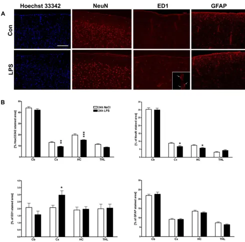
Sepsis induces microglial activation and neuronal cell loss
Histological and immunohistochemical procedures showed that the observed hemodynamic, electrophysio-logical and metabolic changes in the brain were paralleled by cell loss in the cortex and hippocampus (Figure 3A and 3B). Additionally, staining for the neuronal marker NeuN revealed a reduction in the cortical layers and the hippoc-ampus (Figure 3A and 3B). ED1-reactive microglial cells were found to be increased in LPS-treated animals, mostly located in close vicinity to brain blood vessels (Figure 3A and 3B), but also found with round to oval appearance scattered throughout the brain parenchyma (data not shown). We were not able to detect changes in GFAP immunoreactivity within 24 h after LPS administration (Figure 3A and 3B).
LPS-induced changes of inflammatory markers
RT-PCR of whole brain lysates showed that intraperito-neal injection of LPS caused an upregulation of various pro- and anti-inflammatory mediators within the brain, with the chemokine monocyte chemoattractant protein 1 (MCP-1) showing the strongest response, followed by the cytokines interleukin 1 beta (Il-1β), transforming growth factor beta (TGF-β) and tumor necrosis factor alpha
Changes of hemodynamics and electroencephalography in response to sepsis induction Sample figure title
Figure 1
Reduction of cerebral glucose uptake in septic encephalopathy
Figure 2
Reduction of cerebral glucose uptake in septic encephalopathy. Shown are five representative transversal [18
F]FDG-PET brain slices of rats treated with vehicle (Con, first row) or bacterial lipopolysaccharide (LPS, second row) at 24 h. Corre-sponding region-of-interest (ROI) masks are displayed below. B, Quantification of [18F]FDG uptake (relative to cerebellar (CB)
Discussion
Sepsis-induced brain dysfunction receives increasing attention since it directly causes brain damage [9,10] and correlates well with morbidity and mortality rates of sys-temic septicemia [2]. Despite this fact, the established
treatment protocols for patients suffering from sepsis or septic shock lack a specific neuroprotective approach, and the therapeutic strategy mainly focuses on antimicrobial drugs and stabilization of cardiovascular parameters. A prerequisite for the development of improved treatment
Histological analysis of regional LPS induced changes of cell number, micro- and astroglial activation
Figure 3
approaches is the availability of appropriate animal mod-els that use tools and techniques relevant to the clinical setting. In this study we aimed to investigate the mutual relation of CBF changes, EEG and brain metabolism in experimentally induced sepsis in rats. We correlated our findings to inflammatory gene transcription and histolog-ical analysis of neuronal loss as well as micro- and astro-glial activation.
Induction of experimental sepsis by LPS did not signifi-cantly alter systolic blood pressure, heart rate or the calcu-lated shock index at 24 h. Thus, LPS application did not cause a profound septic shock syndrome but rather resulted in slight changes reminiscent of a hyperdynamic circulatory status which can be observed in early stages of sepsis, where a marked peripheral vasodilatation is offset by a substantial increase in cardiac output resulting in lit-tle or no change of mean arterial blood pressure [11]. Of note, a mean arterial blood pressure within normal limits may resemble more closely the situation of patients who are treated with vasopressants if septic shock is present.
Since the lower limit of cerebral autoregulation in rats is around 50 mmHg [12,13], it seems unlikely that the CBF reduction found in the present study is caused by systemic cardiovascular changes, but rather results from impaired cerebral microcirculation as recently reported in a similar model [14].
During human sepsis, EEG changes are common and the degree of EEG abnormalities is associated with the clinical severity and the prognosis of SE [5]. Although EEG changes found in septic rats were not as pronounced as those described in septic patients [5], our study yielded comparable results, revealing a generalized slowing of overall EEG activity and a significant decrease of alpha activity. These findings confirm previous observations that LPS-induced sepsis in rats reduced EEG activity in fre-quency bands ≥ 8 Hz [15] up to 12 h after LPS administra-tion. In the latter study, a significant increase for frequencies between 2 and 4 Hz was only observed within the first 6 h of the experiment, as in our study where no significant changes of low frequency bands were detected at 24 h. Interestingly, both serotype and LPS concentra-tion of the latter study were different to our experimental protocol, suggesting that the observed effects on EEG activity are independent from these factors. Paralleling the LPS-induced EEG changes, microPET in vivo analysis of cerebral glucose uptake, a metabolic process that has been linked to neuronal activity in rodents [16], was signifi-cantly reduced in all cortical areas examined. Explorative data analysis showed a significant correlation of slowing of the EEG activity and the decrease of regional CBF found in septic animals. Reduction of regional CBF might there-fore, at least in part, be causative for the observed EEG-changes considered to reflect SE. Alternatively, the induc-tion of sepsis may have caused substantial brain dysfunc-tion through both, systemic release [17] as well as local generation of inflammatory molecules by peripheral immune cells or locally activated microglia. Such an acti-vation of microglial cells and astrocytes, which both can serve as major source of inflammatory molecules has been well documented in rodent models of SE [18] and brains of septic patients [9]. In this study, we found that micro-glial activation in the cortex was associated with a signifi-cant increase of inflammatory gene transcription of Il-1β, TNFα, TGF-β, iNOS and MCP-1. In addition, we observed a reduction of total and neuronal cell number in the cor-tex and hippocampus, even so we can not exclude that this may be caused by swelling of the brain. Of note, both cytokines, TNF-α and Il-1β, have been reported to affect neuronal function [4,19,20] or survival [21,22]. Similarly, iNOS-derived NO can down regulate neuronal activity [23,24], and it has been shown that neuronal viability is remarkably sensitive to sustained iNOS dependent NO generation [25-27]. Thus, inflammatory molecules in concert with inflammation-triggered NO generation may
Reduction of cerebral glucose uptake in septic encephalopa-thy
Figure 4
directly impair neuronal function and cause neurodegen-eration during SE. Neuronal cell death and a reduction of neuronal activity – as evidenced by reduced cerebral glu-cose utilization and EEG slowing, in turn, may negatively regulate the local CBF since the latter is directly coupled to the activity of the neighboring neurons [28,29]. It is there-fore likely that during SE, cerebral microcirculatory fail-ure, systemic and local inflammation and neuronal activity mutually influence and promote each other.
Conclusion
Together, the present study highlights the complexity of these changes and the early impairment of neuronal metabolism and activity during SE.
Recent data suggest that sepsis causes long-term behavio-ral changes in rodents [30,31], and minor cognitive impairment might well be present in human sepsis survi-vors [3]. Since our model mimics key aspects of human SE, it might be used to develop brain specific therapeutic strategies for patients suffering from sepsis
Competing interests
The authors declare that they have no competing interests.
Authors' contributions
AS carried out most of the EEG and cerebral CBF experi-ments and generated a first draft of the manuscript. SH carried out the small animal PET experiments. FM partici-pated in the EEG measurements. MW conducted the immunohistological analysis. SP carried out the qPCR analysis. TO participated in the EEG and CBF measure-ments. MS oversaw the small animal PET and interpreted the data. MPK helped to draft the manuscript and partici-pated in the coordination. TG helped to draft the manu-script. MTH conceived of the study, and participated in its design and coordination and helped to draft the script. All authors read and approved the final manu-script.
Acknowledgements
This study was supported by the Interdisciplinary Clinical Research Center (Central project group 4b; Hen3/003/06) Münster, Germany.
References
1. Pine RW, Wertz MJ, Lennard ES, Dellinger EP, Carrico CJ, Minshew
BH: Determinants of Organ Malfunction Or Death in
Patients with Intra-Abdominal Sepsis – A Discriminant-Analysis. Archives of Surgery 1983, 118:242-249.
2. Sprung CL, Peduzzi PN, Shatney CH, Schein RMH, Wilson MF, Shea-gren JN, et al.: Impact of Encephalopathy on Mortality in the Sepsis Syndrome. Critical Care Medicine 1990, 18:801-806. 3. Wilson JX, Young GB: Progress in clinical neurosciences:
Sep-sis-associated encephalopathy: Evolving concepts. Canadian Journal of Neurological Sciences 2003, 30:98-105.
4. Pickering M, Cumiskey D, O'Connor JJ: Actions of TNF-alpha on glutamatergic synaptic transmission in the central nervous system. Experimental Physiology 2005, 90:663-670.
5. Young GB, Bolton CF, Archibald YM, Austin TW, Wells GA: The Electroencephalogram in Sepsis-Associated Encephalopa-thy. Journal of Clinical Neurophysiology 1992, 9:145-152.
6. Soehle M, Heimann A, Kempski O: On the number of measure-ment sites required to assess regional cerebral blood flow by laser-Doppler scanning during cerebral ischemia and reper-fusion. Journal of Neuroscience Methods 2001, 110:91-94.
7. Schafers KP, Reader AJ, Kriens M, Knoess C, Schober O, Schafers M:
Performance evaluation of the 32-module quadHIDAC small-animal PET scanner. Journal of Nuclear Medicine 2005,
46:996-1004.
8. Boashash B: Estimating and Interpreting the Instantaneous Frequency of A Signal .1. Fundamentals. Proceedings of the Ieee
1992, 80:520-538.
9. Sharshar T, Annane D, de la Grandmaison GL, Brouland JP, Hopkin-son NS, Gray F: The neuropathology of septic shock. Brain Pathology 2004, 14:21-33.
10. Nguyen DN, Spapen H, Su FH, Schiettecatte J, Shi L, Hachimi-Idrissi S, et al.: Elevated serum levels of S-1000 protein and neuron-specific enolase are associated with brain injury in patients with severe sepsis and septic shock. Critical Care Medicine 2006,
34:1967-1974.
11. Parrillo JE: Mechanisms of Disease – Pathogenetic Mecha-nisms of Septic Shock. New England Journal of Medicine 1993,
328:1471-1477.
12. Tonnesen J, Pryds A, Larsen EH, Paulson OB, Hauerberg J, Knudsen GM: Laser Doppler flowmetry is valid for measurement of cerebral blood flow autoregulation lower limit in rats. Exper-imental Physiology 2005, 90:349-355.
13. Rosengarten B, Hecht M, Kaps M: Carotid compression: Investi-gation of cerebral autoregulative reserve in rats. Journal of Neuroscience Methods 2006, 152:202-209.
14. Rosengarten B, Hecht M, Auch D, Ghofrani HA, Schermuly RT, Grim-minger F, et al.: Microcirculatory dysfunction in the brain pre-cedes changes in evoked potentials in endotoxin-induced sepsis syndrome in rats. Cerebrovascular Diseases 2007,
23:140-147.
15. Lancel M, Mathias S, Schiffelholz T, Behl C, Holsboer F: Soluble tumor necrosis factor receptor (p75) does not attenuate the sleep changes induced by lipopolysaccharide in the rat dur-ing the dark period. Brain Research 1997, 770:184-191.
16. Kornblum HI, Araujo DM, Annala AJ, Tatsukawa KJ, Phelps ME, Cherry SR: In vivo imaging of neuronal activation and plastic-ity in the rat brain by high resolution positron emission tom-ography (microPET). Nature Biotechnology 2000, 18:655-660. 17. O'Dwyer MJ, Mankan AK, Stordeur P, O'Connell B, Duggan E, White
M, et al.: The occurrence of severe sepsis and septic shock are related to distinct patterns of cytokine gene expression. Shock 2006, 26:544-550.
18. Semmler A, Okulla T, Sastre M, Dumitrescu-Ozimek L, Heneka MT:
Systemic inflammation induces apoptosis with variable vul-nerability of different brain regions. Journal of Chemical Neuro-anatomy 2005, 30:144-157.
19. Tancredi V, Darcangelo G, Grassi F, Tarroni P, Palmieri G, Santoni A,
et al.: Tumor-Necrosis-Factor Alters Synaptic Transmission in Rat Hippocampal Slices. Neuroscience Letters 1992,
146:176-178.
20. Kelly A, Lynch A, Vereker E, Nolan Y, Queeman P, Whittaker E, et al.:
The anti-inflammatory cytokine, interleukin (IL)-10, blocks the inhibitory effect of IL-1 beta on long term potentiation – A role for JNK. Journal of Biological Chemistry 2001,
276:45564-45572.
21. de Bock F, Derijard B, Dornand J, Bockaert J, Rondouin G: The neu-ronal death induced by endotoxic shock but not that induced by excitatory amino acids requires TNF-alpha. European Jour-nal of Neuroscience 1998, 10:3107-3114.
22. Venters HD, Dantzer R, Kelley KW: A new concept in neurode-generation: TNF alpha is a silencer of survival signals. Trends in Neurosciences 2000, 23:175-180.
23. Mori K, Togashi H, Ueno K, Matsumoto M, Yoshioka M: Aminogua-nidine prevented the impairment of learning behavior and hippocampal long-term potentiation following transient cer-ebral ischemia. Behavioural Brain Research 2001, 120:159-168. 24. Wang QW, Rowan MJ, Anwyl R: beta-amyloid-mediated
Publish with BioMed Central and every scientist can read your work free of charge "BioMed Central will be the most significant development for disseminating the results of biomedical researc h in our lifetime."
Sir Paul Nurse, Cancer Research UK
Your research papers will be:
available free of charge to the entire biomedical community
peer reviewed and published immediately upon acceptance
cited in PubMed and archived on PubMed Central
yours — you keep the copyright
Submit your manuscript here:
http://www.biomedcentral.com/info/publishing_adv.asp
BioMedcentral
inducible nitric oxide synthase and superoxide. Journal of Neu-roscience 2004, 24:6049-6056.
25. Boje KM, Arora PK: Microglial-Produced Nitric-Oxide and Reactive Nitrogen-Oxides Mediate Neuronal Cell-Death. Brain Research 1992, 587:250-256.
26. Leist M, Fava E, Montecucco C, Nicotera P: Peroxynitrite and nitric oxide donors induce neuronal apoptosis by eliciting autocrine excitotoxicity. European Journal of Neuroscience 1997,
9:1488-1498.
27. Heneka MT, Loschmann PA, Gleichmann M, Weller M, Schulz JB, Wullner U, et al.: Induction of nitric oxide synthase and nitric oxide-mediated apoptosis in neuronal PC12 cells after stim-ulation with tumor necrosis factor-alpha lipopolysaccharide. Journal of Neurochemistry 1998, 71:88-94.
28. Ances BM: Coupling of changes in cerebral blood flow with neural activity: What must initially dip must come back up. J Cereb Blood Flow Metab 2004, 24(1):1-6.
29. Chaigneau E, Tiret P, Lecoq J, Ducros M, Knopfel T, Charpak S: The relationship between blood flow and neuronal activity in the rodent olfactory bulb. Journal of Neuroscience 2007, 27:6452-6460. 30. Barichello T, Martins WR, Reinke A, Feier G, Rifter C, Quevedo J, et al.: Cognitive impairment in sepsis survivors from cecal liga-tion and perforaliga-tion. Critical Care Medicine 2005, 33:221-223. 31. Semmler A, Frisch C, Debeir T, Ramanathan M, Okulla T,
Klock-gether T, et al.: Long-term cognitive impairment, neuronal loss and reduced cortical cholinergic innervation after recovery from sepsis in a rodent model. Experimental Neurology 2007,

![A bis[pentaiodobismuthate(III)] salt of 4 hydroxypyridinium](data:image/gif;base64,R0lGODlhAQABAIAAAP///wAAACH5BAEAAAAALAAAAAABAAEAAAICRAEAOw==)