www.fm.viamedica.pl
Address for correspondence: J. Dzierżanowski, Department of Neurosurgery, Medical University of Gdańsk, Dębinki 7, 80–957 Gdańsk, Poland, tel: +48 58 349 23 31, fax: +48 58 345 58 88, e-mail: [email protected]
Morphometry of the pterional
and pterional-orbitozygomatic approaches
to the basilar artery bifurcation by the use
of neuronavigation systems:
a new technical concept
J. Dzierżanowski, P. Słoniewski, M. Rut
Department of Neurosurgery, Medical University of Gdańsk, Poland
[Received 2 June 2008; Accepted 23 August 2008]
Much attention has been paid in the neurosurgical literature to optimising the approaches to intracranial pathology. The aims of the innovations reported are to increase the safety of operations by reduction of brain retraction and to improve exposure of the neurovascular structure in the operating area. It was our intention to investigate whether an image-guided frameless stereotactic system is suitable in morphometric studies based on the analysis of the pterional and the cranio-orbi-tozygomatic approaches to the basilar artery bifurcation (BAB).
We analysed 60 virtual models of pterional craniotomy and the same number of those extended by orbitozygomatic osteotomy, created using computer tomogra-phy in the neuronavigation system. It was decided to calculate the percentage change of the cranial area of exposure, the depth of the surgical corridor and the angle of view to the bifurcation of the basilar artery. Three positions of the BAB (normal, high and low) were examined for each model of craniotomy.
In the material analysed, after the extension of the pterional craniotomy by orbitozy-gomatic osteotomy, the cranial area of exposure for 60 models of cranio-orbitozy-gomatic craniotomies increased by a mean of 39.28% (from 30.89% to 48.06%). The decrease in the depth of the surgical corridor for a normal-lying BAB was 19.16%, for a high-lying BAB 19.09% and for a low-lying BAB 19.12%. The mean changes in the individual BAB locations did not differ significantly in statistical terms (F = 0.011; p = 0.99). The mean increase in the cranial angle of attack for a normally located BAB was 10.72°, for a high-lying BAB 11.1° and for a low-lying BAB 10.31°. The post-hoc test showed significant differences in the angle of attack between a normal and a low-lying BAB (p = 0.034) and a high and a low-lying BAB (p = 0.00007). Neuronavigation systems, already well-known for their intraoperative use, can also be useful in morphometric studies, and the advantages of this method are the practically unlimited number of results which can be analysed in detail and the repeatability of the technique. The pterional-orbitozygomatic approach compared to the pterional increases the working area, minimises retraction of the brain, shortens the working distance, enables instruments to be used more easily, widens the angle of view and improves the visibility of the anatomical structures in the working area, especially for a high-lying BAB. (Folia Morphol 2008; 67: 267–272)
INTRODUCTION
Much attention has been paid in the neurosurgi-cal literature to optimising surgineurosurgi-cal approaches [12, 13, 15, 16, 18, 21, 24]. Changes in such para-meters as the working area and the angle of view into anatomical structures after extension by orbitozy-gomatic osteotomy are aspects of this that are ana-lysed most frequently. The published measurements for the neurosurgical approaches are based on the assumption that quantification will be useful for the surgeon in deciding which approach is the best for any particular case.
The aim of this anatomical study is to present a new concept using an image-guided frameless ste-reotactic system in vivo for analysing morphometric surgical approaches on the basis of the pterional (PT) and pterional-orbitozygomatic (POZ) approach-es to the basilar artery bifurcation (BAB) by means of preoperative computer tomography (CT) images. We have measured the percentage change in “the cranial area of exposure”, the depth of the “cranial surgical corridor” and “the cranial angle of attack” in degrees, when a pterional craniotomy was extend-ed to an orbitozygomatic osteotomy. Our goal was to measure the parameters of a surgical approach in relation to high, normal and low-lying bifurcations.
MATERIAL AND METHODS
Sixty virtual models of PT and POZ craniotomies were analysed using CT scans and a neuronaviga-tion system. All images were made with the GE Medical System and the neuronavigation Stealth Station, Medtronic Treonä with Cranial 5 software. Virtual models of craniotomies were created by precise studies of approaches on the skulls as shown in Figure 1A and 1B. Extreme points of craniotomy were marked and their solid location then studied. Analogous points were marked on the bone recon-struction in navigation. After the connection of these points we obtained a broad outline of the PT, which was increased by the orbitozygomatic part. The model of the surgical approach defined is clear and simple to use in further calculations (Fig. 2). We anal-ysed only the benefits after extensive craniotomy, with no absolute values of the surgical window or the angle of attack.
The change in the depth of the cranial surgical corridor was measured along the trajectory of the approach to the BAB along the lesser wing of the sphenoid bone. For each model of craniotomy we examined three locations of the BAB. The BAB was considered normal when was located at the level of
the line connecting the anterior and posterior cli-noids, a high bifurcation being more than 5 mm above this line and a low bifurcation more than 5 mm below this line on the basis of other authors’ experience [1, 22].
The increase in the cranial angle of attack was measured along the wing of the sphenoid bone. This is the angle between the lower planes of PT and POZ craniotomies.
Statistical analysis
The statistical analysis was performed using STATISTICA 7.0. software. Statistical comparisons be-tween the various measurements, such as the crani-al window of exposure, the cranicrani-al angle of attack and the depth of the cranial corridor, were per-formed using repeated-measurement analysis of variance followed by the Kolmogorov-Smirnov, Lilliefors and Lavenea tests and post-hoc analysis
Figure 1. The pterional craniotomy (A) and pterional-orbitozygomatic craniotomy (B); extreme points of the craniotomy were marked and their solid location was analysed. The results were used to create the virtual craniotomies in the neuronavigation station.
A
(ANOVA). In all cases p £ 0.05 was considered sig-nificant.
RESULTS
The cranial area of exposure or the superficial external window of exposure for all the models of craniotomies was larger for the POZ approach by a mean of 39.28% (from 30.89% to 48.06%), and differences in the working window were statistical-ly significant (Fig. 3)
The mean decrease in the depth of the cranial surgical corridor (the distance from the surface of the craniotomy to the BAB) for the normal-lying BAB was 19.16%, for a high-lying BAB it was 19.09% and for a low-lying BAB 19.12% (Fig. 4).
The mean changes in the depth of the cranial sur-gical corridor in the individual BAB locations (high, normal and low) did not differ in a statistically signi-ficant way F (2.117) = 0.011; p = 0.991) (Fig. 4, 5).
In converting a PT approach to a POZ approach we found that the increase in the cranial angle of
Figure 2. Image showing the building of a virtual model of craniotomies using the bone reconstruction of patients’ heads in the neuronavi-gation system. We defined and connected the extreme points of these two approaches in three projections: sagittal, coronal and axial.
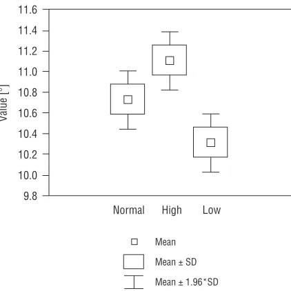
attack for a normally located BAB was a mean of 10.72°, for a high-lying BAB 11.10° and for a low-lying BAB 10.31° (Fig. 5). With the use of a post-hoc NIR test significant differences were demonstrated between the angles of attack of the normal and low-lying BABs (p = 0.034) and the high and low-low-lying BABs (p = 0.001)
DISCUSSION
The history of neuronavigation systems (frame-less stereotactic systems) is short and dates back to the early 1990s. Their utilisation provides for con-tinuous monitoring of the position of microsurgical instruments in the working area with reference to the nerve structures. It also facilitates decision-mak-ing about the total excision of the pathology, espe-cially when it is located in the neighbourhood of “the eloquent structures”. Because of this, the risk is reduced of complications associated with exten-sion of the approach, the manipulation of micro-surgical instruments, bleeding decreases and the duration of the operation [3, 5, 10, 14, 25].
The majority of morphometric studies have been performed in vivo using cadaveric heads [6, 7, 11, 19, 20, 22]. Although they seem to have faithfully rendered operative conditions, some authors reveal their limitation in anatomical studies [7, 11]. This arises for the following reasons. Cadaveric heads are
chemically fixed, which causes a difference in the stiffness and resistance of tissues [7, 11]. Spatial relationships change due to the lack of blood circu-lation or cerebrospinal fluid. Either the range of os-teotomy or the retraction of the brain with spatulas in reaching the aneurism can be maximal and al-most limitless, which cannot be repeated in in vivo conditions. The limitation is also in the small num-ber of anatomical preparations [11], which, with some variability in anatomical structure, determines the type of statistical analysis.
The advantage of this study is the practically unlimited number of results which can be analysed in detail. The measurements were not performed in vivo for three reasons. Firstly, an additional adjust-ment with the active microprobe could cause com-plications for patients and might extend the opera-tive time. Secondly, the working of the system is linked to errors. The registration error is not more than 2–3 mm, which is calculated by a computer at the beginning of the work after the registration of 47 chosen points from the surface of the patient’s head. Additionally, the error may be increased by any interoperative changes connected with the ro-tation of the head, which is immobilised in the three-point headholder during the craniotomy [14], brain oedema or brain shift because of the force of grav-ity or after the release of cerebrospinal fluid [17].
Figure 4. Graph of the percentage decrease in the depth of the cranial surgical corridor after extension pterional craniotomy of the orbitozygomatic part for high, normal and low-lying basilar bifurcations.
Thirdly, it would be impossible to calculate how much the parameters (the cranial angle of attack, the cranial area of exposure, the depth of the crani-al surgiccrani-al corridor) have changed in relation to the extent of the craniotomy with respect to an indivi-dual patient.
The results obtained show that the increase in the cranial area of exposure for the pterional craniotomy after orbitozygomatic osteotomy is between 30.89% and 48.06% (mean 39.28%). We emphasise that this value should not be interpreted as an increase in the real working area, which, being situated deeply in the surgical corridor is also limited by neurovascular structures. The orbitozygomatic osteotomy to the skull base causes only the possibility of better visual-isation. The same concerns the depth and angle of attack of the surgical corridor or area. Similar results were obtained by Schwarz et al. [20], who have shown that the increase in the area of exposure in approach-ing the basilar tip target in orbitozygomatic osteoto-my was 39–51%. Unfortunately, the authors do not disclose what the relation was of the BAB to the pos-terior clinoid process.
In ideal conditions the depth of the surgical ap-proach corresponds to the distance from the sur-face of the craniotomy to the neck of the aneurysm. However, during the operation the surgeon’s hands are situated above the brain surface. The relation-ship between the side of the approach and the left-or right-handedness of the surgeon is also impleft-or- impor-tant. The following factors increase the depth of the surgical approach, so that the term “depth of the surgical corridor” as presented in previous studies is relative. This is why we have introduced the term “cranial surgical corridor,” the distance from the low-er edge of both craniotomies (the skull base) to the BAB moving along the trajectory of approach (the wing of the sphenoid bone). In the calculation pre-sented the decrease in the cranial surgical corridor is about 19%. According to Andaluz et al. [2], who measured the morphometric dependence of pte-rional and orbitoptepte-rional craniotomies, the orbito-pteronial approach shortened the distance from 62 mm to 54 mm, with a mean of 13%. The authors concluded that a shorter distance to the target point results in improved illumination and observation for more precise surgical movements [2].
The mean increase in the cranial angle of attack for a normally located BAB was 10.72°, for a high-lying BAB 11.10° and for a low-high-lying BAB 10.31°. We found that the increase in cranial angle of at-tack is statistically significant for high and
low--lying BABs and for normal and lowlow--lying BABs. Sin-dou et al. [23] in an anatomical study using 11 ca-daveric heads demonstrated that the orbitozygo-matic osteotomy in combination with a pterional craniotomy increased the angle of view by 10° in reaching the basilar bifurcation along the sylvian fis-sure. Chanda and Nanda [4] (test performed with 5 preparations) reported a 13°–14° increase in the angle of view for the BAB. Unfortunately the change in the angle value with regard to the anatomical va-riety of the BAB location in relation to the posterior clinoid process has not been analysed in any previ-ous study. This is similar to the results found by Gonzalez et al. [11], who quantitatively assessed dif-ferent variations of the pterional, orbitozygomatic and maxillar approaches to determine the working area and the angle of attack in three virtual triangles in the anterior cranial fossa. They showed an increase in the angle of attack for the anterior clinoid process of 10.2 ± 0.7° after extending the pterional craniotomy of an orbitozygomatic osteotomy. Figueiredo et al. [8] compared the angle of approach and area of expo-sure to the ACoA offered by the pterional, orbitopte-rional and orbitozygomatic approaches before and after gyrus rectus resection. The vertical angle, de-fined as the main craniocaudal axis of the approach perpendicular to the cranial base, increased signifi-cantly between the PT and POZ approaches by 10.2° before gyrus rectus resection [8].
CONCLUSION
Neuronavigation systems, already well-known for their intraoperative use, can also be useful in mor-phometric studies, and the advantages of this meth-od are the practically unlimited number of results which can be analysed in detail and the repeatabil-ity of the technique.
Morphometric studies provide much information that is useful for the analysis of surgical approach-es, but the clinical value of precise measurements with descriptive statistics needs careful judgment. There are in vivo studies that are linked to errors which do not take into consideration all aspects of the operation.
REFERENCES
1. Abdel-Aziz KM, van Loveren HR, Tew JM (1999) The Kawase approach to retrosellar and upper clival basi-lar aneurysms. Neurosurgery, 44: 1225–1236. 2. Andaluz N, von Loveren HR, Keller JT (2003) Anatomic
and clinical study of the orbitopterional approach to anterior communicating artery aneurysms. Neurosur-gery, 52: 1140–1149.
3. Benardete EA, Leonard MA, Weiner HL (2001) Com-parison of frameless stereotactic systems: accuracy, precision and application. Neurosurgery, 49: 1409– –1416.
4. Chanda A. Nanda A (2002) Anatomical study of the orbitozygomatic transsellar-transcavernous-transcli-noidal approach to the basilar artery bifurcation. J Neu-rosurg, 97: 151–160.
5. Dorward NL, Alberti O, Palmer JD (1999) Accuracy of true frameless stereotaxy. J Neurosurg, 90: 160–168. 6. Erturk M, Kayalioglu G, Ozer MA (2003) Morphome-try of the anterior third ventricle region as a guide for the subfrontal (translaminaterminalis) approach. Neu-rosurgical Rev, 26: 249–252.
7. Evans JJ, Hwang YS, Lee JH (2000) Pre- versus post-anterior clinoidectomy measurements of the optic nerve, internal carotid artery and opticocarotid trian-gle: a cadaveric morphometric study. Neurosurgery, 46: 1018–1023.
8. Figueiredo EG, Zabramski JM, Deshmukh P, Crawford NR, Spetzler RF, Preul MC (2006) Comparative analysis of anterior petrosectomy and transcavernous approaches to retrosellar and upper clival basilar artery aneurysms. Neurosurgery, 58 (ONS suppl. 1): ONS13–ONS21. 10. Germano IM, Villalobos H, Silvers A (1999) Clinical use
of the optical digitizer for intracranial neuronaviga-tion. Neurosurgery, 45: 261–270.
11. Gonzalez LF, Crawford NR, Horgan M (2002) Working area and angle of attack in three cranial base ap-proaches: pterional, orbitozygomatic and maxillary extension of the orbitozygomatic approach. Neuro-surgery, 50: 550–557.
12. Hakuba A, Liu S, Nishimura S (1986) The orbitozygo-matic infratemporal approach: a new surgical tech-nique. Surg Neurol, 26: 271–276.
13. Heros RC, Lee SH (1993) The combined pterional/an-terior temporal approach for aneurysms of the upper basilar complex. Technical note. Neurosurgery, 33: 244–251.
14. Hill DL, Maurer Cr Jr, Maciunas RJ (1998) Measure-ment of intraoperative brain surface deformation un-der a craniotomy. Neurosurgery, 43: 514–528. 15. Kato Y, Sano H, Behari S (2002) Surgical clipping of
basilar aneurysms: Relationship between the different approaches and the surgical corridors. Minim Invas Neurosurg, 45: 142–145.
16. Krisht AF, Kadri P (2005) Surgical clipping of complex basilar apex aneurysms: a strategy for successful out-come using the pterional transzygomatic transcavern-ous approach. Neurosurgery, 56 (ONS suppl. 2): ONS261–ONS273.
17. Nabavi A, McL Black P, Gering DT (2001) Serial intra-operative magnetic resonance imaging of brain shift. Neurosurgery, 48: 787–798.
18. Sano K (1980) Temporopolar approach to aneurysms of the basilar artery and around the distal bifurcation. Neurol Res, 2: 361–367.
19. Sato S, Sato M, Oizumi T (2001) Removal of anterior clinoid process for basilar tip aneurysm: clinical and cadaveric analysis. Neurol Res, 23: 298–303.
20. Schwartz MS, Anderson GJ, Horgan MA (1999) Quan-tification of increased exposure resulting from orbital rim and orbitozygomatic osteotomy via the frontotem-poral transsylvian approach. J Neurosurg, 91: 1020– –1026.
21. Sekhar LN, Kalia KK, Yonas H (1994) Cranial base ap-proaches to intracranial aneurysms in subarachnoid space. Neurosurgery, 35: 472–483.
22. Seoane E, Tedeschi H, de Oliviera E (2000) The pre-temporal transcavernous approach to the interpedun-cular and prepontine cisterns: microsurgical anato-my and technique application. Neurosurgery, 46: 891–899.
23. Sindou MP, Alaywan M (1994) Fronto-temporal ap-proaches with removal of the orbital rim and/or the zygomatic arch: surgical technique, microsurgical anat-omy and clinical applications. In: Samii M (ed) Skull base surgery: anatomy, diagnosis and treatment. Basel, Karger, pp. 739–741.
24. Sindou M, Emery E, Acevedo G (2001) Respective indi-cations for orbital rim, zygomatic arch and orbito-zy-gomatic osteotoomies in the surgical approach to cen-tral skull base lesions. Critical, retrospective review in 146 cases. Acta Neuroch, 143: 967–975.