This is an open access journal, and articles are distributed under the terms of the Creative Commons Attribution-Non Commercial-ShareAlike 4.0 License, which allows others to remix, tweak, and build upon the work non-commercially, as long as appropriate credit is given and the new creations are licensed under the identical terms.
© 2018 Journal of Advanced Pharmacy Education & Research | Published by SPER Publication
24
Cortical activation after constraint induced movement therapy in
stroke patients: A randomized controlled trial
Gehan M. Ahmed
1, Ebtesam M. Fahmy
2, Saly H. Elkholy
3, MoatazEl Semary
1, Abeer A. Mohammed
1*,
Wanees M. Badawy
11 Department of Neuromuscular Disorder, Faculty for Physical Therapy, Cairo University, Cairo, Egypt, 2 Department of Neurology, Faculty of Medicine, Cairo University,
Cairo, Egypt, 3 Clinical Neurophysiology Unit, Faculty of Medicine, Cairo University, Cairo, Egypt.
Correspondence: Abeer A. Mohammed, Assistant Lecturer at the Department of Neuromuscular Disorder, Faculty for Physical Therapy, Cairo University. Address: 7
Ahmed El-Zayat Street, Dokki, Giza, Egypt. Telephone: +201121606559, Fax: 202 37482979. Postal Code: 12613. E-mail: [email protected]
ABSTRACT
Background: Constrained induced movement therapy (CIMT) has been a neurorehabilitation method that proved to improve arm function and induce neural plasticity after stroke. However, there have been limited controlled trials discussing the cortical activity changes after CIMT in chronic stroke. Objective: This study aimed to scrutinize the effect of adding a constraint induced movement therapy to the intensive physical therapy program on cortical activity and functional outcome of the upper extremity in patients with chronic stroke. Subjects and Methods: Forty stroke patients were included. They were randomly assigned into two equal groups; study group received functional training with CIMT in addition to intensive physical therapy program, and the control group received intensive physical therapy program only. The outcome measures included: the motor function of the upper extremity using the Wolf Motor Function Test (WMFT), and the cortical activity of the affected cerebral hemisphere using the Quantitative Electroencephalogram (QEEG). Results: There was a significant increase in WMFT mean scores in the study and control groups post treatment (P<0.0001, 0.015 respectively), with remarkable improvement in the study group (P= 0.0496). A statistically significant increase in alpha wave frequency, relative and maximum power post treatment in the study group was only (p<0.05). A statistically significant decrease in theta wave, relative and maximum power was also detected in the study group post treatment (P=0.0091, 0.0176 respectively). A statistically significant difference was observed between both groups regarding post treatment of mean values of alpha and theta waves variables (p<0.05), except for theta wave frequency (p=0.308). Conclusion: CIMT improved the motor function of upper extremity and was associated with increased cortical activation of the affected hemisphere in patients with chronic stroke.
Keywords: Stroke, Constrained induced movement therapy, Quantitative EEG.
Introduction
Stroke has been the most common cause of long-term disability. Functional impairment of upper extremity has been commonly encountered after stroke. Thirty-three to 66% of the patients with arm paresis showed minimal recovery of function six months after the stroke [1, 2]. Even after recovery, about 20% of
the stroke survivors cannot independently complete different activities of daily livings (ADLs); such as feeding and personal care. Those patients tend to rely on their unaffected arm to perform ADLs, leading to “nonuse” with progressive suppression of movements [1, 3].
Reducing disability and improving the functional outcomes of the upper extremity have been the major goals in stroke rehabilitation. Constraint-induced movement therapy (CIMT) and modified CIMT have been the common approaches that have been used for upper limb rehabilitation in mild and moderate motor dysfunction after stroke. CMIT has been primarily developed to overcome learned nonuse phenomena, and improve motor functions of the affected upper extremity [4]. It was also proved to induce cortical reorganization. This therapeutic approach has been based on two main principles: the forced use of the affected arm by constraining of the unaffected
Access this article online
Website: www.japer.in E-ISSN: 2249-3379
How to cite this article: Gehan M. Ahmed, Ebtesam M. Fahmy, Saly H. Elkholy, MoatazEl Semary, Abeer A. Mohammed, Wanees M. Badawy.
Cortical activation after constraint induced movement therapy in stroke patients: A randomized controlled trial. J Adv Pharm Edu Res 2018;8(3):24-29.
one, and the massed practice of the involved arm in a functional task through a shaping method [5, 6].
Review studies reported a lack of homogeneity of the studies investigating the effects of CIMT, and provided variable results concerning the effects of CIMT on motor impairment, functions and disability levels of arm and hand. Understanding the cortical changes which accompany improvement of motor function after CIMT still needs a lot of research [4, 6].
This study was designed to investigate the effect of combined constraint induced movement therapy and the intensive physical therapy program on cortical activity and functional outcome of the upper extremity in patients with chronic stroke.
Patients and Methods
Study design
The study was a randomized, single-blind, pre–post-test, controlled trial. It was approved by the official ethical committee at the Faculty of Physical Therapy, Cairo University (No: P.T.REC/012/00976) and followed the Guidelines of Declaration of Helsinki on the conduct of the human research.
Participants
Fifty-seven patients, of both sexes, with chronic ischemic stroke were screened for the study eligibility. Patients were recruited from the Outpatient Clinics of Cairo university hospitals, and Faculty of Physical Therapy, Cairo University, between June 2016 and November 2017.
Inclusion criteria were:
• Age range between 45 and 60 years.
• Duration of illness between six months to two years.
• Motor deficit involving one arm with the proper use of the unaffected hand for ADLs.
• Ability to perform at least 20° wrist extension and 10° finger extension of the affected upper limb.
• Action Research Arm Test (ARAT) score <50 in the affected arm [7].
• Adequate balance and safety while wearing the restraint [8].
• Mild spasticity of the affected upper limb with a score of (1) or (1+) according to Modified Ashworth’s Scale (MAS) [9].
Exclusion criteria were: aphasia, perceptual problems like apraxia or unilateral spatial neglect, visual or auditory defects, recurrent strokes, ataxia or sensory impairment, shoulder pain or musculoskeletal problems that could interfere with training, shoulder-hand syndrome, cognitive dysfunction and EEG abnormalities or epilepsy.
Randomization
Informed consent was obtained from all the patients after detailed explanation of the study details. The participants were
also informed about the privacy of all the obtained information and their right to refuse or withdraw at any time. All data were coded to ensure anonymity. The patients were randomly assigned to two groups: the study group (n=20) and the control group (n=20). Randomization was performed by a blinded and an independent research assistant using a computer-generated randomization cards saved in sealed envelopes.
Physical therapy intervention:
One experienced physical therapist having 14 years of clinical experience in the field of neurorehabilitation treated all the patients. All the patients were treated for two hours, 5 days per week for successive four weeks (5X4=20 therapeutic sessions) [10].
The study group received a training task with CIMT in addition to an intensive physical therapy program [10]. The CIMT focused on the restriction of the unaffected hand, with a mitt, and intensive training of the affected upper extremity with functional activities. The activities included gross and fine motor skills training such as throwing a ball, folding clothes, using a spoon, writing and picking the object with specific grasps. Shaping techniques were also considered to change and increase the level of task difficulty according to the patients’ ability.
The control group received only the intensive physical therapy program which included: active resisted strengthening exercises for weak upper limb muscles, stretching exercises for tight muscles of the affected upper limb, postural control, balance training, and finally a proprioceptive Neuromuscular Facilitation (PNF) training.
Assessment and outcome measures
The following outcome measures were assessed before and after the end of the treatment intervention:
The measurement of the motor function of upper extremity using the WMFT:
This scale was used based on the report of Morris et al, [11] which indicated that WMFT has been a useful measure for the effectiveness of CIMT in patients with stroke. It has been a time-based scale composed of 15 functional tasks that have been divided into; performance time and functional ability [12]. Studies showed good reliability, validity, and responsiveness of the test in patients with chronic stroke [13]. An excellent inter-test and inter-rater reliability, and internal consistency and stability had been reported for both the performance time and Functional Ability rating scale measures (FAS). The intra-class correlation coefficient (ICC) values were between0.88 to 0.98, with most of the values being near to 0.95 [11-13].
The measurement of cortical activity changes utilizing the QEEG:
Before the evaluation, all the participants were informed about all the instructions by the same technician who made the EEG examination for all the patients. Data analysis was performed by an experienced electroencephalographer for the data collected over the sites O1 and O2 which represented the integration areas of the alpha wave of left and right hemispheres, respectively. Frequency and relative power of alpha wave were analyzed for the affected hemisphere for all the participants [14, 15].
Data analysis and statistical design
- The collected data were statistically analyzed using the
SPSS program for Windows, version 18 (SPSS, Inc., Chicago, IL) for:
- The arithmetic mean was an average description of central
tendency for the observations.
- The standard deviation was a mean of dispersion of results. - Mann Whitney test was used to analyze the data of WFMT - Paired T-test was used to compare the mean of alpha wave
variables pre and post treatment within each group.
- Unpaired t-test was used to compare the mean of alpha
wave variables pre and post treatment of two independent groups.
- The statistical significance was set with the value of P ≤
0.05.
Results
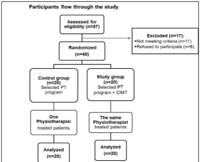
A total of 57 patients with stroke were eligible for inclusion (figure 1). Forty patients who were included and randomized into the study group (n=20) received functional training with CIMT in addition to intensive physical therapy program, and the control group (n=20) received only intensive physical therapy program. Figure (1) demonstrated the flow chart of the participants throughout the study. At baseline, both groups were matched (P > 0.05) in their general characteristics (table 1).
Figure 1: Participants flow chart. PT=physical therapy.
General Characteristics of Subjects:
The clinical characteristics of included subjects are represented in table (1).
Table 1: Demographic and clinical characteristics of subjects.
General Characteristics
Control Group Study Group Comparison Mean ±SD Mean ±SD t-value P-value
Age(yrs.) 54.30±5.401 52.60± 4.773 1.055 0.2982
Weight(Kg) 76.15±4.534 73.00± 9.222 1.371 0.1785 Height (cm) 175.1±5.370 172.2±6.940 1.478 0.1477 BMI (Kg/m2) 24.85±1.359 24.57±2.282 0.4733 0.6387
Duration of
illness(months) 12.40±4.784 11.60± 5.345 0.4988 0.6208
SD: standarddeviation, yrs.: years, Kg: kilogram, cm: centimeter, BMI: body mass index, P: probability, Significance :( P<0.05).
WMFT scores:
A significant increase in the scores of WMFT was found in the study and control groups post treatment (P<0.0001, 0.015 respectively). Moreover, a significant difference was detected between both groups post treatment with remarkable improvement in the study group (P= 0.0496)Table (2)
QEEG Parameters:
Within groups
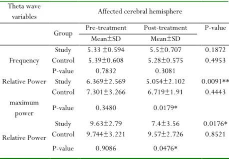
The comparison of the mean values of alpha wave variables on the affected side in pre and post treatment revealed a statistically significant increase in alpha wave frequency, relative and maximum power post treatment only in the study group (p=0.0160, 0.016 and 0.0039 respectively), as can be seen in Table (3). A statistically significant decrease in theta wave relative and maximum power was also detected in the study group post treatment (P=0.0091, 0.0176 respectively), while there was no significant difference in theta wave frequency post treatment in the study group (p=0.1872), as shown in Table (4) The comparison of the mean values of alpha and theta waves variables within the control group revealed no significant differences (p<0.05)., as shown in Tables (3, 4).
Table 2: Comparison of WMFT scores pre and post treatment within and between both groups.
WMFT score
Pre-treatment Post-treatment
P- value
M
in
im
um
Max
imu
m
Me
di
an
IQR
M
in
im
um
Max
imu
m
Me
di
an
IQR
Study group 11.00 67 39.50 27.5 26.00 71 50.50 24 <0.0001**
Control
group 12.00 66 37.50 10.50 11.00 59 41.50 18.50 0.0155*
P- value 0.5154 0.0496*
SD: standard deviation, P-value: probability value, *Significant, ** highly significantI QR (interquartile range)
Between groups
both groups (p<0.05) except for theta wave frequency(p=0.308), as can be seen in Tables (3, 4).
Table 3: Comparison of alpha wave variables pre and post treatment within and between both groups. Alpha wave variables Affected cerebral hemisphere
Group Pre-treatment Post-treatment P-value Mean±SD Mean±SD
Frequency Control 8.611Study 9 ±±0.686 0.6978 8.83 9.444±±0.618 0.922 0.0160*0.2151 P-value 0.1009 0.0255*
Relative Power
maximum Power
Study 8.991±4.227 11.57±3.486 0.016*
Control 9.427±4.428 8.576±4.14 0.3590 P-value 0.7643 0.0248*
Relative Power
Study 18.05±7.399 22.1±9.16 0.0039** Control 16.52±7.074 15.46±7.14 0.2832 P-value 0.5300 0.0208*
SD: standard deviation, P-value: probability value, *Significant, ** highly significant
Table 4: Comparison of theta wave variables pre and post treatment within and between both groups. Theta wave
variables Affected cerebral hemisphere
Group Pre-treatment Post-treatment Mean±SD Mean±SD P-value
Frequency
Study 5.33 ±0.594 5.5±0.707 0.1872 Control 5.39±0.608 5.28±0.575 0.4953 P-value 0.7832 0.3081
Relative Power
maximum power
Study 6.369±2.569 5.054±2.102 0.0091** Control 7.301±3.266 6.719±1.91 0.4443 P-value 0.3480 0.0179*
Relative Power
Study 9.63±2.79 7.4±3.56 0.0176* Control 9.744±3.221 9.57±2.726 0.8521 P-value 0.9086 0.0476*
SD: standard deviation, P-value: probability value, *Significant, **highly significant
Discussion and Conclusion
Functional impairment of the upper extremity remains a challenge in the field of stroke rehabilitation. In many studies, the CIMT has been commonly investigated as a single approach for upper extremity rehabilitation, and showed positive results [13, 16]. From another perspective, combining therapeutic approaches would provide different sensory input and consequently could facilitate the motor functions of upper extremities in a different way [17].
The results of this study highlighted the efficacy of the additional CIMT to the intensive rehabilitation program in improving cortical excitability, and functional performance of upper extremity in chronic stroke patients. This improvement was remarkable in the study group and can be explained considering the effect of CIMT in which repetitive task practices induced neural plasticity and increased the cortical map of the affected limb [18], and consequently improved the limb awareness and functional activities [18, 19]. These findings were matched with the results of Singh and Pradhan, [20] and Lin et al, [13] which showed a significant improvement in the motor function of upper
extremity after CIMT measured by WFMT, [12] and Fugl Meyer scale [21]. On the contrary, Sirtori et al, [22] revealed a limited effect of CIMT on motor impairment of upper extremity. This contradiction can be attributed to variations in participant selection criteria, sample size, the design of the study, and the outcome measures.
In the current study, QEEG was specifically used to monitor changes in brain pathophysiology in patients with chronic stroke [23]. The researchers focused on assessing reorganization in cortical occipital areas, (O1) and (O2), because changes of activation in these areas were related to alpha wave integration and stroke recovery [24]. After the stroke, there was a decrease in the power of faster frequency brain bands (alpha and beta) and an increase in the slow frequency bands (delta and theta). The disturbing power was associated with reduced brain metabolism (CMRO2) that influenced brain activation and function [25]. Further, Schleiger et al., [24] reported that alpha slowing the following ischemic stroke can inform prognoses of stroke. Consequently, changes in alpha frequency and relative power were the main variables of interest in this work aiming to detect the post-treatment effect of the user intervention on cortical reorganization. The results of this study showed a significant improvement in alpha wave variables in the study group compared to the controls. The improvement of cortical activation in this study can explain the remarkable change in the motor function of the affected arm in the study group who received CIMT. This explanation agreed with Carrion et al., [26] who stated that delta and alpha bands were the signs of recovery process; the higher the alpha power, the better the patients' outcome. They also stated that relative QEEG power in alpha bands had a tendency to increase with improvement in motor performance and activities of daily livings (ADLs). The results also came in agreement with Szaflarski et al., [27] who concluded that the increased use of the affected arm during CIMT induces cortical reorganization, as measured by QEEG. Additionally, the authors reported that the amount of cortical reorganization was positively related to the degree of increase in affected arm use and ability.
Limitations
One limitation of the current study was that the primary outcomes were the measurement of the upper extremity motor function and cortical activity changes as direct effects of the use of CIMT. However, other outcomes as muscle strength, postural control and balance function, were not a target of this work. Another limitation could be lacking after-treatment effects. Consequently, it could be recommended to evaluate the long-term effects of the additional CIMT on both motor function of upper extremity and cortical activity.
With the limitations of this study, it could be concluded that adding CIMT to intensive physical therapy program has been valuable and effective in enhancing cortical reorganization and motor functions of the upper extremity in chronic stroke. Future research is still needed for better understanding and analysis of the relation between cortical activity changes and improvement of motor function. Future research is also recommended to evaluate the after-effect of CIMT on arm motor functions, ADLs, and the quality of life.
Acknowledgment
The authors would like to thank all the patients who participated in the study.
Financial support and sponsorship
Nil.Conflicts of interest
There were no conflicts of interest.References
1. Albert SJ, Kesselring J (2012). Neurore habilitation of stroke. Journal of Neurology; 259(1):817-32.
2. Castellanos NP, Leyva I, Buldu JM et al. (2011). Principles of recovery from traumatic brain injury: reorganization of functional networks. NeuroImage; 55: 1189-99.
3. Hammer A, Lindmark B. (2009). Effects of forced use on arm function in the subacute phase after stroke: a randomized, clinical pilot study. Physical Therapy; 89(6):526-39.
4. Thrane G, Askim A, Stock R, et al. (2015). Efficacy of Constraint induced movement therapy in early stroke rehabilitation: A randomized controlled multisite trial. Neurorehabilitation and Neural Repair; 29: 517-25. 5. Wu, CY, Chuang, LL, Lin, KC, et al. (2011). Randomized
trial of distributed constraint-induced therapy versus bilateral arm training for the rehabilitation of upper-limb motor control and function after stroke. Neurorehabilitation and Neural Repair; 25:130-39.
6. Kwakkel G, Veerbeek JM, van Wegen EEH, et al. (2015). Constraint-Induced Movement Therapy after Stroke. The Lancet Neurology; 14 (2):224-34.
7. Kitago T, Liang J, Vincent S. et al. (2012). Improvement after Constraint-Induced Movement Therapy: Recovery of Normal Motor Control or Task-Specific Compensation. Neurorehabilitation and Neural Repair; 27(2) 99–109. 8. Wolf SL, Winstein CJ, and Miller JP, et al. (2006). Effect
of constraint-induced movement therapy on upper extremity function 3 to 9 months after stroke: The EXCITE randomized clinical trial. JAMA; 296:2095–104.
9. Ansari NN, NaghdiS, ArabTK, et al. (2008). The interrater and intrarater reliability of the Modified Ashwer Scale in the assessment of muscle spasticity: limb and muscle group effect. Neurorehabilitation; 23(3):231-7.
10. Myint JM, Yuen GF, Yu TKet al. (2008). A study of constraint-induced movement therapy in subacute stroke patients in Hong Kong. ClinRehabil; 22(2):112–24. 11. Morris DM, Uswatte G, Crago JE, et al. (2001). The
Reliability of the Wolf Motor Function Test for Assessing Upper Extremity Function after Stroke. Arch Phys Med Rehabil; 82:750‐755.
12. Wolf SL, Catlin PA, Ellis M, et al. (2001). Assessing Wolf Motor Function Test as Outcome Measure for Research in Patients after Stroke. Stroke; 32:1635‐1639.
13. Lin KC, Huang YH, Hsieh YW, et al. (2009). Potential predictors of the motor and functional outcomes after distributed constraint-induced therapy for patients with stroke. Neurorehabil Neural Repair; 23:336–342. 14. Niedermeyer E. (2005). Cerebrovascular disorders and
EEG, in Electroencephalography: Basic Principles, Clinical Applications, and Related Fields, 3rd EdsNiedermeyer E., Lopes da Silva F. editors. (Philadelphia, PA: Lippincott Williams and Wilkins), 339–362.
15. Schleiger E, Sheikh N, Rowland T, et al. (2014). Frontal EEG delta/alpha ratio and screening for post-stroke cognitive deficits: the power of four electrodes. Int. J. Psychophysiol; 94: 19–24.
16. Wu CY, Chen YA, Lin KC, et al. (2012). Constraint-induced therapy with trunk restraint for improving functional outcomes and trunk-arm control after stroke: a randomized controlled trial. Phys Ther; 92: 483–492. 17. Aman JE, Elangovan N, Yeh I-L, Konczak J. (2014). The
effectiveness of proprioceptive training for improving motor function: a systematic review. Frontiers in Human Neuroscience; 8: 1075.
18. Wolf SL, Winstein CJ, and Miller JPet al. (2008). Retention of upper limb function in stroke survivors who have received constraint-induced movement therapy: The EXCITE randomized trial. Lancet Neurol; 7:33–40. 19. Peurala SH, Kantanen MP, Sjogren T, et al. (2012).
20. Singh P, Pradhan B. (2013). Study to assess the effectiveness of modified constraint-induced movement therapy in stroke subjects: A randomized controlled trial. Annals of Indian Academy of Neurology; 16 (2):180-84.
21. Crow JL, Harmeling-van der Wel BC. (2008). Properties of the motor function sections of the Fugl-Meyer assessment scale for people after stroke: a retrospective study. Physical Therapy; 88: 1554-67.
22. Sirtori V, Corbetta D, Moja L, et al. (2009). Constraint-induced movement therapy for upper extremities in people with stroke. Systemic Review; (4).
23. Finnigan S, Van Putten MJ. (2013). EEG in ischaemic stroke: Quantitative EEG can uniquely inform subacute prognoses and clinical management. ClinNeurophysiol; 124: 10-9.
24. Schleiger E, Wong A, Read S, et al., (2017). Poststroke QEEG informs early prognostication of cognitive impairment. Psychophysiology; 54: 301–09.
25. Jordan KG. (2004). Emergency EEG and continuous EEG monitoring in acute ischemic stroke. J ClinNeurophysiol; 16: 341–52.
26. Carrion J L, Rodriguez JF, Lopez JD, et al. (2009). Delta-alpha ratio correlates with level of recovery after neurorehabilitation in patients with acquired brain injury. ClinNeurophysiol; 120(6): 1039–45.
27. Szaflarski, Jerzy P, Page, et al. (2006). Cortical reorganization following modified constraint-induced movement therapy: a study of 4 patients with chronic stroke. Archives of physical medicine and rehabilitation; 87(8):1052-58.