This is an open access journal, and articles are distributed under the terms of the Creative Commons Attribution-Non Commercial-ShareAlike 4.0 License, which allows others to remix, tweak, and build upon the work non-commercially, as long as appropriate credit is given and the new creations are licensed under the identical terms.
© 2019 Journal of Advanced Pharmacy Education & Research | Published by SPER Publication
91
Evaluation of the effect of flavonoids isolated from
Spinacia
oleracea
leaves on pituitary-adrenal ovarian axis in mice
treated with doxorubicin
Huda F. Hasan
Physiology and Pharmacology, College of Veterinary Medicine, University of Baghdad, Iraq.
Correspondence:Huda F. Hasan, Physiology and Pharmacology, College of Veterinary Medicine, University of Baghdad, Iraq. E-mail: Dr.hudaalqaraghuli@ yahoo.com.
ABSTRACT
This research was done to evaluate the effect of flavonoids isolated from Spinacia oleracea leaves on the pituitary-adrenal ovarian axis in mice treated with doxorubicin. Forty female mice were distributed into four groups; the first group (G1) was given a daily orally dose of Spinacia oleracea leaves extract (100 mg/kg), the second group (G2) was given a daily oral dose of flavonoids (100 mg/kg), the third group (G3) was given distilled water, and the fourth group (G4) was left without any treatment. At the beginning of the first week, G1, G2, and G3 were intra-peritoneally injected with doxorubicin (0.15 mg/kg) twice weekly. G1 and G2 showed a significant reduction (P<0.05) in ACTH, Cortisol, FSH, and LH levels with a significant increase (P<0.05) in the levels of glutathione peroxides, superoxide dismutase, and an enhancement in the fertility index compared to G3. Histopathological section of the ovary in G3 showed degeneration of epithelial cells, ovary section in G2 showed a presence of multiple mature follicles contained oocyte. Ovary in G1 showed the presence of mature follicle. The histopathological section in the adrenal cortex of G3 showed the congestion of blood vessels. The adrenal cortex of G2 showed moderate vacuolization and mononuclear cells infiltration. The adrenal cortex of G1 showed a mild vacuolization. From this study, we concluded that the flavonoids isolated from Spinacia oleracea leaves have the ability to attenuate the side effects of chemotherapy drugs by enhancing antioxidant and improving fertility.
Keywords:Spinacia oleracea, flavonoids, doxorubicin, adrenal ovarian axis.
Introduction
Some drugs such as chemotherapy drugs can adversely influence fertility and may lead to ovarian insufficiency at therapeutic doses [1]. Anthracycline (Doxorubicin) is commonly used to treat various types of cancer including breast cancer and leukemia and it has been demonstrated to generate toxicity in the reproductive system. In addition, the use of traditional medical herbs has increased among the general public to remove reproductive system dysfunction [2]. Spinacia oleracea is a vital plant rich in numerous vigorous antioxidant constituents such as
flavonoids [1]. Spinacia oleracea is also considered a source of iron, apocynin folate, fatty acid (Omega 3), as well as vitamins A and C, which are antioxidants with the ability to prevent and scavage free radicals lead to boost fertility [3]. Flavonoids are complexes that act as antioxidants via the oxidative stress scavenging possessions of their hydroxyl group [4]. Moreover, they have anti-thrombotic, anti-allergic, antiviral, anticarcinogenic and anti-inflammatory effects. Spinacia oleracea flavonoids have a good percentage of quercetin, which plays an important role in promoting female fertility by inducing ovaries folliculogenesis [5, 6]. Furthermore, flavinoids of Spinacia oleracea include Kaempferol and 7-tri-hydroxyflavone that are able to regulate female hormones and fertility [7]. Thus, the aim of this research was to evaluate the effect of flavonoids isolated from
Spinacia oleracea leaves on the pituitary-adrenal ovarian axis in
mice treated with chemotherapy drugs.
Materials and Methods
Access this article online
Website: www.japer.in E-ISSN: 2249-3379
Extraction and isolation of flavonoids from
Spinacia oleracea leaves:
Spinacia Oleracea was extracted according to the method
recorded by Shaheen et al., [8] with mild modification. The leaves were washed and dried at room temperature, after which the leaves were ground then sifted. Then approximately every 300g of the powdered plant was soaked in about 1500 ml of 90% methanol and then placed on a magnetic stirrer at 45 °C for 72 hours. Subsequently, the total mixture underwent a coarse filtration using a clean white cotton material. After that, the extract was filtered by using Whatman filter paper. Alcohol was removed from extract via rotary evaporator at 50 °C for 4 hours. The concentrated extract was poured into a petri dish and put in the incubator at 45 °C until complete drying. The dried extract was stored at -20 °C until use. The extraction process was repeated several times to obtain sufficient crude extract for testing. Prior to concentrating, the crude extract was extracted using diethyl ether, ethyl acetate, and petroleum ether (PE) to isolate flavonoids using the methods documented by Yadav and Kumar [9]. The PE method was excluded because it became rich in fatty materials. Ether was used to extract free flavonoids. As well as, ethyl acetate was supplemented and hydrolyzed additionally within H2SO4 (7%) for 24 hours and then the extraction was repeated using ethyl acetate. The obtained flavonoids were repetitively rinsed with distilled water and dried.
Doses and administration of Spinacia
oleracea leaves and flavinoids:
The crude extract of Spinacia Oleracea was orally administered to mice at a dose of 100 mg/kg B.W, which was prepared by dissolving one gram of crude extract in distilled water until it reached the volume of 100 ml according to Luka et al. [10]. Flavonoids were administered to mice at a dose of 100 mg/kg and the concentration was 10 mg/ml according to Zhenna et al. [11].
Experimental design:
Forty female mice were equally distributed into four groups. The period of treatment was four weeks (February 2018). The 1st group (G1) was treated with Spinacia oleracea leaves extract at a daily dose of 100 mg/kg administered orally, the 2nd group (G2) was orally given a daily dose of flavonoids 100mg/kg, the 3rd group (G3) was daily treated with distilled water (positive control group), while the 4th group (G4) received no treatment (negative control group). At the first week of treatment, G1,
G2, and G3 were treated with 0.15 mg/kg of doxorubicin given (I/P) two times per week.
Adrenocorticotropic
hormone
(ACTH),
Cortisol, follicles stimulating hormone (FSH)
and luteal hormone (LH):
Hormones magnitudes were prepared according to steps founded in commercial Radioimmunoassay (RIA) kits. The analysis was done in the clinical laboratory of the radioactive isotope.
Antioxidant SOD and GTP:
Superoxide dismutase and Glutathione peroxidase levels were performed following the niceties founded in Bio-diagnostic kit
instructions [12], whereas GTP was determined
spectrophotometrically following the technique of Paglia and Valentine [13].
Fertility index:
This parameter was calculated according to Oberlander et al. [14]. Fertility index: number of pregnant mice/the total number
of mated female mice × 100.
Histopathological changes (ovaries and
adrenal glands):
At the end of the treatment period, mice were sacrificed. The adrenal glands and ovaries were impassive the fat and connective tissues were separated and then they were kept in 10% formalin until histologically evaluation according to Luna [15].
Statistical analysis:
Data were subjected to analysis using SAS software [16]. One-way ANOVA and least significant difference (LSD) post-hoc test were used to assess the significant differences between the means obtained. Also, the Chi-square test was used to assess differences between fertility index percentages. p <0.05 was considered significant.
Results
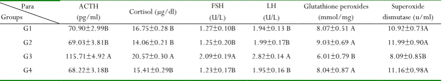
The levels of glutathione peroxides in G1 (8.07 mmol/mg), G2 (9.03 mmol/mg), and G4 (8.04 mmol/mg) were significantly (p< 0.05) higher than in the G3 (6.01mmol/mg) (Table 1). A similar trend was shown concerning the superoxide dismutase as the means were 10.92, 11.99, and 11.16 u/ml in G1, G2, and G4, respectively while the mean in G3 was 8.09 u/ml. As shown in Table 2, the differences between the fertility indexes (FI) were significant (P<0.0001). The highest FI was detected in G4 (100%), followed by 80% for G1 and G2. The lowest FI was shown in G3 (10%).
Table 2: The effect of Spinacia oleracea leaves extract and flavonoids of Spinacia oleracea on Fertility Index (%) of
female mice treated with doxorubicin. Parameter
Groups Fertility index%
G1 80%
G2 80%
G3 10%
G4 100%
Chi-square value 21.31 p <0.0001
The histopathological variations in the ovary of G3 revealed the occurrence of atretic follicles, which had a thin wall of atrophied and degenerated lining epithelial cells (granulosa cells) as shown in (Figure 1), while the ovarian segment in G2 showed the presence of several mature follicles that contained oocyte in addition to the presence of corpus luteum and proliferation progression in their lining epithelial cells with regeneration of atretic follicles (Figure 2). Concerning the histopathological section of the ovary in G1, the results showed the presence of mature follicle and fewer deteriorated follicles with a delicate wall (Figure 3). The histopathological section of the adrenal cortex of animals treated with distilled water and doxorubicin showed necrosis, pyknotic nuclei, and congestion of blood vessels in the cortical adrenal gland (Figure 4). Whereas, the histopathological section in the adrenal cortex of animals treated with flavonoids and doxorubicin showed a moderate vacuolization of adrenal cortex and mononuclear cells infiltration (Figure 5), while histopathological section of the adrenal cortex of animals treated with Spinacia oleracea leaves extract and doxorubicin showed the mild vacuolization of adrenal cortex (Figure 6) as compared with adrenal gland of mouse from negative control group (Figure 7).
Figure 1: Histopathological section of the ovary of a mouse treated with distilled water and doxorubicin (H&EX40).
Figure 2: Histopathological section in the ovary of a mouse treated with flavonoids and doxorubicin (H&EX40).
Figure 3: Histopathological section in the ovary of a mouse treated with Spinacia oleracea leaves extract and doxorubicin (H
& E stain, 40X).
Figure 4: Histopathological section in the adrenal cortex of animal treated with distilled water and doxorubicin showed necrosis, pyknotic nuclei, and congestion of blood vessels in the
Figure 5: Histopathological section in the adrenal cortex of the animal treated with flavonoids and doxorubicin showed moderate vacuolization of the adrenal cortex and mononuclear
cells infiltration (H&Estain 40X).
Figure 6: Histopathological section of the adrenal cortex of the animal treated with Spinacia oleracea leaves extract and doxorubicin showed mild vacuolization of the adrenal cortex
(H&Estain 40X).
Discussion
Increased levels of ACTH, cortisol, FSH, and LH levels and also decreasing levels of glutathione peroxides, superoxide dismutase, fertility index, and increased atretic follicles with degenerated lining epithelial cells (granulosa cells) in G3 may be due to doxorubicin, leading to creation of hydrogen peroxide and superoxide resulted in free radicals creation and exhaustion of tissue antioxidants by its ability to inhibit topoisomerase II and cooperation with DNA topo-isomerase, produced DNA strand disruptions and lead to damage of the oocyte straightly and ovarian toxicity in addition to change various types of cells at this organ. Furthermore, cortisol is commonly mentioned as the “stress hormone” and exists in the organs exposed to stress. Moreover, the increase in ACTH and cortisol levels are slightly correlated to the reduction in total antioxidant concentrations and little total antioxidant capacities [17]. Furthermore, doxorubicin, an anthracycline agent, induces
apoptosis in the oocyte. Anthracyclines are able to induce ovarian injury, endovascular damage, germ cell damage, ovarian toxicity, and desensitization to LH and FSH as well as increasing their levels in the circulation. These consequences are in agreement with the results mentioned by Codacci-Pisanelli et al. [18].
Inhibitory effects of flavonoid on the production of ACTH, cortisol, FSH, and LH and the enhancement of glutathione
good percentage of quercetin, which plays an important role in enhancing female fertility by increasing folliculogenesis in the ovaries [6]. Furthermore, flavinoids of Spinacia oleracea include Kaempferol and 7-tri-hydroxyflavone, which has the ability to regulate female hormones and fertility [7].
Decreased levels of ACTH, cortisol, FSH, and LH along with increased levels of glutathione peroxides, superoxide dismutase, fertility index, and the presence of several mature follicles compared to G3 could be attributed to Spinacia oleracea, which has numerous active antioxidant constituents such as uridine,
flavonoids, and coumaric acids that can act synergistically [4]. Moreover, vitamins C and E could positively act as complements for attenuating the adverse effects of chemotherapy. Spinacia oleracea has excessive nutritious substances such as saponins, terpenes, steroids, folic acid, iron, vitamins A, resins, and folate. The extract of this plant is considered as a source of zinc, which has an important role during differentiation and division of cells and is vital for growth of tissues [20].
Conclusion
The results of the present study confirmed that the flavonoids isolated from Spinacia oleracea leaves have the ability to attenuate the side effect of chemotherapy drugs by enhancing antioxidant and improving fertility.
Ethical approval
All applicable international, national, and/or institutional guidelines for the care and use of animals were followed.
Acknowledgments
The author is appreciative to the Veterinary Medicine / University of Baghdad for supporting research apparatus and College of Pharmacy for providing assistance during the extraction.
Conflict of Interests
The author declares no conflict of interests.
References
Science of Food and Agriculture. 2008 Apr 30;88(6):1099-106.
5. Lugast A, Hovari J. Flavonoid aglycons in foods of plant origin I. Vegetables. Acta Alimentaria. 2000 Oct 1;29(4):345-52.
6. Beazley KE, Nurminskaya M. Effects of dietary quercetin on female fertility in mice: implication of transglutaminase 2. Reproduction, Fertility and Development. 2016 Jun 8;28(7):974-81.
7. Kassem ME, Ibrahim LF, Hussein SR, Sharawy R, El-Ansari MA, Hassanane MM, Booles HF. Myricitrin and bioactive extract of Albizia amara leaves: DNA protection and modulation of fertility and antioxidant-related genes expression. Pharmaceutical biology. 2016 Nov 1;54(11):2404-9.
8. Shaheen SM. Phytochemical Profiling and Evaluation of Antioxidant and Antidiabetic Activity of Methanol Extract of Spinach (Spinacia Oleracea L.) leaves. Int J Pharm Sci Scient Res. 2017;3:8-24.
9. Yadav S, Kumar P. Production, isolation and
identification of flavonoids from aerial parts of Hiptage benghalensis. Int J Life Sci Pharma Res. 2012;2(3):1-5. 10. Luka CD, Abdulkarim M, and Adoga GI. (2014).
Anti-anaemic Potential of Aqueous Extract of Spinacia oleracea Leaves in Phenylhydrazine-treated Rats. N Y Sci J, 7(6):14-18.
11. Fu Z, Wei Z, Miao M. Effects of total flavonoids of raspberry on perimenopausal model in mice. Saudi journal of biological sciences. 2018 Mar 1;25(3):487-92. 12. Nishikimi M, Rao NA, Yagi K. The occurrence of
superoxide anion in the reaction of reduced phenazine methosulfate and molecular oxygen. Biochemical and
biophysical research communications. 1972 Jan 31;46(2):849-54.
13. Paglia DE, Valentine WN. Studies on the quantitative and qualitative characterization of erythrocyte glutathione peroxidase. The Journal of laboratory and clinical medicine. 1967 Jul 1;70(1):158-69.
14. Oberländer G, Yeung CH, Cooper TG. Induction of reversible infertility in male rats by oral ornidazole and its effects on sperm motility and epididymal secretions. Reproduction. 1994 Mar 1;100(2):551-9.
15. Luna LG. Manual of histologic staining methods of the Armed Forces Institute of Pathology.1968.
16. SAS. Statistical Analysis System User's Guide. Statistical. Version 9. 1th ed, SAS. Inst. Inc. Cary. N. C. USA. 2012. 17. Limberaki E, Eleftheriou P, Gasparis G, Karalekos E,
Kostoglou V, Petrou C. Cortisol levels and serum antioxidant status following chemotherapy. Health. 2011 Aug 10;3(08):512-7.
18. Codacci-Pisanelli G, Del Pup L, Del Grande M, Peccatori FA. Mechanisms of chemotherapy-induced ovarian damage in breast cancer patients. Critical reviews in oncology/hematology. 2017 May 1;113:90-6.
19. Ohno S, Shinoda S, Toyoshima S, Nakazawa H, Makino T, Nakajin S. Effects of flavonoid phytochemicals on cortisol production and on activities of steroidogenic enzymes in human adrenocortical H295R cells. The Journal of steroid biochemistry and molecular biology. 2002 Mar 1;80(3):355-63.