Ivan M. Petyaev
1, A, C, D, F, Nailya A. Zigangirova
2, B, E, G, Lydia N. Kapotina
2, B, C,
Elena D. Fedina
2, B, C, Nigel H. Kyle
1, B, E, GChlamydia Trachomatis
Promotes 3T3 Cell
Differentiation into Adipocytes
1 Lycotec Ltd, Granta Park Campus, Cambridge, United Kingdom2 Institute of Epidemiology and Microbiology, Ministry of Health, Moscow, Russia
A – research concept and design; B – collection and/or assembly of data; C – data analysis and interpretation;
D – writing the article; E – critical revision of the article; F – final approval of article; G – other
Abstract
Background. There is experimental and clinical evidence showing that some viral and bacterial pathogens are linked to the accumulation of excessive body fat and obesity.
Objectives. The aim of the study was to investigate the ability of C. trachomatis to propagate in the pre-adipocyte cell line and induce its differentiation into fat cells.
Material and Methods. 3T3 L1 pre-adipocytes or McCoy cells were plated and infected with C. trachomatis. The cell monolayers were further studied by immunofluorescent and quantitative RT-PCR methods.
Results. C. trachomatis can efficiently propagate in 3T3 L1 cells, a mouse pre-adipocyte cell line. The morphologi-cal characteristics of chlamydial growth revealed in 3T3 L1 cells with the monoclonal chlamydial MOMP-specific antibody resembled those seen in McCoy cells, a classic cell line used for chlamydial research. The number of chlamydial 16S rRNA copies detectable in the lysates of McCoy and 3T3 cells infected with C. trachomatis was almost identical, suggesting similar efficiency of pathogen propagation in both cell lines. Moreover, there was a significant increase in aP2 mRNA transcript levels as well as moderate induction of SCD-1 mRNA in the total RNA extracted from the infected 3T3 L1 cells 48 h following the pathogen inoculation. The increased expression of the adipogenic markers was also accompanied by lipid droplet accumulation in the C. trachomatis infected 3T3 L1 cells, suggesting their transformation into differentiated adipocytes.
Conclusions. The direct effect of the pathogen on fat cell progenitors observed in this work may explain abnormal fat deposition at the sites of chronic inflammation caused by C. trachomatis (Adv Clin Exp Med 2014, 23, 4, 511–516).
Key words: C. trachomatis, adipocytes, differentiation. Adv Clin Exp Med 2014, 23, 4, 511–516
ISSN 1899–5276
ORIGINAL PAPERS
© Copyright by Wroclaw Medical University
According to WHO statistics, excess weight and obesity have reached epidemic proportions, affecting at least 1.4 bn people worldwide. As a re-sult approximately 2.8 m adults die each year due to obesity-related diseases. Although increased food intake and decreased level of physical activity are considered the most important primary causes of obesity, there is a growing body of evidence that some viral and bacterial pathogens may contribute to the pathogenesis of obesity. It has been shown recently that past adenovirus infection (Ad36) in-creases adiposity and affects glycemic control in humans [1]. Multiple experimental studies show that adenoviral infections – in particular those
caused by the Ad-36 adenovirus – induce changes in insulin sensitivity, glucose uptake and expres-sion of diabetes-related genes. Moreover, there is a traceable epidemiological link between some ad-enoviral infections and obesity [2, 3]. Among other viruses associated with a risk of obesity are canine distemper virus, Rous-associated virus 7, scrapie, Borna disease virus and SMAM-1 [4]. Some bac-terial pathogens are also supposedly related to the development of obesity [5]. It has been reported re-cently that an endotoxin-producing isolate of
En-terobacter derived from the gut of obese humans
agents may cause abnormalities in regional body fat portioning, resulting in lipodystrophy in cases of HIV infection [7]. Although inflammatory me-diators seem to play a major role in the association between infections and adiposity [6], some viral pathogens can directly promote the differentiation of fat cells. In particular, Ad-36 has been shown to accelerate pre-adipocyte transformation into ma-ture fat cells under in vitro conditions [8].
The present paper reports that Chlamydia tra-chomatis, an intracellular bacterial pathogen, can induce differentiation of pre-adipocyte cells into adipocytes.
Material and Methods
Reagents, Cell Lines
and Pathogen
All reagents were purchased from Sigma-Al-drich unless otherwise specified.
McCoy and Hep2 cells were obtained from the European Collection of Cell Cultures (Salis-bury, UK) and grown in 5% CO2 in DMEM
supple-mented with 2 mM glutamine and 5% FCS. 3T3 L1 cells, a mouse embryonic cell line, were purchased from the HPA Culture Collection (catalog number 86052701, United Kingdom) and were grown in DMEM supplemented with 2 mM glutamine and 10% calf serum in 5% CO2 at 37°C until confluence
was reached, according to the supplier’s instruc-tions. To stimulate differentiation into adipocytes, additions of 0.5 mM 3-isobutyl-1-methylxanthine (IBMX), 0.25 µM dexamethasone and 1µg/mL in-sulin were made to the confluent cultures. Strain L2/ /Bu434 of C. trachomatis was kindly provided by Dr. P. Saikku (University of Oulu, Finland). The path-ogen was initially propagated in Hep2 cells and pu-rified by Renografin gradient centrifugation as de-scribed by Galdwell et al. [9]. Chlamydial titers were determined by infecting Hep2 cells with 10-fold di-lutions of thawed stock suspension. Purified ele-mentary bodies (EB) of known titer were suspend-ed in sucrose-phosphate-glutamic acid buffer [9] and used as inoculums for 3T3 or McCoy cells. 3T3 or McCoy cells were infected by inoculating the cell monolayers using stock solution of C. trachomatis
at 1 MOI, with further centrifugation of the infected cells at 1500 rpm for 60 min. Cell monolayers were refed every 48 h. Infected cells were visualized after permeabilization by direct immunofluorescence us-ing FITC-conjugated monoclonal antibody against major outer membrane protein (MOMP) (Near-Medic Plus, RF). Inclusion-containing cells were visualized using a Nikon Eclipse 50i fluorescence microscope at ×1350 magnification.
Assessment of Bacterial Growth
In order to evaluate C. trachomatis growth in 3T3 or McCoy cells, the infected cell monol-ayers were grown in DMEM supplemented with 2 mM glutamine and 10% calf serum in 5% CO2 at
37°C and harvested after 24, 48, 72 and 144 h of the post-infection period. The infected cells were visu-alized with C. trachomatis MOMP-specific mono-clonal antibody. RNA was extracted and analyzed as described below. Lipid inclusions were visual-ized on day 7 of the post-infection period with Sig-ma Sudan Black B Staining System.
RNA Extraction
and Reverse Transcription
RNA was isolated from cell monolayers grown on 6-well plates using TRIZol (Invitrogen). To-tal mRNA pretreated with DNase I (DNA-free™, Ambion) and quantified on a NanoDrop ND- -1000 spectrophotometer (ThermoFisher Scientif-ic, Wilmington, USA) was transformed into cD-NA using random hexamer primers and a Super-Script III First-Strand Synthesis Kit (Invitrogen, Karlsruhe, Germany).
Quantitative Real-Time
Polymerase Chain Reaction
(RT-PCR)
The 16S rRNA was studied as a constitu-tive marker of the chlamydial developmental cy-cle to compare the efficiency of C. trachomatis
mouse GAPDH (forward primer 5’-AACTTTG-GCATTGTGGAAGG-3’, reverse primer 5’-TGT-GAGGGAGATGCTCAGTG-3’). The mRNA ex-pression levels in the host cells were referenced to the CT values in uninfected control cells grown under the same conditions. This reference value was taken as 1.00. Each cDNA sample was tested by PCR at least three times. All experiments were repeated at least twice. Representative sets of re-sults are shown below.
Results
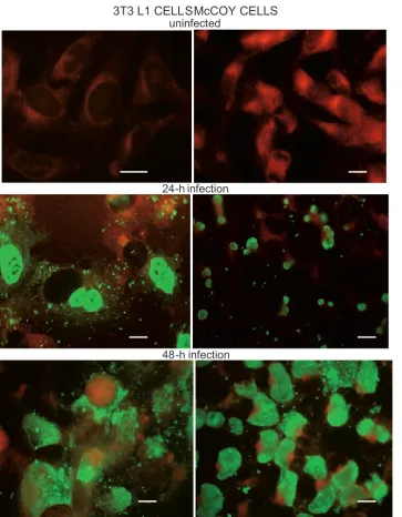
As can be seen from the results of the im-munofluorescence analysis (Fig. 1), inoculation of C. trachomatis into 3T3 L1 and McCoy cell
cultures led to active infection in the cell monolay-ers. In general, the morphology and dynamics of the C. trachomatis infection cycle were very sim-ilar in both cell lines and involved approximately 25–30% of the cells in the cell monolayers. In both cell lines, inclusion bodies started to appear after 24 h of the post-infection period, predominantly in the peripheral zone of the cytoplasm. Inclusion bodies were much larger and more densely stained 48 h after C. trachomatis inoculation and were ac-companied by nuclear dislocation, while some im-munopositive granules were observed outside of the cells. The most intense staining of intracel-lular inclusion bodies was seen after 72 h of the post-infection period, when most of the infected cells were round-shaped and also had multiple im-munopositive extracellular granules surrounding
Fig. 1. Propagation of C. trachoma-tis in 3T3 L1 and McCoy cells. 3T3 L1 and McCoy cells were set up, grown and processed for immunostaining with MOMP-specific monoclonal antibod-ies at the 0-, 24- and 48-h points of the post-infection period, as described in Material and Methods 3T3 L1 CELLSMcCOY CELLS
uninfected
24-h infection
them. Later points in time in the post-infection period were characterized by cytolytic changes in the infected cells, leading to an accumulation of cell debris with densely stained granules and fur-ther spread of the infection to newly infected adja-cent cells (results not shown).
A nucleic acid amplification protocol was used in an attempt to quantify the intensity of chlamyd-ial growth in 3T3 L1 and McCoy cells. As can be seen from Fig. 2, the number of chlamydial 16S rR-NA copies detectable in the lysates of McCoy and 3T3 cells infected with C. trachomatis was almost identical throughout the whole post-infection pe-riod. This implies that the efficiency of propaga-tion in the two host cell lines was equal.
In order to evaluate the effect of C. trachoma-tis on 3T3 cell differentiation, the mRNA adipocyte markers were studied in the total mRNA specimens extracted 48 h after inoculating C. trachomatis into the 3T3 L1 cell monolayers. As can be seen from Ta-ble 1, there was a significant increase in aP2 mRNA transcript levels, as well as moderate induction of SCD-1 mRNA. PPARγ mRNA was almost un-changed at the time point of the experiment shown.
To evaluate changes in lipid content, infected 3T3 L1 cells were double stained with Sudan black and further treated with MOMP-specific monoclo-nal antibodies on day 7 of the post-infection period.
As Fig. 3 shows, there was neither visible lipid nor immunofluorescent staining in the uninfected 3T3 L1 cells. However, as can be seen from the lower panel of Fig. 3, 3T3 L1 cells infected with C. tracho-matis had multiple lipid droplets surrounding the chlamydial inclusion body. A similar aP2 mRNA re-sponse and lipid droplet formation (on day 2 and day 7, respectively) was observed in uninfected 3T3 cells induced with insulin (results not shown).
Discussion
Chlamydia trachomatis infection is the most common sexually transmitted communicable disease in North America and Europe [10] with urogenital, pelvic and ocular manifestations, leading to infertility and preventable blindness if untreated [11]. Howev-er, most cases of C. trachomatis infections are asymp-tomatic, which explains exponential growth of affect-ed individuals around the world. The overall maffect-edian rate of C. trachomatis positivity worldwide is approx-imately 4.7%, with variations from 1.3% to 18.1% in different countries [12]. The existing limitations in treating C. trachomatis infections arise from antibi-otic resistance and insufficient knowledge of the mo-lecular mechanisms underlying the pathogenesis of this disease and its various complications.
Fig. 2. 16S rRNA in 3T3 L1 and McCoy cells infected with C. trachomatis. 3T3 L1 and McCoy cells were set up, grown and harvested at the 0-, 24-, 48- and 72-h points of the post-infection period. 16S rRNA was measured as described in Material and Methods
3T3 L1
McCoy 0
1 2 3 4 5 6 7 8 9
16
S
rR
N
A
C
op
y
N
um
be
rs
/1
00
c
op
ie
s
of
β
-a
c
n 0 hour
24th hour
48th hour
72th hour
Table 1. Fold changes in mRNA expression levels in 3T3 L1 cells infected with C. trachomatis
Experimental Condition GAPDH mRNA aP2 mRNA SCD-1 mRNA PPARγ mRNA
Control: uninfected 3T3 L1 cells 1 1 1 1
Infected 3T3 L1 cells 1 11.4 2.9 1.32
In the present paper it is shown that C. trachom-atis,a bacterial pathogen responsible for most cases of preventable blindness and urogenital infections in the world, can efficiently propagate and fully accom-plish its infectious cycle in a pre-adipocyte cell line: 3T3 cells. This statement is solidly supported by the fact that the number of 16S rRNA copies detectable during the post-infection period in 3T3 cells is almost equal to that measurable in McCoy cells, a classic cell line used for chlamydial research for many decades.
However, the most important finding made in the current study is the fact that propagation of
C. trachomatis in 3T3 cells promotes the differentia-tion of pre-adipocytes into fat cells. This conclusion is well supported by the results of the histochemistry analysis, which reveal the appearance of lipid drop-lets in the cytoplasm of 3T3 cells infected with the chlamydial pathogen, as well as the mRNA changes found in the infected 3T3 cells. According to the find-ings of the current study, infecting the 3T3 L1 cells with C. trachomatis led to an increase in aP2 tran-script numbers with corresponding upregulation
in SCD-1 mRNA which are well known molecu-lar markers of adipocyte differentiation. These clear signs of 3T3 cell differentiation induced by C. tra-chomatis came as a big surprise in the current study, as recently published experiments with C. pneumo-niae and 3T3 cells yielded negative results. Accord-ing to Shi et al. [13], C. pneumoniae infection in 3T3 cells does not lead to the transformation of pre-ad-ipocytes into fat cells under similar conditions to those used in the present study. Therefore, the cur-rent results illustrating the ability of C. trachomatis
to induce adipocyte differentiation represent a novel finding, revealing a striking difference in the outcome of infections caused by two closely related but distinct chlamydial pathogens.
Although the results of the present study do not reveal the molecular mechanism underlying
C. trachomatis-induced pre-adipocyte
differentia-tion, there is a body of both experimental and clin-ical evidence supporting this observation and ex-plaining it to some extent. In particular, it is widely believed [14] that C. trachomatis relies heavily on
Fig. 3. Lipid drop-let accumulation in 3T3 L1 cells infected with C. trachomatis. 3T3 L1 cells were set up, grown and subjected to a double staining protocol (immunostaining with MOMP-specific monoclonal anti-body and/or sudan staining) on day 7 after infection with C. trachomatis. Immunofluorescent images were obtained as described in Material and Methods Sudan Staining Sudan + MOMP Ab Staining
uninfected 3T3 L1 cells
the lipid metabolism of host cells and is capable of redirecting lipid trafficking and biosynthesis in mammalian cells [15]. C. trachomatis inclusions, even in host cells with limited ability to synthesize lipids, such as HeLa cells, have been shown to be surrounded by host lipid droplets with neutral li-pids and tubular structures expressing adipocyte-differentiation-related protein, perilipin, Rab18 and other potential inducers of adipogenesis [16]. Moreover, from a clinical point of view, chronic inflammation caused by C. trachomatis infection is often accompanied by extensive remodelling of urogenital tissues and accumulation of cervico-dorsal fat [17, 18]. Chronic pelvic inflammation is
reportedly often associated with fat tissue depos-its [19]. In particular, besides lymph node enlarge-ment, an extensive mesenteric fat accumulation has been reported in patients with Fitz-Hugh-Cur-tis syndrome, a rare complication of C. trachoma-tis infection [20, 21].
Although at this stage it not clear what contri-bution C. trachomatis infection may make in the pathogenesis of systemic obesity, the direct effect of the pathogen on fat cell progenitors observed in this work may explain abnormal fat deposition at the sites of chronic inflammation caused by C. trachomatis.
Further research is needed to clarify the possible role of C. trachomatis in body fat content regulation.
References
[1] Lin WY, Dubuisson O, Rubicz R, Liu N, Allison DB, Curran JE, Comuzzie AG, Blangero J, Leach CT, Göring H, Dhurandhar NV: Long-Term Changes in Adiposity and Glycemic Control Are Associated With Past Adenovirus Infection. Diabetes Care 2013, 36, 701–707.
[2] Wang ZQ, Cefalu WT, Zhang XH, Yu Y, Qin J, Son L, Rogers PM, Mashtalir N, Bordelon JR, Ye J, Dhurandhar NV: Human adenovirus type 36 enhances glucose uptake in diabetic and nondiabetic human skeletal muscle cells independent of insulin signaling. Diabetes 2008, 57, 1805–1813.
[3] Wang ZQ, Yu Y, Zhang XH, Qin J, Floyd E: Gene expression profile in human skeletal muscle cells infected with human adenovirus type 36. J Med Virol 2012, 84, 1254–1266.
[4] Mitra AK, Clarke K: Viral obesity: fact or fiction? Obes Rev 2010, 11, 289–296.
[5] Pasarica M, Dhurandhar NV: Infectobesity: Obesity of infectious origin. Adv Food Nutr Res 2007, 52, 61–102.
[6] Fei N, Zhao L: An opportunistic pathogen isolated from the gut of an obese human causes obesity in germ free mice. ISME J 2013, 7, 880–884.
[7] Galescu O, Bhangoo A, Ten S: Insulin resistance, lipodystrophy and cardiometabolic syndrome in HIV/AIDS. Rev Endocr Metab Disord 2013, 14, 133–140.
[8] Suplicy H.L, Bornschein A: Infections as the etiology for obesity. Arq Bras Endocrinol Metabol 2009, 53, 159–164.
[9] Galdwell HD, Kromhout J, Schachter J: Purification and partial characterization of the major outer membrane protein of Chlamydia trachomatis. Infect Immun 1981, 31, 1161–1176.
[10] Brunham RC, Rappuoli R:Chlamydia trachomatis control requires a vaccine. Vaccine 2013, 8, 31, 1892–1897.
[11] Manavi K: A review on infection with Chlamydia trachomatis. Best Pract Res Clin Obstet Gynaecol 2006, 20, 941–951.
[12] Jamil MS1, Bauer HM, Hocking JS, Ali H, Wand H, Walker J, Douglas L, Donovan B, Kaldor JM, Guy RJ: Chlamydia screening strategies and outcomes in educational settings: a systematic review. Sex Transm Dis 2014, 41, 180–187.
[13] Shi Y, Liu Y, Murdin A, Raudonikiene-Mancevski A, Ayach BB, Yu Z, Fantus IG, Liu PP: Chlamydophila pneu-moniae inhibits differentiation of progenitor adipose cells and impairs insulin signaling. J Infect Dis 2008, 1, 197, 439–448.
[14] Elwell CA, Engel JN: Lipid acquisition by intracellular Chlamydiae. Cell Microbiol 2012, 14, 1010–1018.
[15] Kumar Y, Cocchiaro J, Valdivia RH: The obligate intracellular pathogen Chlamydia trachomatis targets host lipid droplets. Curr Biol 2006, 22, 16, 1646–1651.
[16] Subtil A: Rerouting of host lipids by bacteria: are you CERTain you need a vesicle? PLoS Pathog 2011, 7, e1002208.
[17] Sales KJ, Katz AA: Inflammatory pathways in cervical cancer – the UCT contribution. S Afr Med J 2012, 23, 102, 493–496.
[18] Tsinzerling AV: Chlamydiosis: diagnosis and role in human pathology. Arkh Patol 1989, 51, 3–9.
[19] Jung SI, Kim YJ, Park HS, Jeon HJ, Jeong KA: Acute pelvic inflammatory disease: diagnostic performance of CT. J Obstet Gynaecol Res 2011, 37, 228–235.
[20] Mesurolle B, Mignon F, Gagnon JH: Fitz-Hugh-Curtis syndrome caused by Chlamydia trachomatis: atypical CT findings. AJR Am J Roentgenol 2004, 182, 822–824.
[21] Shimada N, Honda Y, Sugimoto M: Image diagnosis of abdominal infectious diseases (infectious enterocolitis and Fitz-Hugh-Curtis Syndrome). Nihon Rinsho 2007, 28, 65, Suppl 2, 1, 247–250.
Address for correspondence:
Ivan M. Petyaev
Lycotec Ltd. Granta Park Campus Cambridge, CB21 6GP
United Kingdom Tel.: +44 792 136 37 40
E-mail: [email protected]
Conflict of interest: None declared