O
KTAWIAM
AZANOWSKA1, D
OROTAK
AMIŃSKA1, K
ATARZYNAK
OŚCIELSKA−K
ASPRZAK1,
D
OMINIKAD
RULIS−F
AJDASZ1, M
AGDALENAK
RAJEWSKA1, K
RZYSZTOFF
ALKIEWICZ1,
A
GNIESZKAH
AŁOŃ2, P
AWEŁC
HUDOBA3, W
OJCIECHP
OLAK3, D
ARIUSZP
ATRZAŁEK3,
I
RENAW
IKIERA−M
AGOTT4, M
ARIAB
ORATYŃSKA1, M
ARIANK
LINGER1High Risk of FSGS Recurrence in Kidney Allograft
Recipients Independent of Heterozygous
NPHS2
Mutation*
Duże ryzyko nawrotu ogniskowego segmentalnego stwardnienia
kłębuszków nerkowych u biorców przeszczepu nerki niezależnie
od obecności heterozygotycznej mutacji genu podocyny
1Department of Nephrology and Transplantation Medicine, Silesian Piasts University of Medicine in Wrocław, Poland
2Department of Pathological Anatomy, Silesian Piasts University of Medicine in Wrocław, Poland
3Department of Vascular, General and Transplant Surgery, Silesian Piasts University of Medicine in Wrocław, Poland
4Department of Pediatric Nephrology, Silesian Piasts University of Medicine in Wrocław, Poland Adv Clin Exp Med 2008, 17, 2, 207–212
ISSN 1230−025X
ORIGINAL SCIENTIFIC PAPERS
© Copyright by Silesian Piasts University of Medicine in Wrocław
Abstract
Background.A high recurrence rate of focal segmental glomerulosclerosis (FSGS) is one of the most frequent events after kidney transplantation, with a risk of graft loss in more than 50% of the affected patients, but patients with a homozygous podocin mutation (NPHS2) are at low risk of FSGS recurrence because of proper podocine structure in the allograft. The mechanism of recurrence is still obscure, and a multifactorial origin has been proposed.
Objectives. The purpose of the study was to identify recipients at risk of recurrence of FSGS or FSGS de novoin terms of podocin gene mutations (NPHS2).
Material and Methods.Twelve patients (4 females, 8 males, mean age at transplantation: 35.5 ± 10.4 years) were analyzed. Of these, 5 of the 9 recipients (55%) with pre−transplant FSGS had recurrence of proteinuria and 3 devel− oped de novo FSGS. Delayed graft function was observed in 3 patients with proteinuria and primary non−function with immediate recurrence of heavy proteinuria and graftectomy in one patient. Acute rejection occurred in 5 of the 8 patients with recurrent FSGS and in only one of the 4 without proteinuria. After 1 year the mean serum cre− atinine concentration (1.6 mg/dl) in six patients with significant proteinuria (6.2 ± 1.9 g/d) was higher than in the 4 patients with no proteinuria (0.97 mg/dl). During the observation period, 4 patients lost graft function in an average of 51 months and one patient died.
Results. Mutational analysis of NPHS2 (5’UTR, coding sequences and flanking region) was performed in 10 patients and revealed two heterozygous mutations in exon 5 (R229Q) with recurrence of FSGS during the first month after transplantation in the first and no recurrence in the other recipient.
Conclusions.These observations confirm the high risk of FSGS recurrence after kidney transplantation (55.5%), which was not affected by the presence of a heterozygous NPHS2mutation but is connected with worse graft func− tion after one year (Adv Clin Exp Med 2008, 17, 2, 207–212).
Key words:FSGS, podocin, NPHS2mutation, recurrence after kidney transplantation.
Focal segmental glomerulosclerosis (FSGS) is a glomerular disease with a high recurrence rate after kidney transplantation. Proteinuria appears in approximately 30% of patients within a few days after kidney transplantation with a relatively good response to plasmapheresis (PF), but with graft loss in more than 50% of the affected patients [1–3]. The risk of recurrence differs in children and adults (50% vs. 11%, respectively) [4]. Some patients with no FSGS in native kidneys develop late−onset de novo FSGS (over 6 months) after transplantation. Although recurrence of FSGS negatively impacts kidney allograft survival, it has also been noted that some individuals with pro− teinuria have adequate kidney function for a num− ber of years. The mechanisms of FSGS in native kidneys as well as FSGS recurrence in kidney allo− grafts have not been fully elucidated. It is current− ly believed that primary FSGS is caused by alter− ations in glomerular epithelial cells (podocytes) induced by a circulating vascular permeability fac− tor (VPF) produced by T lymphocytes or by an intrinsic podocyte cellular defect [5, 6].
The discovery of a mutation of podocin, exclusively expressed by podocytes, showed new pathogenic mechanisms of the disease [7, 8]. Mutations of the NPHS2 gene, which encodes podocin, cause a steroid−resistant nephrotic syn− drome (SRNS) in children with autosomal reces− sive inheritance and nonfamilial sporadic FSGS in adults indistinguishable from idiopathic FSGS on
clinical grounds, but in which proteinuria is deter− mined by homozygous mutations of podocin [7, 9–12]. Typically, NPHS2 mutations require two defective alleles for the clinical manifestation of the disease [10, 13]. Among patients with a homozygous NPHS2 mutation, a molecular defect of podocin is considered to be the disease− causing mechanism. Since this mechanism should vanish after kidney transplantation, patients bear− ing homozygous mutations are at low risk of FSGS recurrence, with only the possibility of other proteinuria−inducing factors (i.e. VPF or autoantibodies against unmutated podocin) [14]. In patients with only a single NPHS2 mutation, FSGS does not seem to be related to NPHS2, assuming that a second mutation has not been omitted. However, heterozygous NPHS2 muta− tions, sequence variants, and polymorphisms may play a role in atypical cases of SRNS with a later onset, mild clinical course, and recurrence after kidney transplantation [8, 14]. The recurrence of FSGS in patients carrying homozygous or het− erozygous mutations support the general idea of a multifactorial origin of the primary disease. The clinical course of kidney allografts with recurrence or de novo FSGS varies and it is controversial which factors are of importance in determining the outcome. The aim of the study was to identify kid− ney allograft recipients at risk of FSGS recurrence in terms of NPHS2mutations.
Streszczenie
Wprowadzenie. U chorych z ogniskowym segmentalnym stwardnieniem kłębuszków nerkowych (FSGS – focal segmental glomerulosclerosis) w nerkach własnych często wystepuje nawrót choroby po przeszczepie nerki, z utra− tą czynności przeszczepu u około połowy z nich. Pacjenci z homozygotyczną mutacją genu podocyny (NPHS2) mają jednak małe ryzyko nawrotu ze względu na przeszczepienie narządu z prawidłową strukturą podocyny.
Cel pracy. Chociaż mechanizm nawrotu FSGS jest niejasny, to uważa się, że ma podłoże wieloczynnikowe. Ce− lem pracy była identyfikacja pacjentów z nawrotem FSGS lub FSGS de novopod kątem mutacji genu dla podocy− ny (NPHS2).
Materiał i metody. Przeprowadzono analizę danych 12 pacjentów (4 kobiet i 8 mężczyzn) w wieku 35,5 ± 10,4 lat podczas przeszczepiania nerki. Nawrót FSGS po przeszczepie nerki obserwowano u 5 z 9 chorych (55%), u których przyczyną niewydolności nerek własnych było FSGS oraz u 3 pacjentów z obrazem FSGS de novo. U 3 pacjentów obserwowano opóźnione podjęcie czynności przez przeszczepioną nerkę z obecnością białkomo− czu, a u jednego pacjenta natychmiastowe wystąpienie ciężkiego białkomoczu było przyczyną usunięcia przeszcze− pu. Ostre odrzucanie przeszczepu wystąpiło u 5 z 8 pacjentów z nawrotem FSGS i tylko u 1 z 4 pacjentów bez biał− komoczu. Po roku obserwacji u 6 pacjentów ze znaczącym białkomoczem (6,2 ± 1,9 g/dobę) średnie stężenie kre− atyniny w surowicy wynosiło 1,6 mg/dl, podczas gdy u 4 pacjentów bez białkomoczu wynosiło 0,97 mg/dl. Podczas obserwacji stwierdzono, że u 4 pacjentów przeszczepiona nerka utraciła czynność średnio po 51 miesią− cach, a jeden pacjent zmarł.
Wyniki. Analiza mutacji genu NPHS2 (region 5’UTR, sekwencje kodujące oraz sąsiadujące z nimi obszary intro− nowe) była wykonana u 10 chorych. U żadnego chorgo nie wykazano homozygotycznej mutacji w obrębie genu podocyny. W dwóch przypadkach wykazano natomiast heterozygotyczną mutację w 5 egzonie (R229G), z wczes− nym nawrotem FSGS (w pierwszym miesiącu po przeszczepie u jednej osoby i brakiem nawrotu u drugiej).
Wnioski. Opisane obserwacje potwierdziły duże ryzyko nawrotu FSGS po przeszczepie nerki (55,5%), niezależ− nie od obecności heterozygotycznej mutacji genu dla podocyny (NPHS2). Nawrót FSGS wiązał się z gorszą czyn− nością nerki po roku (Adv Clin Exp Med 2008, 17, 2, 207–212).
Material and Methods
The clinical data of 12 patients (4 females, 8 males, mean age at transplantation: 35.5 ± 10.4 years) with pre−transplant history of FSGS (9 patients) or with no FSGS in the native kidneys but de novo FSGS after transplantation (3 patients) were retrospectively analyzed and mutational analyses of their NPHS2 gene were compared. Three patients underwent a second transplantation with loss of the first graft due to recurrent FSGS in two and surgical complications in the third. The initial immunosuppressive therapy consisted of prednisone, calcineurin inhibitors (cyclosporin A/tacrolimus: 9/3 patients), azathioprine or mycophenolate mophetil (5 and 7 patients, respec− tively), and anti−IL2 receptor antibodies in 3 patients. In recurrent FSGS, the treatment was based mainly on cyclosporin A and plasmaphere− sis in particular patients. Acute rejection episodes were treated with intravenous methylprednisolone pulses. The diagnosis of FSGS in 8 patients was confirmed by kidney allograft biopsy. The remain− ing patients without rejection or proteinuria had no protocol biopsies.
Mutational Analysis
Blood samples for mutational analysis were obtained from the patients after informed consent. The study was approved by the Ethics Committee of Silesian Piasts University of Medicine in Wrocław. Mutational analysis of NPHS2(5’UTR, coding sequences and flanking region) was per− formed in 10 patients. Genomic DNA was extract− ed from peripheral blood samples with a QiaAmp Blood Kit (Qiagen) according to standard proce− dure. Molecular analysis of podocin was per− formed by DNA sequencing. All eight exons of the
NPHS2gene were amplified by PCR using flank− ing intronic primers, subjected to automatic sequence analysis by a dye−terminator reaction, and compared with gene bank sequences for
NPHS2 (NC_000001.9 reg. 177786299− −177811691). Primer sequences and PCR condi− tions are available on request.
Results
The analyzed data revealed that delayed graft function (DGF), lasting a mean of 18 days, was observed in only 3 patients with proteinuria. One recipient of a second kidney allograft with primary graft non−function and immediate onset of heavy NS required graftectomy after 2.5 months. Acute rejection was observed in 6 of the 12 patients (50%)
in the first or second month posttransplant, predom− inantly in patients with recurrent FSGS (5 patients), and was successfully treated with methylpred− nisolone (mean dose: 2.3 g per patient). Nephrotic− −range proteinuria developed in 8 of the 12 patients after kidney transplantation, with a mean observa− tion period of 32 ± 26 months. Five of the 9 patients (55%) with pre−transplant FSGS experienced recur− rence and 3 of the remaining patients developed
de novo FSGS. Proteinuria relapsed in 5 patients at a mean of 42 days (range: 1–180 days), with onset within the first 2 weeks posttransplant in 4 patients and in the 6th month in 1 patient, while de novo FSGS had later onset: after the 6thmonth in two and in the 36thmonth in one patient. Normal serum cre− atinine concentration (< 1.4 mg/dl = 128 µmol/l) was achieved in all patients but one at a mean of 21 days (range: 2–90 days). A follow−up analysis was conducted in 10 recipients after 1 year. Six of them had significant proteinuria (6.2 ± 1.9 g/d) with a mean serum creatinine concentration of 1.6 mg/dl (range: 1.1–2.0 mg/dl) and 4 patients with no pro− teinuria had lower creatinine concentration (0.97 mg/dl, range: 0.7–1.4 mg/dl). During the observa− tion period, 4 patients lost graft function after an average of 51 months (range: 22–82 months) and one patient died due to cerebral stroke.
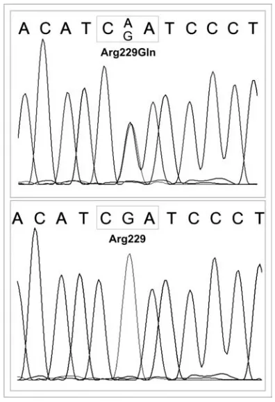
Mutational analysis of NPHS2was performed in 10 patients and revealed solely heterozygous mutations in exon 5 (R229Q) in two of them (Fig. 1). Early recurrence of FSGS (during the first two weeks posttransplant) and a relatively mild course with ultimate loss of the allograft after 7 years was observed in the first patient, while the second has no proteinuria after 6 years despite a second kidney transplant (the first was lost because of surgical complications).
Discussion
higher incidence of R229Q and a milder phenotype was demonstrated by Caridi et al. [8]. The observed R229Q alleles were only heterozygous and R229Q polymorphism is not thought to cause dis− ease when present alone as a heterozygous variant, but in combination with another heterozygous mutation it contributes to autosomal−recessive FSGS [9]. In addition, the R229Q polymorphism was identified in 3.75% of healthy control cohorts, but never in the homozygous state [14]. Tsukagushi et al. found that the R229Q heterozygous variant (with the same frequency as in a normal popula− tion) did not appear to cause FSGS, but rather to enhance the susceptibility to FSGS in association with a second mutant NPHS2 allele [9]. It is believed that patients bearing a homozygous muta− tion of the NPHS2gene are not at risk of recurrence after kidney transplantation [14]. However, delayed recurrence of mild proteinuria after kidney transplantation in some patients with NPHS2muta− tions has been observed by Bertelli et al. [15].
Several pathogenic factors have been postulat−
ed in such cases of recurrent FSGS, among these a yet unidentified lymphokine (VPF) derived from T cells [5, 16]. Evidence for a circulating VPF in FSGS originates from clinical observations of immediate recurrence (within a week) of FSGS after kidney transplantation and good response to plasmapheresis. Under normal conditions, VPF activity is neutralized by serum components such as apolipoproteins. Therefore, loss of inhibitors may play a central role leading to a recurrence of FSGS after transplantation [3, 16]. The estimated frequen− cy of FSGS recurrence after kidney transplantation varies from 20% to 40% (average: 30%) and up to 80% after a second kidney transplantation [1–3]. The patients of the present study developed a high, but allowable, recurrence rate (55%) of FSGS after kidney transplantation, especially since two patients with second kidney allografts and loss of the previ− ous grafts due to FSGS were included. Moreover, there were no patients with a homozygous muta− tion, so the protective mechanism could not be dis− closed. In the 5 relapsed patients, proteinuria appeared later than observed by Pardon et al. (42 days vs. 22 days, respectively) [17]. The differ− ence is probably strongly influenced by the small number of patients. Delayed graft function is likely related to proteinuria early after transplantation and occurred only in recipients with relapse. Similar observations were also reported by others [17, 18]. Acute rejection was observed more often among recipients with relapse than in non−proteinuric patients (5 vs. 1 patient, respectively). Similar data were also reported by Kim et al. but not confirmed by Pardon et al. [17, 18]. Because of the low num− ber of patients, these results have to be interpreted with caution. The four patients of the present study with recurrent FSGS lost their graft function after an average of 51 months, which is like the patients with posttransplant glomerulonephritis reported by Ostrowska et al. [19]. Lack of homozygous NPHS2
mutations affecting both alleles in our small group of kidney transplant patients is consistent with low FSGS recurrence after transplantation, while het− erozygous mutations have no protective impact. In one recipient with a heterozygous R229Q mutation, proteinuria developed early after transplantation and probably arose from circulating VPF and not from mutation. The second one is free of proteinuria after 6 years of observation despite the heterozy− gous mutation.
The authors concluded that the recurrence rate of FSGS after kidney transplantation is high (55%), but it is independent of a heterozygous mutation of the NPHS2gene. Patients with recur− rent FSGS more often experience delayed graft function and acute rejection and have worse graft function after one year than patients without pro−
Fig 1. Mutational analysis of NPHS2(5’UTR, coding sequences and flanking region) with heterozygous R229Q (Arg229Gln) mutations in exon 5 (upper panel) compared with the normal gene sequence (lower panel)
teinuria. The genetic assessment of the mutation should be advised to avoid ineffective treatment of nephrotic syndrome and to refer homozygous patients to kidney transplantation, but in the case
of heterozygosity the course is unpredictable. Patients should be aware of the mutation, but in kidney transplantation a lack of mutation or het− erozygosity should rather be taken into account.
References
[1] Abbot KC, Sawyers ES, Oliver JD, Ko CW, Kirk AD, Welch PG, Peters TG, Agodoa LY:Graft loss due to recurrent focal segmental glomerulosclerosis in renal transplant recipients in United States. Am J Kidney Dis 2001, 37, 366–373.
[2] Weber S, Tönshoff B: Recurrence of focal segmental glomerulosclerosis after renal transplantation: clinical and genetic aspects. Transplantation 2005, 80, S128–S134.
[3] Carraro M, Caridi G, Bruschi M, Artero M, Bertelli R, Zennaro C, Musante L, Candiano G, Perfumo F, Ghiggeri GM: Serum glomerular permeability activity in patients with podocin mutations (NPHS2) and steroid− resistant nephrotic syndrome. J Am Soc Nephrol 2002, 13, 1946–1952.
[4] Senggutuvan P, Cameron JS, Hartley RB, Rigden S, Chantler C, Haycock G, Williams DG, Ogg C, Koffman G: Recurrence of focal segmental glomerulosclerosis in transplanted kidneys: analysis of incidence and risk factors in 59 allografts. Pediatr Nephrol 1990, 4, 21–28.
[5] Shalhoub RJ: Pathogenesis of lipoid nephrosis: a disorder of T−cell function. Lancet 1974, 2, 556–560.
[6] Branchley PEC: Vascular permeability factors in steroid−sensitive nephrotic syndrome and focal segmental glomerulosclerosis. Nephrol Dial Transplant 2003, 18, Suppl 6, vi21–vi25.
[7] Karle SM, Uetz B, Ronner V, Glaeser L, Hildebrandt F, Fuchshuber A:Members of the APN Study Group: Novel mutations in NPHS2 are detected in familial as well as sporadic steroid−resistant nephrotic syndrome. J Am Soc Nephrol 2002, 13, 388–393.
[8] Caridi G, Bertelli R, di Duca M, Dagnino M, Emma F, Onetti Muda A, Scolari F, Miglietti N, Mazzucco G, Murer L, Carrea A, Massella L, Rizzoni G, Perfumo F, Ghiggeri GM: Broadening the spectrum of diseases related to podocin mutations. J Am Soc Nephrol 2003, 14, 1278–1286.
[9] Tsukaguchi H, Sudhakar A, Le TC, Nguyen T, Yao J, Schwimmer JA, Schachter AD, Poch A, Abreu PF, Appel GB, Pereira AB, Kalluri R, Pollak MR: NPHS2 mutations in late−onset focal segmental glomeruloscle− rosis: R229Q is a common disease−associated allele. J Clin Invest 2002, 110, 1659–1666.
[10] Engeler Dusel JA, Burdon KP, Hicks PJ, Hawkins GA, Bowden DW, Freedman BI: Identification of podocin (NPHS2) gene mutations in African Americans with nondiabetic end−stage renal disease. Kidney Int 2005, 68, 256–262.
[11] Boute N, Gribouval O, Roselli S, Bennessy F, Lee H, Fuchshuber A, Dahan K, Gubler MC, Niaudet P, Antignac C: NPHS2, encoding the glomerular protein podocin, is mutated in autosomal recessive steroid−resis− tant nephrotic syndrome. Nat Genet 2000, 24, 349–354.
[12] Caridi G, Bertelli R, Carrea A, di Duca M, Catarsi P, Artero M, Carraro M, Zennaro C, Candiano G, Musante L, Seri M, Ginevri F, Perfumo F, Ghiggeri GM: Prevalence, genetics, and clinical features of patients carrying podocin mutations in steroid−resistant nonfamilial focal segmental glomerulosclerosis. J Am Soc Nephrol 2001, 12, 2742–2746.
[13] Ruf RG, Lichtenberger A, Karle SM, Haas JP, Anacleto FE, Schultheiss M, Zalewski I, Imm A, Ruf E−M, Mucha B, Bagga A, Neuhaus T, Fuchshuber A, Bakkaloglu A, Hildebrandt F,and the Arbeitsgemeinschaft für Pädiatrische Nephrologie Study Group: Patients with mutations in NPHS2 (podocin) do not respond to stan− dard steroid treatment of nephrotic syndrome. J Am Soc Nephrol 2004, 15, 722–732.
[14] Weber S, Gribouval O, Esquivel EL, Morinière V, Tete M-J, Legendre C, Niaudet P, Antignac C: NPHS2 mutation analysis shows genetic heterogeneity of steroid-resistant nephrotic syndrome and low post-transplant recurrence. Kidney Int 2004, 66, 571–579.
[15] Bertelli R, Ginevri F, Caridi G, Dagnino M, Sandrini S, Di Duca M, Emma F, Sanna−Cherchi S, Scolari F, Neri TM, Murer L, Massella L, Basile G, Rizzoni G, Perfumo F, Ghiggeri GM: Recurrence of focal segmen− tal glomerulosclerosis after renal transplantation in patients with mutations of podocin. Am J Kidney Dis 2003, 41, 1314–1321.
[16] Savin VJ, Sharma R, Sharma M, McCarthy ET, Swan SK, Ellis E, Lovell H, Warady B, Gunwar S, Chonko AM, Artero M, Vincenti F: Circulating factor associated with increased glomerular permeability to albumin in recur− rent focal segmental glomerulosclerosis. N Engl J Med 1996, 334, 878–883.
[17] Pardon A, Audard V, Caillard S, Moulin B, Desvaux D, Bentaarit B, Sahali D, Roudot−Troraval F, Lang P, Grimbert P: Risk factors and outcome of focal segmental glomerulosclerosis recurrence in adult renal recipients. Nephrol Dial Transplant 2006, 21, 1053–1059.
[18] Kim EM, Striegel J, Kim Y, Matas AJ, Najarian JS, Mauer SM: Recurrence of steroid−resistant nephrotic syn− drome in kidney transplants is associated with increased acute renal failure and acute rejection. Kidney Int 1994, 45, 1440–1445.
Address for correspondence:
Oktawia MazanowskaDepartment of Nephrology and Transplantation Medicine Silesian Piasts University of Medicine
Traugutta 57/59 50−417 Wrocław Poland
Tel.: +48 71 733 25 02
E−mail: [email protected]
Conflict of interest: None declared