© 2015 AB Atif et al. This is an open access article distributed under the terms of the Creative Commons Attribution License -NonCommercial-ShareAlike Unported License (http://creativecommons.org/licenses/by-nc-sa/3.0/).
Journal of Applied Pharmaceutical Science Vol. 5 (01), pp. 042-047, January, 2015 Available online at http://www.japsonline.com
DOI: 10.7324/JAPS.2015.50108 ISSN 2231-3354
Comparative analysis of the antibacterial, antifungal, antiproliferative
and cyclic response element (CRE) induced expression of downstream
luc gene activities of Monopterus albus and Channa straitus extracts
AB Atif
1, MK Zahri
1, AR Esa
1, BA Zilfalil
2, USM Rao
1, S Nordin
1* 1Faculty of Medicine and Health Sciences, Universiti Sultan Zainal Abidin, Terengganu, Malaysia. 2
Department of Paediatrics and Child Health, School of Medical Sciences, Universiti Sains Malaysia, Malaysia.
ARTICLE INFO ABSTRACT
Article history:
Received on: 20/10/2014 Revised on: 18/11/2014 Accepted on: 09/12/2014 Available online: 30/01/2015
In Malaysia, Monopterus albus is commonly found in rice fields, muddy ponds and swamp areas. Channa
striatus have been widely used as a source of traditional medicines. The extracts of Malaysian local
Monopterus albus and Channa straitus have been reported to have different bioactive properties and these
properties can be used at molecular level as alternative tool for different disorders. The comparative analysis of antibacterial, antifungal, antiproliferative and CRE induced expression of downstream luc gene activities of both the extracts were performed. The bacteriostatic and antibacterial effects of both extracts were revealed beside higher antifungal activity of Channa straitus. The extracts from Monopterus albus showed higher levels of antiproliferative activity as compare to Channastraitus. The results were found supportive towards up regulation of hrluc by Monopterus albus extracts and down regulation by Channa striatus
extracts. This is the first report on comparing the bioactive properties of Monopterus albus and Channa
straitus. The results from this study demands a further research on identifying the bioactive molecules
involve in these actions at a molecular level.
Key words: Monopterus
albus;Channastraitus;
antiproliferative; bacteriostatic;
lucgene.
INTRODUCTION
Country side has been and still is a incessant cradle of medicinal products. When saying this, many scientists might only think at herbal vegetation as spring of medicinal bioactive. In point of fact, animals are a so far ill-reconnoitred source for medicines even though they are well known ingredients for many prevalent medicines, some of them documented by current and/or past pharmacopoeias around the world. Snakes, frogs and other various insects are used in many Asitic Materia Medica; Spanish flies and leeches were listed for a long time in Western Pharmacopoeias and maggots has been recently listed in the US Pharmacopoeia (Root-Berstein and Root Bernstein, 1999; Rubin, 2004). In the preceding epochs, a prodigious devotion has been paid to marine animals mainly sponges, but non-marine animal drugs are still
.
* Corresponding Author
Nordin Simbak, Faculty of Medicine and Health Sciences, Universiti Sultan Zainal Abidin, Malaysia. Email:[email protected]
Tel: +6096275514, Fax: 096275771
principally derelicted by researchers as a source of medicines perhaps because they pose somber complications including complex chemical matrixes, reduced yield on bioactive, ethical glitches, protection by the authorities and sometimes difficulty in finding a reliable and viable supply. Malaysia is, outside any doubt, gifted with profligate flora and fauna species providing to its inhabitants with a exceptional source of stable foods as well as medicinal products.
Native medicinal plants have been a very popular health choice among Malaysians but the traditional knowledge also includes the medicinal use of animal drugs, such as fishes,
insects and others. The Malaysian local swamp eel (Monopterus
albus) and snakehead fish (Channa striatus) are traditionally
consumed as part of local delicacies. It is also known that eels and snakehead fish have been widely used as a source of traditional medicines such as the cure for kidney disease and impotency as well as hastening healing of surgical wounds.
Haruan (Channa striatus) is in great demand in the Malaysian
Detailed knowledge of the genetic diversity and population genetics of C. striatus is needed for sound management, conservation, stock identification, and successful farming of the species. Haruan, the local name for the snakehead C. striatus, is an obligate freshwater fish of the family Channidae that has important economic value as a food fish, and has pharmacological properties as well as medicinal value (Mat Jais, 1991, 2007a, 2007b; Rahim et al., 2009; Jamaluddin et al., 2011).The wound healing process can be delayed by infection of micro-organisms that are normally sequestered at the skin surface which had obtained access to the underlying tissues (Edwards and Harding, 2004).Various studies had been done with the intention to investigate potential healing process of eels and snakehead fish. Previous study had shown that eels and snakehead fish had the ability to provide antimicrobial and antifungal effect. (Bragadeeswaran and Tnangaraj, 2011,
Haniffa et al., 2009). Both were found to have a potential
contribution in assisting the process of wound healing. (Pravin
Kumar et al., 2012, Nik Mohd Ikram and Ridzwan, 2013). Even
though there are studies performed on the effect of eel and snakehead fish alone, there is none involved in comparing both of the species. We thus aim to compare the effect of anti bacterial, anti fungal effect and in addition anti proliferation as well as gene expression effects of the both eel and snakehead fish extracts.
METHODS
Preparation of Monopterus albus and Channa striatus Extracts The extracts were prepared by the method described
previously (Uthayakumar et al., 2012) with slight modifications.
The fishes were acclimatized to artificial cave fitted tank and after the organisms were sacrificed, the flesh along with the skin was cut into small pieces and then meshed to lyophilize. There were two extracts prepared. Extract A: The lyophilized samples of eel and snake head fish (1mg/ml) were suspended in 95% ethanol for 3 times. The ethanol extract was pooled evaporated and suspended in distilled water to get a final volume of 50 ml and extracted with
dichloromethane (Ch2Cl2). Extract B: The lypholized extracts
(Extract A) were dissolved in distilled water and 5% dimethylsulfoxide.
Antibacterial activities of the extracts
The antibacterial activities of the extracts were
determined against two bacterial human pathogens; E.coli and
Vibrio cholera by the methods described previously (Uthayakumar
et al., 2012). This was determined by using standard diffusion
disc plate assay (Lehrer et al., 1991) by determining the
suppression zone of bacterial growth around 3mm diameter well measured in millimeters.
Antifungal activities of the extracts
The antifungal activity of the of the extracts were determined by measuring inhibition zone after 24 hours incubation on potato dextrose agar medium adjusted with McFarland density to obtain final concentration of 104 CFU/ml. The diluted extracts
(5%) were applied on each fungal strain medium and were incubated at 28oC. Four strains were used for this purpose which includes Aspergillusniger, Aspergillusflavus, Candida albicans and Mucor species. Standard antibiotics were included as antifungal control as used previously (Uthayakumar et al., 2012).
Generation time and growth rate of E. coli and V. cholerae A final concentration of E.coli was obtained by diluting an overnight culture in 10 ml of LB broth. The bottles were
incubated in shaker at 200 rpm at 37oC. The culture’s OD was
measured at 600 nm, after every hour post-incubated. A line graph
was obtained by plotting the OD600nm against the calculated CFU.E.
coli bacteria were maintained on non-selective LB agar plates and
were harvested in non selective LB broth. Vibrio cholerae were maintained in LP broth supplemented with polymyxin B
(0.75μg/ml). For the preparation of growth curves, final
concentrations of E. coli were obtained by diluting overnight
cultures of in 10 ml of LB broth. The bottles were incubated at shaking condition (200 rpm) at 37oC. The OD of the culture was measured after every hour post-incubated at 600 nm. A line graph
was obtained by plotting the OD600nm against the calculated CFU.
For the growth curves of V. cholera, overnight culture of V.
cholerae was diluted in 10ml of LB broth with appropriate
supplement (0.75 μg/ml Polymyxin B) so that final concentration
was 1x10⁶cells/ml.
The culture was incubated at 37oC while shaking at 200 rpm. An aliquot of the culture was taken every 1h and its OD was measured at 600 nm. Growth curves were shown as line graphs
where number of cells was plotted against OD600 nm. The growth
rate (K) was calculated by using the formula (K=log 10 Xt – log
10 Xo/0.301 x t. The generation time (tgen)was calculated from the
calculated value of growth rate(K) by using the formula (tgen =
1/K).
Antiproliferative activity of extracts
HeLa cells were seeded into 96-well microtitre plates (Becton Dickinson Franklin Lakes, NJ) at 2.5 x 10⁵ cells/well in
100 μl of DMEM with 10% (v/v) of fetal bovine serum at 5% CO2
concentration. The cells were treated for total of three days with all the four extracts beside a negative untreated HeLa cells lines. The cells were treated on second and third day. At day four, cell viability was measured by conventional MTT assay. A total of
50μl of 5mg/ml of MTT reagent in PBS was added to all treated
and untreated HeLa cells’ wells.
To this, a total of 150 μl of DMSO was added to each
well. This resulted in solublization of invisible purple formazine precipitated which was produced by the reduction of MTT reagent by the viable HeLa cells. This formazine dye was measured at 570 nm. All assays were performed in triplicate. The cytotoxic effect in MTT assay is defined as;
Maintenance of HeLa cell culture and sub-culturing
The HeLa cells were thawed by shifting the vials from nitrogen tank to a water bath at 37oC for 2 minutes. During the warming, the vial’s cap was loose to avoid minor explosion by reducing the pressure in vial. The cells were suspended in total of 5 ml of medium and were transferred to 15 ml sterile falcon tube. The tube was centrifuged at 1,000 rpm for 5 minutes and supernatant was removed. The cell pallet was resuspended in 15 ml of medium. The cell suspension was transferred to a sterile
culture flask to be incubated at 37oC in a humidified atmosphere of
5% CO2 for 24 hours in a CO2 incubator. The medium was
changed every three days (or sometimes depending on the color of medium). The confluency of the cells was observed and the medium was discarded. Using the sterile PBS the cells were washed in order to remove any excessive medium or dead cells from the flask. A total of 2 ml of 5% (v/v) Trypsin-EDTA solution was poured into 25 ml culture flask and the flask was transferred
to 37oC for 5 minutes. The neutralization of trypsin was performed
by adding 2 ml of complete medium followed by transfer of suspension into a sterile 15 ml falcon tube. This suspension was then subjected to centrifugation at 1,000 rpm for 5 minutes at room temperature followed by discarding the supernatant. The re-suspension of cell pallet was done by adding 5 ml of pre-warmed medium followed by cell counting using improved Neubauer. The appropriate volume of medium was added and the cell suspension was transferred into a new sterile flask to be incubated in CO2
incubator.
Co-transfection of reporter gene vectors into HeLa cell lines and Luciferase assays
Transfection of the reporter and control vectors were performed using the commercially available TransFast™ (Promega) kit. The transfection into HeLa cell lines was performed in 6 wells plate. The plating cell density for 6 wells plate was calculated to be 2.5 x 10⁵ cells/ml (growth area for 6 wells plate was 9.2 cm2 and the relative area was 5X). The luciferase assay was performed with Dual Luciferase® Reporter Assay System
(Promega) and luminescence was measured by GloMax® 20/20
Luminometer (Promega). Two test reporter gene vectors; pGL4.74
(hRluc test reporter gene; Invitogen Corp, USA) and
pGL4.74::CRE (pGL4.74 with a 720 bp from proximal promoter of human survival of motor neuron gene with a cyclic response
element (CRE) inserted at EcoRI and Hind III in pGL4.74) were
used with a control vector pGL4.10 (luc2 control reporter gene;
Invitogen Corp, USA) for the co-transfection into HeLa cell lines with the optimization charged ratio of charge ratio of 1:2 of 2 µg/ml pGL4.74 to TransFast which was confirmed. The post transfection incubation time was optimized by transfecting 2 µg/ml pGL4.74 and pGL4.10 (2 µg/ml) into HeLa cell lines and it was determined to be significant (0.731) at two hours (Pearson correlation (r) = -0.213). The RLU values of all the samples were obtained and were normalized for data correction to be presented as Ratio of RLU of reporter (hRluc) to control gene (luc2) (R/F), Normalized fold change (in activity) of hRluc / luc2 for different
extracts and percent activity of luciferase determined in different extracts.
RESULTS
Antibacterial activities of the extracts
The results of antibacterial activity (Table 1) reflects a significant comparative difference (63.6 % and 83.3%) between
the restriction zones for extracts ( A and B) from Monopterus
albus which were higher from the restriction zones observed and
measured for Channa striatus extracts (A and B) respectively.
The restriction zones were found to be higher as compared with the restriction zones observed in ampicillin as a control (5 + 2 for
E.coli and 7+3 for V. cholerae). The growth curves plotted for
E.coli and V. choleraeagainst Monopterus albus and Channa
straitus extracts was used to calculate the generation time and
growth rate of these pathogens and calculations showed the bacteriostatic effect of these extracts (Table 1).
The generation time and growth rate for E.coli and V.
cholerae were found higher in extracts of Monopterus albus as
compared to Channa straitus (Table 1) with the extract B in both
cases to show higher values of generation time and growth rate. These results reflect the bacteriostatic and antibacterial effect of
Monopterus albus and Channastraitus extracts.
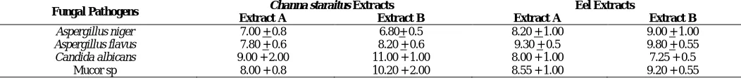
Anifungal activities of the extracts
All the extracts showed higher anti fungal activity as compared to the control (ampicillin). The extract A (ethanol) from
Channa striatus showed maximum antifungal activity as compared
to other abstracts (table 2).
Antiproliferative activity of extracts
The cytotoxicity for all the extracts were measured for HeLa cell lines and maximum toxicity was found for extracts A
(ethanol) for Monopterus albus (45%) and Channa striatus (51 %)
as compared to the extracts B (aqueous) for Monopterus albus (58
%) and Channa striatus (67 %). Overall, the extracts from
Monopterus albus was found higher (11.76 % and 13.43 % for
extract A and B respectively) than extracts from Channa striatus.
The extracts from both animals showed anti proliferative activity
with higher activity for Monopterus albus (figure 1).
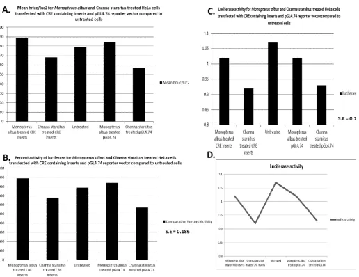
Effects of extracts on gene expression
The mean hrluc/luc2 for for Monopterus albus and
Channastraitus treated HeLa cells transfected with pGL4.74
parental vector (with hrluc) and pGL4.74::Pro720 (with CRE site)
was measured. The cotransfection of pGL4.10 (with luc2) was
performed for the normalization of data for luciferase assay. It was found that the mean RLU for pGL4.74 and pGL4.74::CRE
constructs treated with Monopterus albusextracts were higher
(76.4% and 67.8%) respectively as compared to the mean RLU for
pGL4.74 and pGL4.74::CRE constructs treated with Channa
straitus extracts. The untreated pGL4.74 was used as a negative
negative control for co-transfection. The results suggested the
down regulation of downstream hrluc gene by Channa straitus
extract but upregulation of hrluc gene by Monopterus albus.
Percent activity of luciferase and was calculated as a process of normalization of data for luciferase assay and the results were found consistent with the mean RLU values. The percent activity of luciferase for all the constructs showed to be highest for the transfections of pGL4.74 and pGL4.74::Pro720 (76.4% and
67.8%), which were treated with Monopterus albus extract as
compared to the Channa striatus extracts (figure 2). The results
were found supportive towards up regulation of hrluc by
Monopterus albus extract and down regulation by Channa striatus
extracts. The difference in luciferase activity was
calculated to be maximum for 1.07 (95.32 % of pGL4.74::Pro720
treated with Monopterus albus extract, 85% of pGL4.74::Pro720
treated with Channa striatus extract, 95.32 % of pGL4.74 treated
with Monopterus albus extract and 86.91% of pGL4.74 treated
with Channa striatus extract). The luciferase activity of pGL4.74::
Pro720 treated with Monopterus albus was found higher (14%)
than the luciferase activity of pGL4.74:: Pro720 treated with
Channa striatus (85.98% of Monopterus albus treated). Similarly,
for the pGL4.74 without CRE site, the luciferase activity of
pGL4.74 treated with Monopterus albus was found higher (9%)
than the luciferase activity of pGL4.74treated with Channa
striatus (91% of Monopterus albus treated). The results of
luciferase assays were in accordance with the normalized values and were found significant by Pearson correlation analysis.
Table 1: Antibacterial activity of Monopterus albus extracts (2mg/ml) and Channastraitus extracts (2mg/ml)against E.coli and V. cholera (mean + SD).
Pathogens Monopterus albus extracts Channa staraitus extracts Untreated
Extract A Extract B Extract A Extract B
E. coli
Antibacterial activity 11.00 + 1 12.00 + 2.00 7.00 + 2.00 10.00 + 2.00 Mixed growth
Growth rate (generation/hour) 5.5 4.7 6.6 5.8 3.57
Generation time (Hours/generation) 0.18 0.21 0.15 0.17 0.28
V. cholerae
Antibacterial activity 11.00 + 1 13.00 + 1.00 8.00 + 3.00 10.00 + 1.00 Mixed growth
Growth rate (generation/hour) 4.3 3.8 5.2 4.7 3.31
Generation time (Hours/generation) 0.23 0.26 0.19 0.21 0.30
Table 2: Antifungal activity of Monopterus albus extracts (2mg/ml) and Channastraitus extracts (2mg/ml)against Aspergillusniger, Aspergillusflavus, Candida
albicans, Mucorsp (mean+ SD).
Fungal Pathogens Channa staraitus Extracts Eel Extracts
Extract A Extract B Extract A Extract B
Aspergillus niger 7.00 + 0.8 6.80+ 0.5 8.20 + 1.00 9.00 + 1.00
Aspergillus flavus 7.80 + 0.6 8.20 + 0.6 9.30 + 0.5 9.80 + 0.55
Candida albicans 9.00 + 2.00 11.00 + 1.00 8.00 + 1.00 7.25 + 0.5
Mucor sp 8.00 + 0.8 10.20 + 2.00 8.55 + 1.00 9.20 + 0.55
DISCUSSION
The role of Channa striatus and Monopterus albus in
wound healing, antibacterial activities, antifungal activities has been accepted traditionally in Malaysia and neighboring countries
with strong scientific evidences with Channa striatus being
indigenous to many of such tropical countries (Mohsin et al.,
1983). The flesh is claimed in rejuvenating in recuperation from serious illness and in postnatal diet beside the use in wound
healing (Mat Jais et al., 1994). Many claims have been considered
about the flesh of both animals without any scientific claims. We planned to screen and identify the bioactive molecules in the flesh of these two animals. Instead of targeting a specific organ or component, we aimed in using the whole animal to avoid any loss of any crucial element in future analysis of bioactive molecules by UHPC and LCMS etc. This study was a preliminary study to
compare the effects of Channa striatus and Monopterus albus. The
results showed that the antimicrobial effects (anti bacterial and anti
fungal) of extracts from both animals were markedly high as compare to control (ampicillin) but if, we compare the
antibacterial and antifungal effects of both the animals, extracts
from Monopterus albus showed higher antibacterial and antifungal
activities (table 1 and 2).
The antiproliferative effect on HeLa cell lines reflected again that both the extracts have antiproliferative (by MTT assay)
activity but again the Monopterus albus showed a little more
activity (11.76 % and 13.43 % for extract A and B
respectively)than Channa striatus (figure 1).Previous studies on
the antiproliferative activity of fish protein hydrolysates on human breast cancer cell lines also supports this present piece of work (L.
Picot et al., 2006).For the future plans, as we are looking into the
role of these extracts at molecular level, the effects of these effects were analyzed on the gene expression. We used pGL4.74 vector which has an insert of hrluc in its backbone. Besides hrluc, we
cloned a region from human promoter (promoter region of SMN2
gene in humans) with a CRE binding site as an enhancer. We choose the region with CRE binding site as it is one of the cAMP response element involved in transcriptional control of many of
the human genes (Sarmila et al., 2003). The results of
co-transfection were interesting because likewise in antibacterial and
Fig. 2: Effects of extracts from Monopterus albus and Channa straitus on treated HeLa cells transfected with CRE containing inserts and pGL4.74 reporter
antifungal, where Monopterus albus was more effective, in gene
expression analysis, again the aqueous extract from Monopterus
albus was found more effective in over expression of downstream
gene (hrluc) in the presence and absence of human promoter
region (figure 1 and 2). The results were optimized to normalize
the data (table 3). Interestingly, Channa striatus was found to
down regulate (decrease in expression) the expression of
downstream gene (hrluc) in the presence and absence of human
promoter region (figure 1 and 2). These all findings reflect the
more dominant role of extracts from Monopterus albusover
Channa striatus. These findings also suggest, if the role of Channa
striatus in down regulating the expression of the gene is well
understood using microarray expression analysis then, might the
extracts or bioactive molecules from Channa striatus can be used
as a therapy at molecular level. Similarly, the molecular analysis
of the Monopterus albus in future can help to find some novel
therapies for up regulating the human genes. He Y et al did report the presence of biochemical macromolecules or expression
analysis in Channa striatus (He et al., 2006) and Monopterus
albus but yet, no comparison has been reported among biological
activities of extracts from both of these animals. To the best of our knowledge, this is the first preliminary study on the comparative
analysis of the extracts from Monopterus albus and Channa
straitus.
ACKNOWLEDGEMENTS
We acknowledge the grant from UniSZA for sponsoring this project.
REFERENCES
Edwards R, Harding KG. Bacteria and wound healing. Curr Opin Infect Dis. 2004; 17:91-96.
Haniffa M.A., Dhanaraj, M., Arun Singh, S.v., Muthu Ramakrishnan, C., Manikandaraja, D., Milton, James M. Antibacterial Activity of Skin and Intestinal Mucus of Five Different Freshwater Fish Species Viz., Channa striatus, C. micropeltes, C. marulius, C. Punctatus and C. gachua. Malaysian Journal of Science. 2009; 28: (3).
Jamaluddin, J.A.F., Pau, T.M., Siti-Azizah, M.N. Genetic structure of the snakehead murrel, Channastriata (channidae) based on the cytochrome c oxidase subunit I gene: influence of historical and geomorphological factors. Genet. Mol. Biol. 2011; 34: 152-160.
L. Picota, S.Bordenavea, S. Didelota, I. Fruitier-Arnaudina, F. Sanniera, G.Thorkelssonb, J.P. Bergéc, F. Guérardd, A. Chabeaudd, J.M. Piota, M. N. Nik Mohd Ikram and B. H. Ridzwan. Antiproliferative activity of fish protein hydrolysates on human breast cancer cell lines. International Research Journal of Pharmacy and Pharmacology. 2013; 3: 1-8.
Lehrer RI, Rosenman M, Harwig SSL, Jackson R, Eisenhauer P. Ultrasensitive assays for endogenous antimicrobial peptides. J Immunol Meth. 1991; 137: 167-173.
Mat Jais AM. 1991. Haruan, Channa striatus, Farming in Backyard. Proceedings of the Third Asian Conference of Technolology for Rural Development. 1991;91: 230 - 232.
Mat Jais, AM. Molecular size of the bio-active components from Haruan Channa striatus extract. J. Appl. Sci. 2007a; 7: 2198–2199.
Mat Jais, A.M. Pharmacognosy and pharmacology of Haruan
Channa striatus, amedicinal fish with wound healing properties. Bol.
Latinoam. Caribe Plant. Med. Aromaticas (BLACPMA). 2007b; 6: 52–60. Mat Jais AM, Ross McCulloch, Kevin Croft. Fatty acid and amino acid composition in haruan as a potential role in wound healing. General Pharmacology: The Vascular System. 1994; 25: 947–950.
Mohsin AKM and Ambak MA. Freshwater Fishes of Peninsular Malaysia, Kuala Lumpur, Malaysia, Universiti Pertanian Malaysia press, 1984, Pp282.
Pravin Kumar N, Marimuthu K, Vengkades Rao R., Xavier R, Kathiresan S, Suresh CV, Sreeramanan S. Asian Pacific Journal of Tropical Disease. 2012; 302-305.
Rahim, MHA, Rozila, A, Mat Jais, AM. The physical–chemical and morphological study of Haruan Channa striatus in Peninsular Malaysia. Res. J. Biol. Sci. 2009; 4: 994–1009.
Root-Bernstein R, Root-Bernstein M. Honey, Mud, Maggots and Other Medical Marvels: The sciences behind folks remedies and old wives’ tales by Houghton Mifflin Company, Massachusetts, USA. 1999.
Rubin R. 2004. Maggots and leeches: Good medicine. USA TODAY http://www.usatoday.com/news/health/2004-07-07-leeches-maggots_x.htm [Posted 7/7/2004, Last accessed 23/07/2007]
S. Bragadeeswaranand S. Tnangaraj. Asian Journal of Biological Sciences. 2011; 4: 272-276.
SarmilaMajumder, SaradhadeviVaradharaj, KalpanaGhoshal, UmraoMonani, Arthur H. M. Burghes.Identification of a Novel Cyclic AMP-response Element (CRE-II) and the Role of CREB-1 in the cAMP-induced Expression of the Survival Motor Neuron (SMN) Gene. The journal of biological chemistry, 2004; 279: 14803–1481.
Venkatachalam Uthayakumar, Venkatachalam
Ramasubramanian, Dhanabalan Senthilkumar, Venkatesan Brindha Priyadarisini, Ramasamy Harikrishnan. Biochemical characterization, antimicrobial and hemolytic studies on skin mucus of fresh water spiny eel Mastacembelusarmatus. Asian Pacific Journal of Tropical Biomedicine. 2012;863-869.
How to cite this article: