In order to make all calculations more objective, computer systems that measure digital pictures were used. A digital analysis of the UMC pic-tures, combining different techniques of computer graphics, was approved as a more precise and re-liable measuring method. Hence, the decision to elaborate an application based on modern meth-ods (raster and vector graphics) that could en-sure precision and repeatability of UMC meaen-sure- measure-ments, putting special emphasis on qualitative and quantitative parameters.
The purpose of our research was to introduce a metric classification of the orbital opening. Orbit is the main structure of the upper
cra-niofacial massif (UMC). It is one of the most com-plicated areas of the facial part of the cranium. It is an anatomical structure located in the upper fron-tal part of the face. It takes the shape of an irregu-lar tetrahedronal pyramid or a cone. The bases of both orbits are directed to the front, slightly later-ally. The long axis of both orbits converges to the back and over sella turcica [1, 2].
Classic anthropometric methods used to mea-sure the skull variability are burdened with mis-takes resulting from the construction of measur-ing devices as well as from researcher’s experience.
Tomasz Lepich
1, A–D, F, Józefa Dąbek
2, B–E, Edyta Jura-Szołtys
3, C–F,
Małgorzata Witkowska
3, B, E, F, Mieczysław Piechota
4, D–F, Grzegorz Bajor
5, FOrbital Opening Shape
and Its Alphanumerical Classification
1 Department of Human Anatomy, Medical University of Silesia, Katowice, Poland 2 Department of Cardiology, Medical University of Silesia, Katowice, Poland 3 ENT Department, Medical University of Silesia, Katowice, Poland4 Department of Cardiology, Municipal Hospital, Tychy, Poland
A – research concept and design; B – collection and/or assembly of data; C – data analysis and interpretation;
D – writing the article; E – critical revision of the article; F – final approval of article
Abstract
Background. Orbit is the one of the most complicated areas of the facial part of cranium. The anthropological analysis of the orbits comprises basic measurement characterizing their shape: width and height. Classic anthro-pometric methods used to measure the skull variability are burdened with mistakes resulting from construction of measuring devices as well as from researcher’s experience.
Objectives. The purpose of our research was to introduce a metric classification of the orbital opening.
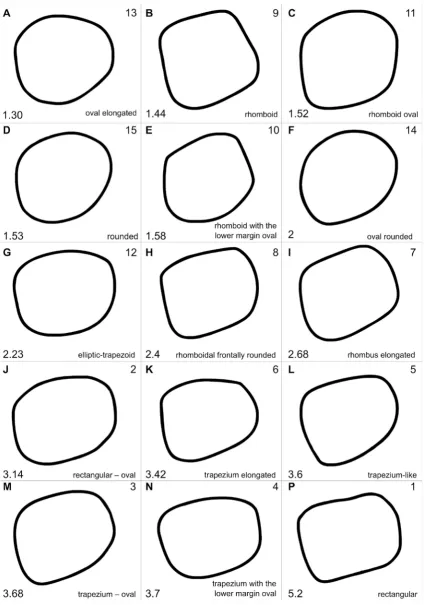
Material and Methods. The study was carried out on 184 skulls. In our study we suggest introducing a clas-sification of the orbital opening shape by calculating a functional for the 15 categories of the orbit shape. Shape categories have been arranged following the increasing value of the functionals. Each shape category of the orbital opening, according to the Piasecki’s descriptive classification, was assigned a letter from the alphabet.
Results. We have observed a greater number of symmetrical skulls in the female group (29.11%) than in the male (23.81%). In both groups the symmetry type AA was the most frequent, it corresponds to the value of functional comprised in the interval from 0 to 1,30. According to the Piasecki’s descriptive classification it was the oval elon-gated type.
Conclusions. Our alphanumerical classification based on the value of functional and on the orbit outline assigned to the value is an objective and useful method of the orbital opening shape analysis (Adv Clin Exp Med 2015, 24, 6, 943–950).
Key words: classification, orbit, skull.
ORIGINAL PAPERS
Adv Clin Exp Med 2015, 24, 6, 943–950
Material and Methods
The study was carried out on 184 skulls in a good state of preservation from early medi-eval ages (10th and 13th century) found in Kije
and Złota Pińczowska sites. The skulls were made available by the Warsaw University Archeolo-gy Institute. The above-mentioned archeological sites were thoroughly described in a monograph by M. Zoll-Adamikowa [8]. The analysis was con-ducted on the remaining parts of the maturus and adultus cranium conserved as a skull without man-dible (calvarium). The age of the skulls was dated on the basis of dentition and suture calcification and it ranged from 20 to 55 years. The skulls were divided into gender groups: male skulls (105) and female skulls (79).
The anthropological analysis of the orbits comprises basic measurement characterizing their shape: width and height.
Orbit width is defined as the distance between mf and ekpoints [3–6]. It may also be measured from the point called dakryon (d) on the inner side to the ekpoint. The anthropometric point d is sit-uated at the junction of the frontal bone, lacri-mal bone and the frontal process of maxilla. If the point d is invisible because of total calcification of the sutures or because of its abnormal localisa-tion, the orbit width is measured from the lacri-male (la) point. This point is situated at the cross-ing of posterior lacrimal crest with frontolacrimal suture [3].
Orbit height is the longest distance between the upper and the lower margin of the orbit. It is measured perpendicularly to the line that marks its width, at mid-width [2].
Orbit indexis equal to the quotient of the or-bit height and width; it is expressed in percent-age (H/S × 100). Its value informs about the orbit shape [2, 7].
Measuring Instruments
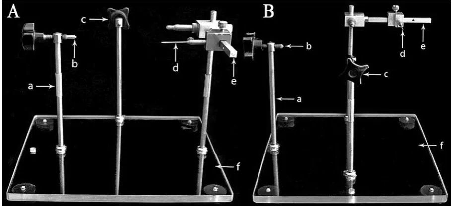
The examined crania were fixed into a cranio-stat. A craniostat (craniosteophor) enables a secure stabilization of the skull and its manipulation in space according to the defined position and sur-face measurements.
In our study the Mollison’s craniostat was used in the author’s modification [9]. Unlike a clas-sic cranoistat, it is constructed on a basis for three stands that are fixed to it. In their upper part the stands are equipped with a screw-grip to hold the cranium. Two of the stands hold up the lateral part of cranium and one infraoccipital grip holds up the posterior part. The grips move horizontally at an unrestricted distance. In addition, in order to po-sition the cranium on the Frankfurt line, the cra-niostat was equipped with an indicator moving in horizontal and vertical surface that enables point-ing out the lower margin of the orbit (Fig. 1).
After fixing them on the Frankfurt line, the photograph of the crania were photographed fron-tally from the distance of 50 cm. Pictures of the cra-nia were taken with a high resolution (2272 × 1704) digital camera Sony. Digital pictures of the orbits were analyzed using the author’s own application Digital Image Cranio-Analyzer 2.0 (DICA 2.0), elaborated at the Silesian Medical University Anat-omy Department, combining bit and raster simul-taneous recording. To analyze the orbit pictures, the non-referring method was used.
In our study we suggest introducing a classifi-cation of the orbital opening shape by calculating a functional for the 15 categories of the orbit shape; the calculation is based on Wierciński and Kapica classification that was modified and published by Piasecki [6, 10]. This scale defines the orbit shape variability. We distinguish the following forms: ar-chimorphic-rectangular and trapezium-like acteristic for white race), diagonal-rhombic (char-acteristic for yellow race) and neomorphic-elliptic and round (typical for black race).
Detailed Characteristics
of the Orbital Opening
Categories by Piasecki [10]
1. Rectangular: the opposite margins of the orbit are parallel to each other, the angles of the orbit are right, the opposite margins are of near-ly equal length,
2. Rectangular – oval: rectangular + all the margins are curved to the exterior, the angles more round,
3. Trapezium – oval: trapezium-like + the margins more curved to the exterior, the angles even more round,
4. Trapezium with the lower margin oval: tra-pezium-like + the lower margin markedly curved, the lower angles round,
5. Trapezium-like: two margins are parallel – external and internal, all the margins are of un-equal length, the internal one is the shortest, the upper angles of the orbit are close to the right an-gle, the lower external angle is acute, the lower in-ternal angle is obtuse,
6. Trapeizum elongated: trapezium-like + the lower external angle of the orbit is more acute and markedly elongated downwards,
7. Rhombus elongated: rhomboid + the low-er extlow-ernal angle of the orbit acute and markedly elongated downwards,
8. Rhomboidal frontally rounded: rhom-boid + the inner margin is markedly curved to the exterior,
9. Rhomboid: the orbit margins are parallel, of corresponding length, all the angles of the orbit are close to the right angle,
10. Rhomboid with the lower margin oval: rhomboid + the lower margin markedly curved, the lower angles more rounded,
11. Rhomboid oval: rhomboid + the margins are more curved to the exterior, the angles more rounded,
12. Elliptic-trapezoid: the form of a trapezium, the upper and lower margin elliptically rounded,
13. Oval elongated: the orbit markedly round-ed, without marked angles, the internal part of the
orbit narrowed, the lower external margin extend-ed downwards,
14. Oval rounded: the orbit markedly round-ed, without marked angles, slightly elongated hor-izontally, equally wide on both sides,
15. Rounded: the orbit markedly rounded, without marked angles, its shape resembles a circle.
Shape categories have been arranged follow-ing the increasfollow-ing value of the functionals. Each shape category of the orbital opening, according to the Piasecki’s descriptive classification, was as-signed a letter from the alphabet. That is how the alphanumerical classification based on the value of a functional was created (Table 1). The value for the functional putting a numeric value to each contour of orbit according:
2 1
)
(
∑
=−
=
ni i sr
r
r
F
In order to analyze orbit symmetry and asym-metry, the values of functionals for the right and left orbit have been compared for each male and female skull. Based on the functional each or-bit was assigned its proper letter from the alpha-bet according to the alphanumerical classification (Fig. 2).
Results
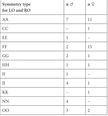
We have observed a greater number of sym-metrical skulls in the female group (29.11%) than in the male one (23.81%). In both groups the sym-metry type AA was the most frequent; it corre-sponds to the value of functional comprised in the interval from 0 to 1.30. According to the Piasecki’s descriptive classification it was the oval elongated type (Table 2).
In the male group there were two dominant types of asymmetry: one corresponding to the FE (EF) configuration found in 6 skulls, the other one was NJ (JN) – found for the same amount of skulls. According to Piasecki’s descriptive classification, the shape types of these skulls are the following: rhomboid rounded at bottom and oval round-ed with rectangular oval and trapezoid roundround-ed at bottom. In the female group the AF (4 skulls) asymmetry type prevailed which corresponds to the oval elongated and oval rounded types.
1 with GG, 1 with HH, 1 with JJ, 1 with KK from all of the 79 female skulls. Inconsiderable differences between the α and β angles were stated. The most significant differences were noted for skulls with the OO-9° symmetry, which – in consistency with
the descriptive classification – is responsible for the rectangular shape of the orbital opening. The slightest differences were observed for the skulls with KK symmetry (less than 1°), which reflects trapezoid elongated shape of the orbital opening.
In male skulls the analysis was carried out on: 7 skulls with AA symmetry, 4 skulls with JJ and NN symmetry, 3 skulls with OO symme-try, 2 skulls with FF and GG symmetry and 1 skull for each of the following types: EE, HH and II. The comparison of angles α and β as well as of the or-bit symmetry in male skulls did not demonstrate such regularity as in female skulls. The most sig-nificant differences were observed for the skulls with the type OO of symmetry and for the type EE (in the descriptive shape classification – rhomboid rounded at bottom). This difference equals 10°. More important differences between the α and β angles were stated in male group (maximal dif-ference equaled 12°). Some exemplary asymmetri-cal skulls were also examined, with special focus on the skulls presenting the most significant dif-ferences between the left and right orbit, on the ba-sis of the value of the functional. In male skulls the differences were more visible (maximal value 14°). In female skulls maximal value was 12°.
Table 1. Orbit classification Classification
based on functional Shape categories according tonominal classification Functional No. from descriptive classification
A oval elongated F < 1.30 13
B rhomboid 1.30 ≤ F < 1.44 9
C rhomboid rounded 1.44 ≤ F < 1.52 11
D rounded 1.52 ≤ F < 1.53 15
E rhomboid rounded at bottom 1.53 ≤ F < 1.58 10
F oval rounded 1.58 ≤ F < 2.0 14
G elliptic-trapezoid 2.23 ≤ F < 2.40 12
H rhomboid frontally rounded 2.40 ≤ F < 2.68 8
I rhomboid elongated 2.68 ≤ F < 3.14 7
J rectangular oval 3.14 ≤ F < 3.42 2
K trapezoid elongated 3.42 ≤ F < 3.60 6
L trapezoid 3.60 ≤ F < 3.68 5
M trapezoid rounded 3.68 ≤ F < 3.70 3
N trapezoid rounded at bottom 3.70 ≤ F < 5.20 4
O rectangular F ≥ 5.20 1
Table 2. Types of symmetry in male and female skulls – alphanumerical classification; n – number of skulls,
♂ – male skull; ♀ – female skull; LO – left orbit; RO – right orbit
Symmetry type
for LO and RO n ♂ n ♀
AA 7 11
CC – 1
EE 1 –
FF 2 15
GG 2 1
HH 1 1
II 1 –
JJ 4 1
KK – 1
NN 4 –
Discussion
The orbit is one of the most complicated struc-tures of the UMC. It is formed by several bones
and its constitution is three dimensional. A thor-ough knowledge of craniometrical parameters de-scribing height (n-pr), width (zma-zma) of the skull; height and width of the orbit, emphasizing
Fig. 2. Arrangement of the orbit inlet classifications: nominal and after the functional
the gender differentiating parameters, allows for a precise reconstruction of bone structures. In our work the parameters describing height of facial cranium such as: n-pr and n-ss were of higher val-ue for male skulls. After normalization through the arithmetical and geometrical modulus, higher val-ues in male skulls were observed for the parame-ter n-ssand in female skulls – for the variable n-pr. The value of the parameter describing orbit width zma-zma and the variables describing orbit width, orbit height, mean and median radius, surface ar-ea and orbit circumference were of higher value in male skulls before normalization of the results. Af-ter normalisation through the arithmetic module, as well as through the geometric module, the values of the above mentioned parameters were higher in female skulls. Female skulls are relatively bigger in comparison to male skulls, which is confirmed in literature [11]. Nagle and al. [12] observed high-er values for the paramethigh-ers describing width and height of the face in male skulls, however their measurements were carried out on living people, not on skulls and the results have not been normal-ized. Radović and al. [13] examined the variability of facial part of cranium in 46 men and 53 wom-en from Croatian population. The craniometric parameters were measured using standard anthro-pometric devices. In male group higher values for the parameters describing height and width of the face n-pr were stated. The correlation between pa-rameters describing height of the face and width of the facial part of cranium has been demonstrated (r = 0.40). Similar values of the correlation coeffi-cient were obtained in our study. Hee-Kyung Park and al. [14] examined craniometric points on 30 human skulls of an unknown gender. The distance separating the points from the lambda point was measured with the use of a laser. The laser mea-surement was compared to the classic one. The width of the right and left orbit has been analyzed. The width of the right orbit (40.65 mm) was great-er than that of the left (40.55 mm). In our study a similar tendency in the orbit size has been ob-served. The right orbit width was 39.11 mm and the left orbit width was 37.84 mm. The differences in the orbit width result from, among others, the fact that the examined skulls were of different pop-ulation origin. In our work broader orbits were ob-served on the right side.
Orbit index along with the skull height index plays an important role in defining the anthropo-logical type. Gładkowska-Rzeczycka [15] tried to reconstruct the orbital opening making plaster casts of bones found in the crematory graves. The author observed that her method makes it possible to define the somatic type of a person, but it is not useful in determining the anthropological type.
There are several orbital opening shape clas-sifications. One of them is descriptive (arbitrary) and based on measuring height of the orbit using a scheme: high orbit, medium-height orbit, quite high and short. These categories are helpful in or-bit characterization, but they do not let to compare results from different researchers.
Martin’s classification distinguishes 3 groups – according to the orbit index: with the orbit in-dex value above 85 – hypsikonch, with the index value ranging between 76 and 84.9 – mesokonch and with the index value equal or inferior to 75.9 – chamaekonch [16]. Kadanov and al. [17] car-ried out research on 412 male skulls from Bulgar-ian population. 15 parameters of the orbital open-ing were studied. The research revealed that in most skulls (63%) the orbit width was greater on the right side, whereas the orbit height was greater on the left side. The orbit index was also observed, which led to the conclusion that: chamaekonch type appeared more often on the right side and hypsikonch on the left side. In our study the type hypsikonch was observed on both sides in male skulls (mean value for the orbit index: right orbit – 85.93, left orbit – 87.97). In female group the re-sults were comparable (the value of the orbit index: right orbit – 88.52, left orbit – 92.13). Another clas-sification of the orbital opening shape based on or-bit index was presented by Patnaik [7]. The oror-bits were divided into 3 groups: megaseme (big) – or-bit index superior to 89, among yellow race mainly, mesoseme (medium) – orbit index between 89 and 83, typical for white race (Europeans – 87), mi-croseme (small) – orbit index below 83.
by Piasecki [14]. This time there were 15 categories. In the above-enumerated works the orbital open-ing shape is compared to a basic geometrical figure. The methodology of the measurement consists of comparing the given feature with a sketch that rep-resents this particular feature in the table of sketch-es. On this basis, the feature is assigned a category number corresponding with the degree the exam-ined feature is developed. This method is burdened with mistakes resulting from a subjective evalua-tion. Piasecki’s nominal orbit classification is more objective, which offers a possibility of comparing results obtained by different researchers.
In our work a new alphanumerical orbital opening classification is presented. The value of functional is the criterion of our classification. It is based on Piasecki’s work [9] and consists in calcu-lating the functional for each of the 15 categories of the orbital opening shape, and next, in arranging results according to their value. The values of func-tionals for each geometrical figure are constant, that is why it is possible to assign only one num-ber to each geometrical figure and thus, determine the orbital opening shape (round, oval or square). The smaller the value of the functional, the round-er gets the shape. This classification gives an im-portant advantage – repeatability.
Our research demonstrated lower values of the functional for both orbits in female skulls and high-er in male skulls. It allows us to classify female or-bit as rounder and male oror-bit as more rectangular. The round orbit shape in female skulls is confirmed
by Patnaik and al. [7] and Wierciński [19]. Accord-ing to Wierciński, the main reason for morpho-logical differences between male and female skulls is the earlier and more accentuated ontgenetic re-tardation. In consequence, female skulls morpho-logically resemble skulls of children that are char-acterised by a rounder orbital opening shape. It is important to emphasize that the value of the func-tional does not depend on the orbit rotation, but on the orbit shape. In the examined female population (29.11%) there were more symmetrical skulls, most frequently of AA configuration (according to the alphanumerical classification based on the value of functional). De Paiva and al. [20] confirm this state-ment; in their research female skulls were less asym-metrical. The most frequent symmetry type in male skulls (23.18%) was the AA configuration as well.
Introduction of a new orbital opening shape classification based on the functional value may become useful for orbit qualitative parameters analysis. Thanks to the combination of the func-tional value with the descriptive classification that we present as our alphanumerical orbit shape clas-sification, it is finally possible to analyze results of different authors, including those who have not used numerical methods in their research. Our al-phanumerical classification based on the value of functional and on the orbit outline assigned to the value is an objective and useful method of the or-bital opening shape analysis. It combines all the advantages that the numerical, descriptive and nominal classifications offer together.
References
[1] Bron A, Tripathi BJ: Wollf`s anatomy of the eye and orbit. Chapmann & Hall, London 1997, 1–17.
[2] Weisman RA: Surgical anatomy of the orbit. Otolaryngol Clin North Am 1988, 21, 1–12.
[3] Lepich T, Dąbek J, Piechota M, Bajor G, Aniszewski Ł, Markowski J: Digital analysis of the orbit using the non-referring method. Arch Med Sci 2014, 24, 182–190.
[4] Picket MM: L’indice orbitaire et l’apreciation de la largeur de l’orbite. Bull Soc Antrop 1954, 10, 100–112.
[5] Reymond J, Piasecki K: Kranioskopia a urazy oczodołów. Czas Stomat 1988, 9, 555–558.
[6] Piasecki K, Reymond J, Wysocki J: Kształt wejścia do oczodołu – metodyka badań. Antropologia a Medycyna i Promocja Zdrowia. Eds.: Malinowski A, Łuczka B, Grabowski J. Wyd. Uniw. Łódzkiego, Łódź 1996.
[7] Patnaik VVG, Bala S, Singla Rajan K: Anatomy of the bony orbit-some applied aspects. J Anat Soc India 2001, 50, 59–67.
[8] Zoll-Adamikowa M: Wczesnośredniowieczne cmentarzyska szkieletowe Małopolski. Wyd. PAN, Wrocław– –Warszawa–Kraków 1996, 61–62.
[9] Lepich T, Dąbek J, Stompel D, Gielecki J: Analysis of the upper massif of the craniofacial with the radial method – practical use. Arch Med Sci 2011, 7, 870–876.
[10] Piasecki K: Las nuevas escalas craneoscopicas observaciones preliminares. Uniwersytet Warszawski, Warszawa 1992.
[11] Brown P, Maeda T: Post Pleistocene diachronic change in East Asian facial skeletons: the size, shape and volume of the orbit. Anthropol Sci 2004, 112, 29–40.
[12] Nagle E, Teibe U, Kapoka D: Craniofacial anthropometry in a group of healthy Latvian residents. Acta Med Lituanica 2005, 12, 47–53.
[13] Radović Z, Muretić Nemirovskij Ž, Gaži-Čoklica V: Craniofacial Variations in a South Dalmatian Population. Acta Stomatol Croat 2000, 34, 399–403.
[15] Gładykowska-Rzeczycka J: „Próba rekonstrukcji wejścia do oczodołu na podstawie fragmentów kości z grobów ciałopalnych. Folia Morphol 1965, 24, 307–310.
[16] Martin R: Lehrbuch der Antropologie t. 2, G. Fisher, Jena, 1928.
[17] Kadanov D, Iordanov I, Aleksandrova N: Symmetry and asymmetry of the orbital opening in Bulgarians. Eksp Med Morfol 1977, 16, 12–18.
[18] Catalina-Herrera CJ: Morphometric study of the orbit’s base in male and female skulls of Spaniards. Bull Assoc Anat (Nancy) 1988, 72, 217–218.
[19] Wiercinski A: Ontogenetic retardations and human evolution. Proc Symp Natur Select Prague 1978, 277–301.
[20] De Paiva LA, Segre M: Sexing the human skull through the mastoid process. Rev Hosp Clin São Paulo 2003, 58, 15–20.
Address for correspondence:
Małgorzata Witkowska ENT Department
Medical University of Silesia Francuska 20-24
40-027 Katowice Poland
E-mail: [email protected]
Conflict of interest: None declared