Development 139, 2903-2915 (2012) doi:10.1242/dev.077081 © 2012. Published by The Company of Biologists Ltd
INTRODUCTION
The specification of the embryonic axes in many organisms is initiated by maternally deposited factors that activate various signaling pathways to establish the body plan. In Drosophila, frogs and fish, asymmetric localization of maternal factors in oocytes and eggs is crucial for correct embryonic patterning (St Johnston, 1995; Grunert and St Johnston, 1996; Mowry and Cote, 1999; Pelegri, 2003; Heasman, 2006; Abrams and Mullins, 2009; Nojima et al., 2010; Lu et al., 2011). In Xenopus, wnt11 RNA is localized in dorsal vegetal cells, and blocking maternal Wnt and -catenin functions leads to dorsal deficiencies (Heasman et al., 1994; Wylie et al., 1996; Schroeder et al., 1999; Heasman et al., 2000; Kofron et al., 2001; Tao et al., 2005). Recent work has also implicated vegetally localized Trim36 ubiquitin ligase in microtubule-dependent cortical rotation and axis formation (Cuykendall and Houston, 2009).
In zebrafish, several experiments have suggested that dorsal determinants are vegetally localized, and are translocated via microtubules to trigger dorsal axis specification (Jesuthasan and Strahle, 1997; Mizuno et al., 1999; Ober and Schulte-Merker, 1999). The molecular nature of the maternal factors is just beginning to be elucidated (Abrams and Mullins, 2009). We previously showed that maternal RNA encoding the Nodal morphogen Squint [Sqt; Nodal-related 1 (Ndr1) – Zebrafish
Information Network] asymmetrically localizes in two cells of 4-cell stage embryos (Gore and Sampath, 2002; Gore et al., 2005), and predicts embryonic dorsal prior to the nuclear accumulation of -catenin in dorsal cells. Removal of sqtRNA-containing (sqt+)
cells and knockdown by antisense morpholino oligonucleotides leads to dorsal deficiencies, suggesting that asymmetrically localized maternal sqtfunctions in dorsal specification.
Paradoxically, females homozygous for the sqt insertion mutations sqtcz35and sqthi975(Heisenberg and Nusslein-Volhard, 1997; Erter et al., 1998; Feldman et al., 1998; Amsterdam et al., 2004) produce embryos with mild dorsal deficiencies and do not manifest the loss of anterior and dorsal structures observed upon depletion of sqt, either by antisense morpholinos or embryological dissections of sqt+cells (Aoki et al., 2002; Bennett et al., 2007; Pei
et al., 2007). Similarly, maternal and zygotic mutations affecting the Nodal co-receptor one-eyed pinhead (oep) (Gritsman et al., 1999) result in embryos similar to zygotic cyc;sqt double mutants (cyc is also known as ndr2 – Zebrafish Information Network) (Feldman et al., 1998; Dougan et al., 2003), but these embryos do not manifest complete loss of anterior or dorsal structures. Therefore, the function of maternal sqthas been a matter of debate (Bennett et al., 2007; Gore et al., 2007).
Using several mutations that disrupt Sqt, we find that sqtRNA has functions independent of Sqt protein in dorsal initiation. Furthermore, our analysis shows that the sqtcz35 and sqthi975 insertion alleles, on which the genetic analysis was based, still express and localize maternal sqttranscripts, similar to wild-type embryos. We demonstrate that mutant RNAs still have dorsal-inducing activity in embryos, and show that this activity is a non-coding function of sqtRNA. Interestingly, the non-coding, dorsal-inducing activity of sqtRNA is dependent on sequences within its 3⬘UTR. We also revisited the activity of sqt morpholinos, and find that sqttranslation initiation site sequences are also required for sqt 1Temasek Life Sciences Laboratory, 1 Research Link, National University of
Singapore, Singapore 117604. 2School of Biological Sciences, Nanyang
Technological University, 60 Nanyang Drive, Singapore 637551. 3Department of
Biological Sciences, National University of Singapore, 14 Science Drive, Singapore 117543. 4Genome Institute of Singapore, 60 Biopolis Street, Genome, Singapore
138672.
*Author for correspondence ([email protected])
Accepted 7 June 2012 SUMMARY
Despite extensive study, the earliest steps of vertebrate axis formation are only beginning to be elucidated. We previously showed
that asymmetric localization of maternal transcripts of the conserved zebrafish TGFfactor Squint (Sqt) in 4-cell stage embryos
predicts dorsal, preceding nuclear accumulation of -catenin. Cell ablations and antisense oligonucleotides that deplete Sqt lead to
dorsal deficiencies, suggesting that localized maternal sqtfunctions in dorsal specification. However, based upon analysis of sqtand
Nodal signaling mutants, the function and mechanism of maternal sqtwas debated. Here, we show that sqtRNA may function
independently of Sqt protein in dorsal specification. sqt insertion mutants express localized maternal sqtRNA. Overexpression of
mutant/non-coding sqtRNA and, particularly, the sqt3⬘UTR, leads to ectopic nuclear -catenin accumulation and expands dorsal
gene expression. Dorsal activity of sqtRNA requires Wnt/-catenin but not Oep-dependent Nodal signaling. Unexpectedly, sqt ATG
morpholinos block both sqtRNA localization and translation and abolish nuclear -catenin, providing a mechanism for the loss of
dorsal identity in sqt morphants and placing maternal sqtRNA upstream of -catenin. The loss of early dorsal gene expression can
be rescued by the sqt3⬘UTR. Our findings identify new non-coding functions for the Nodal genes and support a model wherein sqt
RNA acts as a scaffold to bind and deliver/sequester maternal factors to future embryonic dorsal.
KEY WORDS: 3⬘UTR, Axis formation, Dorsal localization, Dorsal expansion, Maternal factors, Non-coding RNA, Nodal, RNA localization, Squint (Nodal-related 1), Zebrafish
Dorsal activity of maternal
squint
is mediated by a
non-coding function of the RNA
Shimin Lim1,2, Pooja Kumari1,3, Patrick Gilligan1, Helen Ngoc Bao Quach1, Sinnakaruppan Mathavan4and
Karuna Sampath1,2,3,*
D
E
V
E
LO
P
M
E
N
2904
RNA localization. The dorsal-inducing activity of sqt RNA is independent of Sqt/Nodal signaling, but requires functional maternal Wnt/-catenin signaling. Our findings identify novel coding functions for the Nodal genes and reveal new roles for non-coding RNAs in the maternal control of axis specification.
MATERIALS AND METHODS Generation of constructs
pCS2+sqtSTOP, pCS2+FLAGsqt and pCS2+FLAGsqtSTOPwere generated
by PCR-based site-directed mutagenesis. pCS2+sqtSTOP and
pCS2+FLAGsqt were generated using pCS2+sqt (Gore et al., 2005) as PCR template, whereas pCS2+FLAGsqtSTOP was generated from
pCS2+sqtSTOP. pCS2+sqtcz35was generated by amplifying two overlapping
fragments from MZsqtcz35cDNA, followed by overlap extension PCR and subcloning into pCS2+. pCS2+T-sqt was generated using pCS2+sqtcz35as
PCR template, followed by subcloning into pCS2+. Primers are listed in supplementary material Table S1.
Zebrafish strains
Wild-type zebrafish, MZsqtcz35, MZsqthi975, MZdicerhu715, MZoeptz57and homozygous ichp1mutant fish were maintained at 28.5°C and embryos were obtained by natural mating using standard procedures, in accordance with institutional animal care regulations (Westerfield, 2007). The genotype of MZsqtcz35embryos was determined as described (Feldman et al., 1998). Homozygous ichmutant mothers that yielded 100% radialized embryos were used and identified as described (Kelly et al., 2000; Bellipanni et al., 2006).
Quantitative real-time RT-PCR
Total RNA was extracted from embryos using TRIzol reagent (Invitrogen). 250 ng RNA from WT, MZsqthi975and MZsqtcz35embryos and 250 ng RNA from lacZ:glo or lacZ:sqt RNA-injected embryos was used for cDNA synthesis. 1 l first-strand cDNA was used in 10 l PCR reactions. Genomic DNA contamination was checked by PCR to detect actb2 (act), sqt, dharma (dha; bozozok), vox and vent. Primers are listed in supplementary material Table S1. RT-PCR was performed on an ABI 7900HT Fast Real-Time PCR System (Applied Biosystems) using the comparative CTmethod. Control experiments to measure changes in CT
with template dilutions were performed to test whether amplification efficiencies of target (sqt,dha,vox andvent) and control (act) primers were similar. All results were normalized to act.
Capped mRNA synthesis, injections and in situ hybridizations
Capped mRNA was synthesized from linearized plasmids (NotI, NEB) using the SP6 mMessage mMachine Kit (Ambion), and 25 pg aliquots were injected into 1-cell stage embryos. Fluorescent Alexa 488-labeled RNA was synthesized, injected at the 1-cell stage (Gore et al., 2005; Gilligan et al., 2011) and RNA localization scored visually (Gilligan et al., 2011) by two individuals independently.
To target maternal sqt RNA prior to the formation of sqt RNA aggregates that develop upon egg activation (Gore and Sampath, 2002), 10 ng aliquots of control or sqt morpholinos were injected into squeezed wild-type eggs and fertilized with wild-wild-type sperm (Gore et al., 2005; Gore et al., 2007). Injected embryos were fixed in 4% paraformaldehyde/PBS at oblong, sphere, dome, 30% epiboly, 40% epiboly, 50% epiboly, 60% epiboly and at 24 hours post-fertilization (hpf), and processed for whole-mount in situ hybridization to detect goosecoid, chordin,gata2 andno tail (Sampath et al., 1998). Localization of sqtRNA was detected by in situ hybridization using full-length cDNA probes.
In vitro translation and protein detection
The TNT®SP6 Coupled Rabbit Reticulocyte Lysate System (Promega)
was used to transcribe and translate from the following plasmid templates: pCS2+, pCS2+sqtFL:sqt, pCS2+sqtSTOP, pCS2+sqtcz35, pCS2+FLAGsqt,
pCS2+FLAGsqtSTOPand pCS2+T-sqt. 1 g of plasmid DNA was used in
50 l reactions according to the manufacturer’s instructions (Promega). Biotin-labeled protein products were separated by SDS-PAGE and transferred onto Hybond-C Extra membranes (GE Healthcare).
Immunoblotting was performed using avidin and biotinylated HRP (1:200 dilution) (Ultra-sensitive ABC Peroxidase Rabbit IgG Staining Kit, Pierce). Proteins were detected by Kodak Biomax MS film using SuperSignal West Femto Maximum Sensitivity Substrate (Pierce). FLAG epitope-tagged peptides were detected with anti-FLAG M2 mouse monoclonal primary antibody (1:2500, Sigma) and HRP-conjugated anti-mouse IgG secondary antibody (1:5000, DAKO). To detect nuclear -catenin, 512-cell stage embryos were fixed in 4% paraformaldehyde/PBS and processed for fluorescence immunohistochemistry using a rabbit polyclonal anti- -catenin antibody (C2206, Sigma) and Alexa 488-conjugated goat anti-rabbit secondary antibodies (Molecular Probes).
To compare translation efficiencies of Sqt:glo UTR and Sqt:sqt UTR, 1-cell stage wild-type embryos were injected with 20 pg GFP:glo or sqt-GFP:sqt. Approximately 40-50 injected embryos were manually dechorionated and lysed at 50% epiboly in RIPA buffer. Whole embryo lysates (50 g/lane) were separated by SDS-PAGE and transferred onto Hybond-C Extra membranes. Proteins were detected on film as described above. Sqt-GFP was immunoblotted using rabbit polyclonal anti-GFP antibodies (1:2500, Abcam), followed by HRP-conjugated anti-rabbit IgG secondary antibodies (1:5000, DAKO). Tubulin was detected using mouse monoclonal anti-tubulin antibodies (1:2500, Sigma), followed by HRP-conjugated anti-mouse IgG secondary antibodies.
Microscopy
Live embryos injected with fluorescent RNAs or expressing Sqt-GFP fusion protein were manually dechorionated, mounted in 2.5% methylcellulose (Sigma) and visualized using a Zeiss Axioplan2 microscope with a CoolSNAP HQ camera (Photometrics). MetaMorph (Universal Imaging Corporation) and ImageJ (NIH) software packages were used to acquire and process images. Stained embryos from in situ hybridization and immunohistochemistry experiments were mounted in 100% glycerol and imaged using a Zeiss Axioplan2 microscope equipped with a Nikon DXM1200 color camera. Images were acquired using ACT-1 software (Nikon) and cropped using Adobe Photoshop.
For -catenin- and DAPI-stained embryos, images were acquired using a Zeiss LSM 5 Exciter upright confocal microscope. To quantify -catenin-positive nuclei, 15-25 optical sections at 1.76 m intervals starting from the yolk syncytial layer nuclei were examined per embryo. We detected -catenin-positive nuclei in many sections. However, owing to intense membrane and cytoplasmic -catenin staining that obscured nuclear staining upon z-projection of all sections obtained, three serial confocal sections for each embryo were selected, z-projected using LSM Image Browser software, and cropped using Adobe Photoshop.
Measurement of expression domains
Animal pole view images of embryos stained for gscwere used. In ImageJ, we drew a best-fit circle for the circumference of the embryo using the Circle tool. Using the xycoordinates, the diameter of the circle along the x- and y-axes and its center were determined. The Radial Grid tool in ImageJ and the center coordinates were used to mark the center, and using the Angle and Measure tools the angle of gscexpression was determined.
RESULTS
Mutant sqtRNA expands dorsal gene expression in early embryos
To examine whether the insertion mutants had any sqt activity, we first determined if the RNA and protein are expressed. We previously showed that embryos from fish homozygous for the sqtcz35and sqthi975insertions express maternal sqtRNA (Gore et al., 2007). Whole-mount in situ hybridizations show that, similar to wild-type embryos, mutant sqtRNA is localized to two cells at the 4- and 8-cell stage in MZsqt mutant embryos (Fig. 1A-C; genotypes confirmed by PCR, Fig. 1C). Quantitative PCRs show that MZsqt mutant and wild-type embryos have similar levels of maternal sqt RNA at the 1-cell and 4-cell stages, and that a decrease in sqtRNA levels occurs in MZsqt mutant embryos later during gastrulation (Fig. 1D). Similar to wild-type embryos, we
RESEARCH ARTICLE Development 139 (16)
detect non-polyadenylated mutant sqtRNA that is spliced as well as unspliced in pd(N)6-primed cDNA from 4-cell stage embryos,
whereas only spliced sqtRNA is detected using oligo(dT)-primed cDNA (supplementary material Fig. S1A,B). These results show that mutant maternal sqtRNA is expressed and localized to future dorsal cells in MZsqtmutant embryos at levels similar to wild-type embryos. Therefore, the sqt insertion alleles are not maternal transcript nulls, and maternal sqtRNA levels in mutant embryos are similar to those in wild-type embryos.
To test whether the sqtcz35 insertion RNA generates any Sqt protein, we expressed sqtcz35:sqt RNA in a rabbit reticulocyte lysate
expression system. The insertion RNA is predicted to encode a 17 kDa C-terminally truncated peptide lacking any functional ligand. We find that protein expressed from sqtcz35:sqt RNA is the
predicted 17 kDa peptide (Feldman et al., 1998). We also tested synthetic sqtRNA with a stop codon in the first exon (TTG>TAG, which results in Leu11>STOP; sqtSTOP:sqt), a truncated sqtRNA
(T-sqt:sqt) (Bennett et al., 2007), and FLAG epitope-tagged
versions of wild-type Sqt and SqtSTOP(Fig. 1E). Translation from
control and FLAG-tagged sqtRNA showed the expected 44 kDa proteins, whereas translation from sqtSTOP:sqt and T-sqt:sqt RNA
yielded an N-terminally truncated peptide of 39 kDa, lacking the signal sequence (Fig. 1E,F). In this in vitro translation system, the peptide from sqtSTOP:sqt presumably results from utilization of an
internal ATG downstream of the engineered stop.
We then tested whether sqtcz35mutant RNA has any activity in embryos. One-cell stage wild-type embryos were injected with synthetic capped sqtcz35RNA, sqtSTOPor T-sqt RNA, and nuclear
accumulation of -catenin and dorsal expression of goosecoid(gsc) and chordin(chd) was examined at the onset of gastrulation (Fig. 2A-F,L,M,T). Mutant sqt RNA-injected embryos reached developmental landmarks at the same time as control-injected embryos (Fig. 2J,K,Q,R; data not shown), suggesting that morphogenesis or development is not generally delayed. Surprisingly, embryos injected with mutant sqtRNAs (sqtmut:sqt)
showed expansion of nuclear -catenin expression at the 512-cell stage, with ~16 -catenin-positive nuclei (n18 embryos), as compared with control lacZ:glo RNA-injected embryos (glo, Xenopus globin3⬘UTR) that showed about five positive nuclei (n10 embryos; Fig. 2A-D and Table 1). We detected at least 15 -catenin-positive nuclei in more than 50% of sqtmut:sqt RNA-injected
embryos, and three embryos showed more than 25 -catenin-positive nuclei (n18; Table 1 and supplementary material Table S2). The lower borders of the first tier blastoderm cells are not visible (Fig. 2A,C), consistent with yolk syncytial layer (YSL) formation at the tenth mitosis (512-cell to 1000-cell stage) (Kimmel and Law, 1985). Increased numbers of -catenin-positive nuclei were observed both in the blastoderm (red arrows, Fig. 2A-D) and dorsal YSL of sqtmut:sqt-injected embryos (yellow arrows, Fig. 2C,D), whereas in
[image:3.612.52.295.60.522.2]wild-type and control-injected embryos YSL expression of -catenin is not detected until the 1000-cell stage (supplementary material Fig. S2) (Dougan et al., 2003). Thus, injected mutant sqt RNA can substantially increase the dorsal accumulation of nuclear -catenin.
Fig. 1. Mutant sqtRNAs are expressed and localized in MZsqt
mutant zebrafish embryos.(A,B)Whole-mount in situ hybridization to detect localization of maternal sqtRNA (arrowheads) in 8-cell stage wild-type (WT) (A) and MZsqtcz35mutant (B) embryos. (C)The genotype
of wild-type (A) and MZsqtcz35(B) embryos was confirmed by PCR to
detect either wild-type (primer pair 1) or mutant sqt (primer pair 2) alleles. (D)Quantitative PCR to detect sqtRNA shows that maternal sqt transcript levels in MZsqtmutants are similar to those of wild-type embryos at the 1-cell and 4-cell stages, and that reduced sqttranscript levels are observed at gastrula stages in the mutant. Error bars indicate s.d. between three independent experiments. (E) Schematic of constructs to express lacZ, wild-type sqt(WT Sqt), sqtcz35(Sqtcz35), sqt with a terminator codon in exon 1 (SqtSTOP), a 5⬘truncation of sqt (T-Sqt), and FLAG epitope-tagged wild-type sqt(FLAG WT Sqt) and sqtSTOP (FLAG SqtSTOP) in rabbit reticulocyte lysates (as shown in F). sqtcoding sequences are in cyan (exons are indicated as E1-3), with the sqtcz35
insertion in yellow. Black line indicates globin3⬘UTR sequences and the blue line indicates the sqtUTRs. Red octagons indicate the position of the terminator codon in sqtSTOPand FLAG sqtSTOPand black flags mark the position of the FLAG epitope tags. SS, signal sequence. (F) In vitro translation to express Sqt proteins from the constructs described in E showing the expected 44 kDa wild-type Sqt protein, a C-terminus truncated 17 kDa Sqtcz35peptide, and that both sqtSTOPand T-sqt produce the predicted 39 kDa protein from Met35. Scale bar: 100m.
D
E
V
E
LO
P
M
E
N
2906
The sqtmut:sqt-injected embryos show expanded gscexpression
at 30% and 40% epiboly (Fig. 2L; data not shown). Expansion of gsc is only observed along the margin and does not extend animally. By contrast, control embryos injected with lacZ:glo RNA did not show gscexpansion at comparable stages (Fig. 2E; data not shown). Dorsal expansion by sqtmut:sqt RNAs was transient, and
by 60% epiboly gsc expression was indistinguishable from that of control lacZ:glo RNA-injected embryos and uninjected embryos (Fig. 2I,P; data not shown). To quantify dorsal expansion, we measured the angle of gsc expression around the gastrula margin in injected embryos (Fig. 2S). In wild-type embryos and control lacZ:glo-injected embryos, the gsc angle is ~70°, whereas in embryos injected with sqtmut:sqt RNAs the arc of gscexpression is
much broader, resulting in angles ranging between 70° and 150°, with a mean exceeding 95° (Fig. 2T). Similarly, chdexpression at 40% epiboly also expanded significantly (Fig. 2M). Control-injected embryos did not show expanded gsc or chd. We also
observed expanded no tail(ntl) expression around the entire margin (Fig. 2G,N), and reduced ventral gene expression of gata2(Fig. 2H,O) and eve1(not shown), in comparison to control embryos.
[image:4.612.57.516.59.379.2]RESEARCH ARTICLE Development 139 (16)
Fig. 2. Mutant sqtRNAs expand the dorsal domain in early zebrafish embryos.(A-D)Embryos injected with capped lacZ:glo mRNA show -catenin in nuclei of about five cells at the 512-cell stage (A,B), in comparison to mutant sqtRNA-injected embryos which show ~11 -catenin-positive nuclei (C,D). Red arrows indicate -catenin-positive nuclei in the blastoderm, whereas yellow arrows show -catenin-positive nuclei in the yolk syncytial layer (YSL). DAPI staining (B,D) shows all nuclei in blastoderm and YSL. (E-R)Normal expression of gsc(E), chd(F) and ntl(G) in lacZ:glo-injected embryos at 30% epiboly, 40% epiboly and 50% epiboly, respectively, as compared with expanded gsc(L), chd(M) and ntl(N) in mutant sqt:sqt UTR (sqtmut:sqt)-injected embryos. Expression domain of the ventral marker gata2(brackets in H,O) is reduced in sqtmut:sqt RNA-injected embryos (O), in comparison to controls (H). At 60% epiboly, gscexpression is similar in lacZ:glo-injected (I) and mutant sqt:sqt-injected (P) embryos. Embryos injected with sqtmut:sqt RNA reach developmental landmarks such as sphere (J,Q) and 60% epiboly (K,R) at the same time as control-injected embryos (J,K). Red arrowheads (E,F,L,M) mark the extent of gscand chdexpression; yellow asterisks (K,R) mark the shield. (S)Schematic showing measurement of gscangle (). The best-fit circle is indicated in red, green squares mark xand ycoordinates, and the magenta ‘X’ marks the center. (T)Angle of gscexpression in sqtmut:sqt RNA-injected or control lacZ-injected embryos at 30% epiboly. Each blue dot represents a single embryo. N indicates the number of injected embryos for each RNA and(°) shows the mean gscangles. A-D, dorsal views; E-I,K-P,R,S, animal pole views with dorsal to the right; J,Q, lateral view. Scale bars: 25m in A; 100m in E.
Table 1. Quantification of -catenin-positive nuclei in injected embryos
Number of -catenin-positive nuclei (% embryos)
Injected RNA/MO 0-2 3-5 6-9 10-15 >15 Total (N)
lacZ:glo 0 100 0 0 0 10
sqtmut:sqt 0 0 16.7 33.3 50 18
Con MO 0 90 10 0 0 10
sqt MO 100 0 0 0 0 12
Shown is the percentage of embryos that exhibit particular numbers of -catenin-positive nuclei. In lacZ:glo RNA- and control MO-injected embryos, we typically detect four to five -catenin-positive nuclei, and only one embryo showed six positive nuclei. By contrast, mutant sqtRNA-injected embryos show substantially increased numbers of -catenin-positive nuclei, whereas sqt MO-injected embryos
have reduced or no -catenin-positive nuclei.
D
E
V
E
LO
P
M
E
N
[image:4.612.313.564.626.690.2]Thus, mutant sqtRNA that is incapable of supporting functional Sqt protein synthesis or generating the classical ligands can still expand dorsal and reduce ventral gene expression.
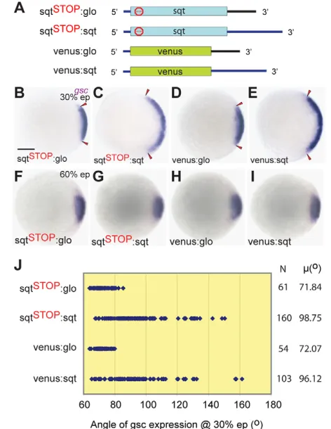
Dorsal activity of sqtRNA is dependent on sequences in the 3⬘UTR
Since localization of sqtRNA to dorsal progenitors depends on sequences in its 3⬘UTR, we tested whether dorsal expansion by overexpression of sqtRNA also requires the 3⬘UTR. Embryos injected with sqtSTOPmutant RNA fused to sqt3⬘UTR (sqtSTOP:sqt)
were compared with those injected with mutant RNA fused with Xenopus globin3⬘UTR (sqtSTOP:glo; Fig. 3A). Whereas sqtSTOP:sqt
RNA injection expanded gscexpression at 30% epiboly (Fig. 3C), injection of sqtSTOP:glo had no discernible effect (Fig. 3B,J), and
injected embryos were indistinguishable from control or uninjected embryos. Expanded gsc expression in sqtSTOP:sqt RNA-injected
embryos was transient and by 60% epiboly all injected embryos were similar to uninjected (not shown) or control-injected embryos (Fig. 3F-I). Therefore, the 3⬘UTR is required for transient dorsal expansion by the sqtRNA.
We then examined whether the sqt 3⬘UTR is sufficient for dorsal expansion. Embryos were injected with RNA encoding the fluorescent protein Venus fused with either sqt 3⬘UTR (venus:sqt) or globin3⬘UTR (venus:glo). Consistent with the evidence that the sqt3⬘UTR is required for dorsal expansion, venus:sqt RNA-injected embryos showed transient expansion of gsc ranging up to 160° and with a mean of 96°. Control venus:glo-injected embryos showed a ~72° gscangle (Fig. 3J), similar to uninjected embryos. Quantitative real-time RT-PCRs to detect expression of sqt,dha,voxand ventin lacZ:sqt-injected embryos showed that endogenous sqtand dhatranscript levels increase transiently at sphere stages and revert to normal levels by 30% epiboly (supplementary material Fig. S3), as compared with control lacZ:glo-injected embryos. Conversely, voxand vent levels are transiently reduced initially and subsequently revert to normal (supplementary material Fig. S3). Thus, the sqt3⬘UTR is both necessary and sufficient to transiently expand dorsal and reduce ventral gene expression.
Activity of sqt3⬘UTR in dorsal is independent of Sqt protein activity
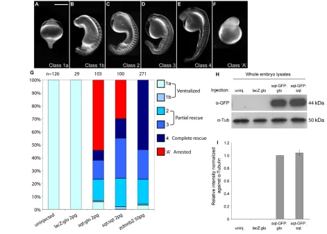
Overexpressing sqt3⬘UTR expands dorsal gene expression, but only transiently, raising the question of whether its dorsalizing activity is developmentally relevant to the embryo. To address this, we used ichabod (ich) mutant embryos, which lack all dorsal structures (Kelly et al., 2000). Embryos from homozygous ichmothers can be rescued by sqtRNA injections (Kelly et al., 2000; Gore et al., 2005). We compared the effect of sqtversus globin3⬘UTR sequences on the ability of Sqt to rescue ich mutant embryos. Capped synthetic mRNA encoding Sqt fused to either sqt(sqt:sqt) or to globin(sqt:glo) 3⬘UTR was injected into ichembryos. In these experiments, Sqt protein would be generated from both RNAs, with the UTRs providing the only difference in activity.
Interestingly, we find that sqt:sqt induces a complete axis and rescues more efficiently than sqt:glo at comparable doses (Fig. 4A-G). Nearly 70% of sqt:sqt-injected embryos show rescue of dorsal structures to varying extents (15% show complete rescue and 53% partial rescue; n100 embryos; Fig. 4G) (Gore et al., 2005). By contrast, only 37% of sqt:glo-injected ichembryos show any rescue of dorsal structures. Furthermore, ~55% of sqt:glo-injected ich embryos (n103 embryos) manifest early gastrula arrest, as compared with ~30% of sqt:sqt injections (n100 embryos), which is likely to be due to unregulated Sqt signaling from mislocalized sqt:glo in contrast to localized Sqt from sqt:sqt. SDS-PAGE to detect Sqt protein in whole embryo lysates showed that the expression levels of Sqt from sqt:sqt versus sqt:glo are similar (Fig. 4H,I), indicating comparable translation efficiencies. These results show that the sqt3⬘UTR confers more efficient activity to sqtin forming dorsal structures. Therefore, the sqt3⬘UTR has biological activity in dorsal specification that is distinct from any activity of Sqt protein.
Dorsal activity of sqtRNA requires canonical Wnt signaling but not Nodal signaling
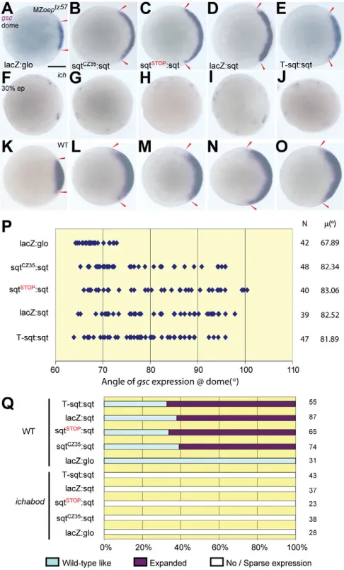
We then tested whether dorsal expansion by sqtRNA requires Nodal signaling. Maternal and zygotic mutations affecting the Nodal co-receptor One-eyed pinhead (MZoep) are thought to cause a complete lack of Nodal signaling (Gritsman et al., 1999). In MZoepmutant embryos, gscexpression is detected at dome stages (Fig. 5A) and is not detected at mid-gastrula stages (Gritsman et al., 1999). MZoepembryos injected with sqtcz35:sqt, sqtSTOP:sqt,
lacZ:sqt and T-sqt:sqt RNA show expanded gscexpression at dome stages (Fig. 5A-E,P), and the gsc angle shows a range from
65°-Fig. 3. The sqt3⬘UTR is necessary and sufficient for dorsal activity of sqtRNA.(A)Schematic of constructs used to express mutant sqtfused with globin3⬘UTR (black; sqtSTOP:glo), sqt3⬘UTR (blue; sqtSTOP:sqt), venus (green) fused with globin3⬘UTR (venus:glo) or sqt3⬘UTR (venus:sqt). (B-I)Expression of gscat 30% epiboly (B-E) is expanded in zebrafish embryos injected with sqtSTOP:sqt (C) or venus:sqt (E), but not with sqtSTOP:glo (B) or venus:glo (D). Dorsal expansion by the sqt3⬘UTR is transient and is not detected at 60% epiboly (F-I). (J)Angle of gscexpansion in injected embryos at 30% epiboly. Each blue dot represents a single embryo. N indicates the number of injected embryos for each RNA and(°) shows mean gscangles. Scale bar: 100m.
D
E
V
E
LO
P
M
E
N
[image:5.612.54.290.63.368.2]2908
100°, with a mean of ~88°, in comparison to control lacZ:glo-injected embryos that show gsc expression ranging from 63°-70°, with a mean of ~66°. Therefore, expansion of dorsal by sqtRNA is Oep-independent, and this function of sqtdoes not require Nodal signaling.
We then tested the requirement of maternal Wnt/-catenin signaling, which is known to be essential for dorsal specification in frogs and fish (Kelly et al., 2000; Tao et al., 2005). To test whether dorsal activity of sqtRNA is mediated via canonical Wnt signaling, we injected mutant sqt RNAs into ichembryos and examined gscexpression at early gastrula. Expression of gscis not expanded in ichembryos injected with sqtmut:sqt RNA (Fig.
5F-J,Q), in contrast to injected wild-type embryos (Fig. 5K-O,Q). Thus, expansion of dorsal by the sqt3⬘UTR requires Wnt/-catenin signaling.
Antisense morpholinos that target sqtATG sequences block sqtRNA localization
The dorsal activity of maternal sqtRNA appears to be mediated by the 3⬘UTR and is independent of Sqt protein or Nodal signaling. However, these findings raise the question of how a sqt ATG-targeting morpholino [sqtMO1 (Gore et al., 2005; Gore
et al., 2007)] leads to loss of dorsal structures (Gore et al., 2005). sqtMO1 spans the translational start site sequence (Feldman and Stemple, 2001) and is presumed to block Sqt protein synthesis. Indeed, co-injection of sqtMO1 with mRNA encoding Sqt-GFP fusion protein leads to substantially reduced expression of Sqt-GFP in the blastoderm (38%, n97), as compared with co-injection of Sqt-GFP with a sqt ATG mismatch control morpholino (ConMO, 76%, n114; Fig. 6B-E). Thus, Sqt protein expression is disrupted by the ATG morpholino, but not by control morpholinos. Interestingly, we found that a substantial number of sqtMO1-injected embryos showed Sqt-GFP fluorescence in the yolk (~62% yolk expression, n97; arrowhead in Fig. 6D), suggesting a localization defect. So, we revisited our sqt morpholino injection experiments (Gore et al., 2005) and tested whether sqtMO1 also affects other functions pertaining to sqt, such as RNA localization and/or maintenance. To examine RNA levels, we performed RT-PCRs and found that sqtRNA levels are unchanged in sqtMO1-injected embryos at least until the 8-cell stage (supplementary material Fig. S4). Therefore, injection of the sqt ATG morpholino does not lead to degradation of sqtRNA at these stages.
[image:6.612.54.525.62.400.2]RESEARCH ARTICLE Development 139 (16)
Fig. 4. Rescue of ichabodembryos by Sqt is more effective with the sqt3⬘UTR. (A-F)Uninjected and lacZ:glo RNA-injected ichabod (ich) embryos are completely radialized (class 1a; A), whereas sqtor zctnnb2[zebrafish (z) beta catenin 2] RNA injection rescues anterior and dorsal structures to varying extents (class 1b-4; B-E), or causes early arrest (class ‘A’; F). (G)Percentage embryos of each class. Injection of sqt:sqt and zctnnb2RNA is more effective in rescuing ichembryos than sqt:glo. (H)Sqt-GFP protein (44 kDa) expressed from embryos injected with sqt-GFP:sqt or sqt-GFP:glo, and lysates from uninjected embryos and lacZ:glo-injected embryos as negative controls, are shown. Tubulin (50 kDa) provides a loading control. (I)Relative intensities of Sqt-GFP bands, normalized against Tubulin, show comparable translation efficiencies of sqt-GFP:sqt and sqt-GFP:glo in whole embryo lysates. Error bars indicate s.d. between two independent experiments. Scale bar: 100m.
D
E
V
E
LO
P
M
E
N
To test whether sqtMO1 affects sqtRNA localization, we co-injected fluorescently labeled sqtRNA with either sqtMO1 or ConMO, and examined the embryos for localization at the 4-cell stage (Gore et al., 2005; Gilligan et al., 2011). Remarkably, sqtMO1 nearly abolishes sqtRNA localization at the 4-cell stage (90%, n127), and in ~35% of the embryos the injected fluorescent sqt RNA was detected as aggregates in the yolk (Fig. 6G,I,J). Similarly, sqtMO2 (which spans exon 2/intron 2; see Fig. 6A) also affects sqtRNA localization (data not shown). By comparison,
ConMO and control target protector morpholinos [TPcontrol
(Giraldez et al., 2005)] do not significantly affect localization (Fig. 6F,H,J), whereas a morpholino targeting the dorsal localization element (DLE MO; see Fig. 6A) reduces sqtlocalization (Fig. 6J) (Gilligan et al., 2011). These results suggest that sequences surrounding the translational start site and splice junctions are also required for sqt RNA localization. Therefore, in addition to disrupting the translation/splicing of sqtRNA, the sqt morpholinos also unexpectedly affect sqtRNA localization.
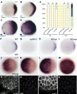
Disruption of sqtRNA localization leads to ectopic dorsal expansion
We find that blocking the translational start site disrupts sqtRNA localization (Fig. 6). However, sqt RNA localization is also dependent on sequences in the 3⬘UTR (Gore et al., 2005; Gilligan et al., 2011), and dorsal expansion also requires the 3⬘UTR. We therefore tested whether dorsal expansion by the 3⬘UTR depends on sqtRNA localization. We injected DLE MO, which disrupts sqt RNA localization (Gilligan et al., 2011). Morpholinos targeting a different region of the sqt3⬘UTR (TPcontrolMO) and miR430 target
protector morpholinos [TPmiR430MO (Giraldez et al., 2005)] were
used as controls. Embryos injected with the DLE MO showed lateral expansion of dorsal gscexpression even at low doses (2 ng; Fig. 7B,E), similar to embryos injected with non-coding sqtRNA (see Fig. 2F and Fig. 3C,E). At higher doses (10 ng), gsc expression extended further towards the animal pole (Fig. 7C,E) or even covered the entire blastoderm (Fig. 7D,E). By contrast, TPcontrolMO-injected and TPmiR430MO-injected embryos did not
manifest gscexpansion at 2 ng doses, and only a few TPmiR430
MO-injected embryos showed mild expansion of gscat 10 ng (Fig. 7E). These experiments show that blocking the dorsal localization element in endogenous sqtRNA with DLE MO leads to expanded or ectopic gscexpression. Thus, dorsal expansion by sqtRNA is not dependent on the DLE in the sqt3⬘UTR.
We also examined the expression of early dorsal markers in sqt ATG morphant embryos. Injection of sqt morpholinos into eggs to target maternal sqtRNA prior to the formation of aggregates in the yolk upon egg activation (Gore and Sampath, 2002) causes loss of dorsal specification. We find that gscexpression is not detected in these embryos, consistent with the loss of dorsal structures (Fig. 7G). This is in contrast to MZsqt and MZoep mutant embryos, in which early gsc expression is detected at comparable stages (Fig. 7H,I and Fig. 5A). Furthermore, -catenin fails to accumulate in dorsal nuclei of sqtMO1-injected embryos, in contrast to ConMO-injected embryos (Fig. 7N-Q, Table 1 and supplementary material Table S2). Ventral markers such as gata2 are concomitantly expanded in sqtMO1-injected embryos (Fig. 7J-M). Therefore, the sequences spanning the translational start site in sqt RNA are required for localization, and disruption of endogenous sqtfunction by sqtMO1 leads to loss of dorsal gene expression, probably by blocking both maternal sqtRNA localization and translation. These results also show that maternal sqtRNA functions prior to nuclear -catenin accumulation.
The sqt3⬘UTR rescues anterior and dorsal structures in sqt morphant embryos
We then tested whether the sqt3⬘UTR is capable of restoring early dorsal gsc expression in sqt morphant embryos. In comparison to control morpholino-injected embryos (Fig. 8A), MZsqtand MZoepembryos (Fig. 7H,I and Fig. 5A), injection of the sqt morpholinos into eggs results in embryos with severe dorsal deficiencies (Gore et al., 2005; Gore et al., 2007) and loss
Fig. 5. Dorsal activity of sqtRNA requires Wnt/-catenin but not Nodal signaling.(A-O)Expression of gscin dome stage MZoep zebrafish embryos (A-E) shows that, compared with lacZ:glo RNA (A,F,K), injection of sqtcz35:sqt (B,G,L), sqtSTOP:sqt (C,H,M), lacZ:sqt (D,I,N), or T-sqt:sqt (E,J,O) RNA expands the dorsal domain (B-E), similar to that in wild-type embryos (K-O). By contrast, ichembryos (F-J) show no/very sparse gsc expression for all injected RNAs. Arrowheads (A-E,K-O) mark the extent of gscexpression. A-O, animal pole views; A-E, K-O, dorsal to the right. (P)The angle of gscin dome stage MZoep embryos after RNA injections. Each blue dot represents a single embryo. N indicates the number of injected embryos and(o) shows mean gsc angles. (Q)Percentage embryos that manifest gscexpression at 30% epiboly after sqtRNA injections in control or ichmutant embryos. Scale bar: 100m.
D
E
V
E
LO
P
M
E
N
[image:7.612.52.295.62.464.2]2910
of gsc expression (Fig. 8C). Co-injection of lacZ:sqt or sqtSTOP:sqt RNA with sqtMO1 or sqtMO2 rescued early gsc
expression in sqt morphant embryos (Fig. 8E and supplementary material Fig. S5), whereas co-injection of lacZ:glo RNA with sqtMOs did not restore gscexpression (supplementary material Fig. S5). At prim-5 stages, sqt morpholino-injected embryos manifest loss of anterior and dorsal structures, whereas co-injection of the sqtMOs with sqtmut RNA or lacZ:sqt RNA
produces phenotypes that are strikingly similar to that of MZsqtcz35mutant embryos (Fig. 8D,F,H,I). The sqt morpholinos prevent endogenous Sqt protein translation and endogenous sqt RNA localization, but cannot target lacZ:sqt. Therefore, the rescue by the sqt3⬘UTR sequences is very significant. Thus, sqt RNA, and specifically the sqt 3⬘UTR, is sufficient to initiate dorsal gene expression in early embryos. These findings indicate that the biological activity of maternal sqt RNA in dorsal specification is likely to reside within its 3⬘UTR.
DISCUSSION
Asymmetric localization of sqtRNA in presumptive dorsal cells shows that dorsoventral asymmetry in the blastoderm is established prior to zygotic transcription, during cleavage stages. Based upon cell ablations and antisense morpholino injections, we proposed that asymmetrically localized sqt RNA and associated factors specify dorsal identity (Gore et al., 2005). However, studies using the insertion mutants sqtcz35 and sqthi975 suggested that early specification of the dorsoventral axis might not require the activity of maternal Sqt (Aoki et al., 2002; Bennett et al., 2007; Pei et al., 2007). Therefore, the function of maternal sqtwas unclear. We find that sqtRNA has functions that are independent of Sqt protein in dorsal initiation. Furthermore, we show that the sqtcz35and sqthi975 insertion alleles are not maternal transcript nulls: sqtcz35RNA is expressed in MZsqtat similar levels to sqtin wild-type embryos at early stages and also localizes to two cells in 4- and 8-cell stage embryos. We observed a reduction in sqt RNA levels during
[image:8.612.57.532.57.414.2]RESEARCH ARTICLE Development 139 (16)
Fig. 6. Morpholinos targeting the sqtATG disrupt sqtRNA localization.(A)The zebrafish sqtgenomic locus (not to scale) indicating positions of the sqt ATG morpholino (MO1), sqt intron 2 morpholino (MO2), sqt DLE morpholino (DLE MO), sqt miR430 target protector morpholino (TPmiR430MO), and target protector control morpholino (TPcontrolMO). Introns (I and II), exons (E1, E2 and E3; cyan boxes) and UTR (dark blue line) are indicated. The dorsal localization element is highlighted in green. (B-D)Embryos injected with sqt-GFP RNA show asymmetric expression of Sqt-GFP fluorescent protein in the blastoderm of 512-cell stage embryos (C), in comparison to uninjected embryos (B) and embryos co-injected with sqtMO1 and sqt-GFP that show Sqt-GFP fluorescence in the yolk (D). (E)Numbers (N) and percentage of embryos showing no expression, expression of Sqt-GFP in the blastoderm, yolk, or both. (F-I)Localization of injected fluorescent control lacZor sqt:sqt RNA in 4-cell stage embryos co-injected with control morpholinos, sqtMO1, TPcontrolMO or DLE MO. (J)Percentage and number of embryos (top) showing sqtRNA localized (F,H), not localized (G), or as aggregates in the yolk (I). Lateral views at 512-cell stage (B-D) or 4-cell stage (H,I), or animal pole views at 4-cell stage (F,G). Scale bars: 100m.
D
E
V
E
LO
P
M
E
N
gastrulation, consistent with a previous report of reduced sqt mutant transcripts in late blastula embryos (Bennett et al., 2007). However, Bennett et al. (Bennett et al., 2007) also stated that MZsqtcz35embryos at the 8-cell stage contain no detectable sqt RNA using sqt-specific RT-PCR primers, and Pei et al. (Pei et al., 2007) reported that sqtRNA is reduced or absent in MZsqthi975 embryos based on RT-PCR using oligo(dT)-primed cDNA. By RNA sequencing and RT-PCR, we find that maternally deposited sqtRNA is non-polyadenylated and unspliced, and that sqtmRNA is detected during cleavage stages (supplementary material Fig. S1A,B) (Gore et al., 2007), providing an explanation for the Pei et al. (Pei et al., 2007) observation. However, our findings differ from the lack of maternal sqtRNA reported by Bennett et al. (Bennett et al., 2007), which we detect in early MZsqt embryos by RT-PCR, quantitative PCR and whole-mount in situ hybridization.
We previously reported that injection of sqt splice-blocking morpholinos into wild-type eggs leads to aberrantly spliced sqt RNA (Gore et al., 2007). The presence of pre-mRNAs has been reported in oocytes in other metazoans as well (Hachet and Ephrussi, 2004). In Xenopus eggs, many maternal pre-mRNAs are also known to be non-polyadenylated or have very short poly(A) tails, which presumably prevent precocious protein translation (Sagata et al., 1980; McGrew et al., 1989; Paris and Philippe, 1990; Varnum and Wormington, 1990). Our findings are also consistent with the recent genome-wide transcriptome analysis in zebrafish by Aanes et al. (Aanes et al., 2011), who showed by RNA
sequencing that a large cohort of maternal transcripts in zebrafish eggs are non-polyadenylated, and become polyadenylated during early embryogenesis (Aanes et al., 2011). Thus, the sqtinsertion alleles are not maternal transcript nulls and not bona fide functional null alleles.
Mutant sqtRNA can expand dorsal gene expression in wild-type embryos, with a concomitant reduction in ventral gene expression. The transient gscexpansion by sqtmut:sqt RNAs and
[image:9.612.52.358.60.435.2]the sqt3⬘UTR is unlikely to be due to a morphological delay as the embryos are not generally delayed. Moreover, the gsc expansion is restricted to the gastrula margin and does not extend animally, in contrast to the broad gscexpression domain reported in early embryos (Schulte-Merker et al., 1994). We also observed expansion of chdand substantially increased numbers of -catenin-positive nuclei in embryos injected with non-coding sqtRNA. Furthermore, dhaand endogenous sqttranscript levels transiently increase, with a concomitant reduction in vox and venttranscripts, providing the basis for the transient increase in chd, gscand ntland reduction in ventral expression of gata2 (Hammerschmidt et al., 1996; Erter et al., 1998; Rebagliati et al., 1998; Yamanaka et al., 1998; Imai et al., 2001; Gilardelli et al., 2004). Thus, sqt RNA, and specifically the sqt 3⬘UTR, can nucleate a complex of factors that are sufficient to expand dorsal gene expression. We also find that mutant/non-coding sqtRNA injections into sqt morphants can phenocopy MZsqt mutant embryos. Taken together, these findings suggest that it is the
Fig. 7. The sqt DLE MO and ATG MO
differentially affect dorsal gene expression.( A-E)gscexpression expands upon injection of the DLE MO even at low doses, in comparison to injections of TPmiR430MO or TPcontrolMO. (E)The percentage and number of embryos and the extent of gsc expression (classes I-IV, A-D) in injected embryos at sphere stages. (F-I)Expression of gscis abolished in sqtMO1-injected embryos at the sphere stage (G), in comparison to control wild-type (F), MZsqt (H) or MZoep(I) embryos. (J-M)Expression of the ventral marker gene gata2is expanded to varying extents in sqtMO1-injected embryos (K-M), in comparison to control embryos (J). (N-Q)Nuclear -catenin (N,P) in dorsal cells is not detected in sqtMO1-injected embryos at the 512-cell stage (P,Q), in contrast to ConMO-injected embryos (arrows, N,O). DAPI staining (O,Q) shows the presence of nuclei. (A-D,F-M) Animal pole views; (N-Q) dorsal views. Scale bars: 100m in A; 25m in N.
D
E
V
E
LO
P
M
E
N
2912
activity of the mutant sqttranscripts in the maternal sqtinsertion mutants that leads to initial dorsal expression in mutant embryos, and MZsqt mutants do not exhibit the severe loss of dorsal identity manifested by sqt morphants in which maternal sqtRNA localization and activity are both disrupted.
Interestingly, we never detected any ectopic sites of gscor chd in the mutant sqtRNA-injected embryos, unlike those injected with wild-type sqtRNA (Erter et al., 1998; Rebagliati et al., 1998). Thus, although mutant sqtRNAs and the sqt3⬘UTR can expand the endogenous dorsal domain, these sqtsequences do not induce dorsal at ectopic locations, presumably because these RNAs harbor the dorsal localization element. By contrast, injection of sqt DLE MO to target endogenous sqtRNA leads to ectopic gscexpression, probably owing to mislocalization and misexpression of endogenous Sqt.
Mutant sqt RNAs transiently expand the dorsal domain in wild-type embryos, but this expansion is not sustained, and by mid-gastrula stages the embryos appear to have regulated dorsal gene expression levels back to that observed in control embryos. This transient expansion is consistent with the normal gsc expression observed at 70% epiboly by Bennett et al. (Bennett et al., 2007) in T-sqt-injected embryos. Similar changes in early patterning with no overt later consequences have also been observed in other contexts/organisms. For example, extra copies of bicoid+in Drosophilalead to transient oversized head regions in embryos, which develop into apparently normal adults (Berleth et al., 1988).
Dorsal gsc expression is also expanded in MZoep embryos, which are presumed to lack all Nodal signaling (Gritsman et al., 1999). However, the expansion is not sustained. Initiation of dorsal gsc expression and its subsequent loss has been reported previously incyc;sqtcompound mutant embryos (Dougan et al., 2003). Thus, maintenance of gscexpression during gastrulation requires Nodal signaling. Although sqtRNA can initiate and expand dorsal gene expression independently of Sqt protein or Oep-dependent Nodal signaling, the sustained expression of dorsal genes requires the signaling functions of Sqt mediated via Oep.
So how does sqtRNA function to initiate and expand dorsal? This is likely to be a non-coding function of sqt, as the sqt3⬘UTR even when fused to heterologous reporter genes (lacZor venus) can expand dorsal gene expression in wild-type embryos and rescues early dorsal gene expression in sqt morphants. Furthermore, coding sqtsequences fused to the sqt3⬘UTR rescue ich embryos more efficiently than sqtfused to globinUTR sequences, demonstrating that the activity of the sqt3⬘UTR is distinct from the Sqt coding sequences. Taken together, these findings suggest that sqtRNA, and specifically the sqt3⬘UTR, functions in dorsal specification. Our current evidence is based upon overexpression and knockdown strategies, which have their limitations (Robu et al., 2007; Eisen and Smith, 2008). Although it is imperative to determine whether a sqt mutant that lacks maternal sqtRNA expression also lacks dorsal identity, such a mutant is not yet available despite our attempts using zinc-finger nuclease (ZFN) (Doyon et al., 2008; Meng et al., 2008) technology.
The sqt 3⬘UTR harbors microRNA (miRNA) target sites (Giraldez et al., 2006; Choi et al., 2007), raising the possibility that dorsalization by the sqt 3⬘UTR might be mediated via miRNAs. However, this seems unlikely because sqt3⬘UTR with mutations in target site sequences of three predicted miRNAs still expands the dorsal domain, as does sqtRNA injection into MZdicerembryos (supplementary material Fig. S6). Although it remains possible that there are other unidentified dicer -independent small RNA targets in the sqt 3⬘UTR, our experiments show that the sqt3⬘UTR functions independently of the miRNAs tested and of dicer. Rather, we surmise that sqt RNA might translocate some factor(s)/protein(s) that bind to the UTRs to the future dorsal side. In Drosophila, oskarRNA has
[image:10.612.52.293.61.515.2]RESEARCH ARTICLE Development 139 (16)
Fig. 8. Loss of dorsal specification due to sqt morpholinos is rescued by the sqt3⬘UTR.(A-H)Early gscexpression is not detected in sqtMO-injected (C), in contrast to ConMO-injected (A), zebrafish embryos. Expression of gsc is rescued in sqt morphants by co-injection of lacZ:sqt (E), but not with lacZ:glo (see supplementary material Fig. S4). Embryos co-injected with sqtMO and lacZ:sqt (F,I) or sqtMO and sqtSTOP:sqt (I) are rescued partially and are similar to MZsqtcz35mutant
embryos (H). Images in F,H were acquired in different focal planes for the rostral and caudal regions of the embryo and subsequently assembled. (A,C,E,G) 30% epiboly, animal pole views with dorsal to the right; (B,D,F,H) prim-5 stage. Arrowheads (A,E) mark the extent of gsc expression. (I)Extent of rescue of sqtMO-injected embryos by co-injection of lacZ:sqt or sqtSTOP:sqt, versus control lacZ:glo RNA. Scale bars: 100m.
D
E
V
E
LO
P
M
E
N
functions that are independent of Oskar protein in early oogenesis (Jenny et al., 2006). Similarly, Xenopus veg-TRNA has a scaffolding function that is independent of the later functions of Veg-T protein in germ layer specification (Zhang and King, 1996; Kloc et al., 2005). Since sqtRNA is normally present in limiting amounts in early embryos (Rebagliati et al., 1998; Gore et al., 2005), even small increases in the amount of sqt3⬘UTR could lead to an amplified effect of any UTR-bound factors (see model in Fig. 9) in the future embryonic dorsal side. These factor(s) might function via the canonical Wnt/-catenin pathway, as the sqt 3⬘UTR by itself is unable to expand the dorsal domain in the context of ich embryos, which are deficient in -catenin signaling. It is conceivable that the factor(s) binding to the sqt3⬘UTR are components of the Wnt/-catenin pathway. In Xenopus, RNA encoding Xwnt11, which is required for dorsal specification and functions via -catenin (Tao et al., 2005), is localized to dorsal vegetal cells of early embryos. The factors that bind and localize Xwnt11RNA are not known. In zebrafish, embryos from ichmutant mothers show that maternal -catenin function is required for dorsal specification, and maternal wnt8a RNA is likely to activate Wnt/-catenin signaling (Lu et al., 2011). Asymmetric localization of sqtRNA in the blastoderm by the 4-cell stage precedes dorsal accumulation of nuclear -catenin at the 128-cell stage (Dougan et al., 2003; Gore et al., 2005).
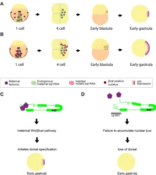
[image:11.612.52.373.60.419.2]The sqt5⬘UTR and 3⬘UTR both affect sqtRNA localization, but somewhat differently. Whereas the sqt ATG morpholino results in sqtRNA being mislocalized diffusely or stuck in the yolk as aggregates, the DLE MO causes sqt RNA to be mislocalized diffusely in the blastoderm. The phenotypes caused by the morpholinos targeting these two regions are also distinct in that the ATG morpholino causes loss of nuclear -catenin and loss of gscexpression, whereas the 3⬘UTR DLE MO causes mislocalized and ectopic gsc, probably owing to mislocalized endogenous Sqt in the blastoderm. This suggests that the sqt 5⬘UTR and 3⬘UTR sequences might function in distinct steps of sqtRNA localization and activity. Moreover, our finding that the sqt ATG morpholino disrupts both sqt RNA localization and translation suggests that the sqt5⬘UTR and 3⬘UTR sequences might interact with each other, perhaps via binding of a protein complex, to regulate sqtRNA localization (see model in Fig. 9). The sqt morpholinos cause loss of dorsal nuclear -catenin and gscexpression, which might underlie the severe loss of dorsal structures in the morphants. Our results suggest that localized maternal sqtRNA functions prior to the accumulation of nuclear -catenin, and raise the possibility that sqt RNA acts as a scaffold to bind and deliver/sequester maternal factors, which are likely to be intracellular component(s) of the Wnt pathway, to future dorsal. Our findings identify novel functions for the Nodal genes and suggest a new role for non-coding RNAs in the control of axis specification.
Fig. 9. Model of sqtRNA as a scaffold.(A)In wild-type zebrafish embryos, sqtRNA (green) localizes to dorsal and delivers/sequesters maternal factors (purple polygons) bound to it, to specify dorsal (red, -catenin-positive nuclei; purple, arc of gscexpression). (B)Upon overexpressing sqtRNA (magenta), the localized RNA and the factors it binds increase, thereby increasing the number of -catenin-positive nuclei in the blastoderm and YSL, expanding dorsal gsc expression. (C)sqtRNA is likely to act as a scaffold that binds and delivers maternal factor(s), initiating dorsal specification in a maternal Wnt/-catenin-dependent manner. (D)In sqtMO-injected embryos, maternal sqtRNA and the associated factors fail to localize to dorsal and -catenin does not accumulate in dorsal nuclei, resulting in loss of dorsal gene expression.
D
E
V
E
LO
P
M
E
N
2914
Acknowledgements
We thank members of the K.S. laboratory, the Singapore fish community, Tom Carney, Bill Chia, Steve Cohen, Ray Dunn, Aniket Gore, Masahiko Hibi, Greg Jedd, Mithilesh Mishra and Mohan Balasubramanian for discussions and suggestions; Shingo Maegawa and Eric Weinberg for ichfish; and the Temasek Life Sciences Laboratory sequencing and fish facilities.
Funding
S.L. was supported by a pre-doctoral fellowship from the Singapore Millennium Foundation and Temasek Life Sciences Laboratory; S.M. is supported by the Genome Institute of Singapore; work in the laboratory of K.S. is supported by Temasek Life Sciences Laboratory.
Competing interests statement
The authors declare no competing financial interests.
Supplementary material
Supplementary material available online at
http://dev.biologists.org/lookup/suppl/doi:10.1242/dev.077081/-/DC1
References
Aanes, H., Winata, C. L., Lin, C. H., Chen, J. P., Srinivasan, K. G., Lee, S. G., Lim, A. Y., Hajan, H. S., Collas, P., Bourque, G. et al.(2011). Zebrafish mRNA sequencing deciphers novelties in transcriptome dynamics during maternal to zygotic transition. Genome Res. 21, 1328-1338.
Abrams, E. W. and Mullins, M. C.(2009). Early zebrafish development: it’s in the maternal genes. Curr. Opin. Genet. Dev. 19, 396-403.
Amsterdam, A., Nissen, R. M., Sun, Z., Swindell, E. C., Farrington, S. and Hopkins, N.(2004). Identification of 315 genes essential for early zebrafish development. Proc. Natl. Acad. Sci. USA 101, 12792-12797.
Aoki, T. O., Mathieu, J., Saint-Etienne, L., Rebagliati, M. R., Peyrieras, N. and Rosa, F. M.(2002). Regulation of nodal signalling and mesendoderm formation by TARAM-A, a TGFbeta-related type I receptor. Dev. Biol. 241, 273-288.
Bellipanni, G., Varga, M., Maegawa, S., Imai, Y., Kelly, C., Myers, A. P., Chu, F., Talbot, W. S. and Weinberg, E. S.(2006). Essential and opposing roles of zebrafish beta-catenins in the formation of dorsal axial structures and neurectoderm. Development133, 1299-1309.
Bennett, J. T., Stickney, H. L., Choi, W. Y., Ciruna, B., Talbot, W. S. and Schier, A. F.(2007). Maternal nodal and zebrafish embryogenesis. Nature 450, E1-E2.
Berleth, T., Burri, M., Thoma, G., Bopp, D., Richstein, S., Frigerio, G., Noll, M. and Nusslein-Volhard, C.(1988). The role of localization of bicoid RNA in organizing the anterior pattern of the Drosophila embryo. EMBO J. 7, 1749-1756.
Choi, W. Y., Giraldez, A. J. and Schier, A. F.(2007). Target protectors reveal dampening and balancing of Nodal agonist and antagonist by miR-430. Science318, 271-274.
Cuykendall, T. N. and Houston, D. W.(2009). Vegetally localized Xenopus trim36 regulates cortical rotation and dorsal axis formation. Development136, 3057-3065.
Dougan, S. T., Warga, R. M., Kane, D. A., Schier, A. F. and Talbot, W. S.
(2003). The role of the zebrafish nodal-related genes squint and cyclops in patterning of mesendoderm. Development130, 1837-1851.
Doyon, Y., McCammon, J. M., Miller, J. C., Faraji, F., Ngo, C., Katibah, G. E., Amora, R., Hocking, T. D., Zhang, L., Rebar, E. J. et al.(2008). Heritable targeted gene disruption in zebrafish using designed zinc-finger nucleases. Nat. Biotechnol. 26, 702-708.
Eisen, J. S. and Smith, J. C.(2008). Controlling morpholino experiments: don’t stop making antisense. Development135, 1735-1743.
Erter, C. E., Solnica-Krezel, L. and Wright, C. V.(1998). Zebrafish nodal-related 2 encodes an early mesendodermal inducer signaling from the extraembryonic yolk syncytial layer. Dev. Biol. 204, 361-372.
Feldman, B. and Stemple, D. L.(2001). Morpholino phenocopies of sqt, oep, and ntl mutations. Genesis30, 175-177.
Feldman, B., Gates, M. A., Egan, E. S., Dougan, S. T., Rennebeck, G., Sirotkin, H. I., Schier, A. F. and Talbot, W. S.(1998). Zebrafish organizer development and germ-layer formation require nodal-related signals. Nature 395, 181-185.
Gilardelli, C. N., Pozzoli, O., Sordino, P., Matassi, G. and Cotelli, F.(2004). Functional and hierarchical interactions among zebrafish vox/vent homeobox genes. Dev. Dyn. 230, 494-508.
Gilligan, P. C., Kumari, P., Lim, S., Cheong, A., Chang, A. and Sampath, K.
(2011). Conservation defines functional motifs in the squint/nodal-related 1 RNA dorsal localization element. Nucleic Acids Res. 39, 3340-3349.
Giraldez, A. J., Cinalli, R. M., Glasner, M. E., Enright, A. J., Thomson, J. M., Baskerville, S., Hammond, S. M., Bartel, D. P. and Schier, A. F.(2005). MicroRNAs regulate brain morphogenesis in zebrafish. Science308, 833-838.
Giraldez, A. J., Mishima, Y., Rihel, J., Grocock, R. J., Van Dongen, S., Inoue, K., Enright, A. J. and Schier, A. F.(2006). Zebrafish MiR-430 promotes deadenylation and clearance of maternal mRNAs. Science312, 75-79.
Gore, A. V. and Sampath, K.(2002). Localization of transcripts of the zebrafish morphogen Squint is dependent on egg activation and the microtubule cytoskeleton. Mech. Dev. 112, 153-156.
Gore, A. V., Maegawa, S., Cheong, A., Gilligan, P. C., Weinberg, E. S. and Sampath, K.(2005). The zebrafish dorsal axis is apparent at the four-cell stage. Nature438, 1030-1035.
Gore, A. V., Cheong, A., Gilligan, P. C. and Sampath, K.(2007). Gore et al. reply to J. T. Bennett et al. Nature450, E2-E4.
Gritsman, K., Zhang, J., Cheng, S., Heckscher, E., Talbot, W. S. and Schier, A. F.(1999). The EGF-CFC protein one-eyed pinhead is essential for nodal signaling. Cell97, 121-132.
Grunert, S. and St Johnston, D.(1996). RNA localization and the development of asymmetry during Drosophila oogenesis. Curr. Opin. Genet. Dev. 6, 395-402.
Hachet, O. and Ephrussi, A.(2004). Splicing of oskar RNA in the nucleus is coupled to its cytoplasmic localization. Nature428, 959-963.
Hammerschmidt, M., Serbedzija, G. N. and McMahon, A. P.(1996). Genetic analysis of dorsoventral pattern formation in the zebrafish: requirement of a BMP-like ventralizing activity and its dorsal repressor. Genes Dev. 10, 2452-2461.
Heasman, J.(2006). Maternal determinants of embryonic cell fate. Semin. Cell Dev. Biol. 17, 93-98.
Heasman, J., Crawford, A., Goldstone, K., Garner-Hamrick, P., Gumbiner, B., McCrea, P., Kintner, C., Noro, C. Y. and Wylie, C.(1994).
Overexpression of cadherins and underexpression of beta-catenin inhibit dorsal mesoderm induction in early Xenopus embryos. Cell79, 791-803.
Heasman, J., Kofron, M. and Wylie, C.(2000). Beta-catenin signaling activity dissected in the early Xenopus embryo: a novel antisense approach. Dev. Biol. 222, 124-134.
Heisenberg, C. P. and Nusslein-Volhard, C.(1997). The function of silberblick in the positioning of the eye anlage in the zebrafish embryo. Dev. Biol. 184, 85-94.
Imai, Y., Gates, M. A., Melby, A. E., Kimelman, D., Schier, A. F. and Talbot, W. S.(2001). The homeobox genes vox and vent are redundant repressors of dorsal fates in zebrafish. Development128, 2407-2420.
Jenny, A., Hachet, O., Zavorszky, P., Cyrklaff, A., Weston, M. D., Johnston, D. S., Erdelyi, M. and Ephrussi, A.(2006). A translation-independent role of oskar RNA in early Drosophila oogenesis. Development133, 2827-2833.
Jesuthasan, S. and Strahle, U.(1997). Dynamic microtubules and specification of the zebrafish embryonic axis. Curr. Biol. 7, 31-42.
Kelly, C., Chin, A. J., Leatherman, J. L., Kozlowski, D. J. and Weinberg, E. S.(2000). Maternally controlled (beta)-catenin-mediated signaling is required for organizer formation in the zebrafish. Development127, 3899-3911.
Kimmel, C. B. and Law, R. D.(1985). Cell lineage of zebrafish blastomeres. II. Formation of the yolk syncytial layer. Dev. Biol. 108, 86-93.
Kloc, M., Wilk, K., Vargas, D., Shirato, Y., Bilinski, S. and Etkin, L. D.
(2005). Potential structural role of non-coding and coding RNAs in the organization of the cytoskeleton at the vegetal cortex of Xenopus oocytes. Development132, 3445-3457.
Kofron, M., Klein, P., Zhang, F., Houston, D. W., Schaible, K., Wylie, C. and Heasman, J.(2001). The role of maternal axin in patterning the Xenopus embryo. Dev. Biol. 237, 183-201.
Lu, F. I., Thisse, C. and Thisse, B.(2011). Identification and mechanism of regulation of the zebrafish dorsal determinant. Proc. Natl. Acad. Sci. USA 108, 15876-15880.
McGrew, L. L., Dworkin-Rastl, E., Dworkin, M. B. and Richter, J. D.(1989). Poly(A) elongation during Xenopus oocyte maturation is required for translational recruitment and is mediated by a short sequence element. Genes Dev. 3, 803-815.
Meng, X., Noyes, M. B., Zhu, L. J., Lawson, N. D. and Wolfe, S. A.(2008). Targeted gene inactivation in zebrafish using engineered zinc-finger nucleases. Nat. Biotechnol. 26, 695-701.
Mizuno, T., Yamaha, E., Kuroiwa, A. and Takeda, H.(1999). Removal of vegetal yolk causes dorsal deficencies and impairs dorsal-inducing ability of the yolk cell in zebrafish. Mech. Dev. 81, 51-63.
Mowry, K. L. and Cote, C. A.(1999). RNA sorting in Xenopus oocytes and embryos. FASEB J. 13, 435-445.
Nojima, H., Rothhamel, S., Shimizu, T., Kim, C. H., Yonemura, S., Marlow, F. L. and Hibi, M.(2010). Syntabulin, a motor protein linker, controls dorsal determination. Development137, 923-933.
RESEARCH ARTICLE Development 139 (16)
Ober, E. A. and Schulte-Merker, S.(1999). Signals from the yolk cell induce mesoderm, neuroectoderm, the trunk organizer, and the notochord in zebrafish. Dev. Biol. 215, 167-181.
Paris, J. and Philippe, M.(1990). Poly(A) metabolism and polysomal
recruitment of maternal mRNAs during early Xenopus development. Dev. Biol. 140, 221-224.
Pei, W., Williams, P. H., Clark, M. D., Stemple, D. L. and Feldman, B.(2007). Environmental and genetic modifiers of squint penetrance during zebrafish embryogenesis. Dev. Biol. 308, 368-378.
Pelegri, F.(2003). Maternal factors in zebrafish development. Dev. Dyn. 228, 535-554.
Rebagliati, M. R., Toyama, R., Fricke, C., Haffter, P. and Dawid, I. B.(1998). Zebrafish nodal-related genes are implicated in axial patterning and
establishing left-right asymmetry. Dev. Biol. 199, 261-272.
Robu, M. E., Larson, J. D., Nasevicius, A., Beiraghi, S., Brenner, C., Farber, S. A. and Ekker, S. C.(2007). p53 activation by knockdown technologies. PLoS Genet. 3, e78.
Sagata, N., Shiokawa, K. and Yamana, K.(1980). A study on the steady-state population of poly(A)+RNA during early development of Xenopus laevis. Dev. Biol. 77, 431-448.
Sampath, K., Rubinstein, A. L., Cheng, A. M., Liang, J. O., Fekany, K., Solnica-Krezel, L., Korzh, V., Halpern, M. E. and Wright, C. V.(1998). Induction of the zebrafish ventral brain and floorplate requires cyclops/nodal signalling. Nature395, 185-189.
Schroeder, K. E., Condic, M. L., Eisenberg, L. M. and Yost, H. J.(1999). Spatially regulated translation in embryos: asymmetric expression of maternal Wnt-11 along the dorsal-ventral axis in Xenopus. Dev. Biol. 214, 288-297.
Schulte-Merker, S., Hammerschmidt, M., Beuchle, D., Cho, K. W., De Robertis, E. M. and Nusslein-Volhard, C.(1994). Expression of zebrafish goosecoid and no tail gene products in wild-type and mutant no tail embryos. Development120, 843-852.
St Johnston, D.(1995). The intracellular localization of messenger RNAs. Cell 81, 161-170.
Tao, Q., Yokota, C., Puck, H., Kofron, M., Birsoy, B., Yan, D., Asashima, M., Wylie, C. C., Lin, X. and Heasman, J.(2005). Maternal wnt11 activates the canonical wnt signaling pathway required for axis formation in Xenopus embryos. Cell120, 857-871.
Varnum, S. M. and Wormington, W. M.(1990). Deadenylation of maternal mRNAs during Xenopus oocyte maturation does not require specific cis-sequences: a default mechanism for translational control. Genes Dev. 4, 2278-2286.
Westerfield, M.(2007). The Zebrafish Book. A Guide for the Laboratory Use of Zebrafish (Danio rerio), 5th edn. Eugene, OR: University of Oregon Press.
Wylie, C., Kofron, M., Payne, C., Anderson, R., Hosobuchi, M., Joseph, E. and Heasman, J.(1996). Maternal beta-catenin establishes a ‘dorsal signal’ in early Xenopus embryos. Development122, 2987-2996.
Yamanaka, Y., Mizuno, T., Sasai, Y., Kishi, M., Takeda, H., Kim, C. H., Hibi, M. and Hirano, T.(1998). A novel homeobox gene, dharma, can induce the organizer in a non-cell-autonomous manner. Genes Dev. 12, 2345-2353.
Zhang, J. and King, M. L.(1996). Xenopus VegT RNA is localized to the vegetal cortex during oogenesis and encodes a novel T-box transcription factor involved in mesodermal patterning. Development122, 4119-4129.