Journal of Chemical and Pharmaceutical Research, 2015, 7(2): 932-939
Research Article
CODEN(USA) : JCPRC5
ISSN : 0975-7384
Preliminary formulation and characterization of solid lipid
nanoparticles containing chloroquine and a P-glycoprotein
inhibitor: Influences of lipid-surfactant ratios
Ifeanyi T. Nzekwe
a*, Valentine I. Azodo
a, Chukwuma O. Agubata
b, Brendon Naicker
c,
Vincent Okore
dand Charles O. Esimone
ea
Department of Pharmaceutics and Pharmaceutical Technology, Nnamdi Azikiwe University, Awka,
Anambra State, Nigeria
b
Department of Pharmaceutical Technology and Industrial Pharmacy, University of Nigeria, Nsukka,
Enugu State, Nigeria
c
Polymers and Composites, Materials Science and Manufacturing, Council for Scientific and Industrial
Research, Pretoria, South Africa
d
Department of Pharmaceutics, University of Nigeria, Nsukka, Enugu State, Nigeria
eDepartment of Pharmaceutical Microbiology and Biotechnology, Nnamdi Azikiwe University, Awka,
Anambra State, Nigeria
_____________________________________________________________________________________________
ABSTRACT
Chloroquine, a once useful therapy, currently faces problems of plasmodial resistance mediated through a number of mechanisms, such as P-glycoprotein (P-gp) mediated drug efflux, which makes attainment of adequate drug levels impossible. In this work, the inclusion of a P-gp inhibitor, chlorpheniramine, and chloroquine in a lipid-based nanoparticle carrier is proposed, with the aim of ensuring that adequate drug levels are attained, so as to overcome drug resistance. Methods: The nanoparticles were prepared by a simple method based on hot pre-emulsion. Physicochemical characterization involved determination of particle size and zeta potential, drug loading, entrapment efficiency and in vitro drug release. Results: The particle sizes varied with ratio of surfactant to lipid and also total excipients concentration. Drug encapsulation was higher than 50 % in all cases. Equal lipid-surfactant systems achieved higher loading than unequal ratios. The nanoparticle dispersion exhibited biphasic drug release in buffer. Conclusions: We conclude that, pending the outcome of in vivo trials and toxicological tests, co-formulation of choroquine and chlorpheniramine in lipid-based nanoparticles is feasible using a simple hot emulsion-dilution method.
Keywords: chloroquine, resistance, P-gp, co-formulation, nanoparticle
_____________________________________________________________________________________________
INTRODUCTION
Chloroquine is known to interfere with the polymerization of haeme to insoluble haemozoin in the digestive vacuole of the protozoan. The accumulation of the drug to different extents by chloroquine-sensitive and resistant phenotypes points to drug efflux mechanisms. Simply put, drug influx and efflux kinetics or, the amount of chloroquine entering and leaving the cell per unit time plays a role in chloroquine resistance. Vacuolar membrane transporters, namely P-gh1 and chloroquine resistant transporter (CRT), coded for by the Pfmdr1 and the Pfcrt genes respectively, are believed to play important roles in drug efflux in resistant phenotypes, ensuring that they accumulate a smaller amount of drug than sensitive ones. P-gh1 is a homologue of Pgp, which is implicated in multidrug resistance in cancer [3, 4].
The underlying philosophy for the use of drugs in combination is that this reduces the probability of resistance. In cancer chemotherapy, where resistance due to drug efflux is also encountered, chemosensitizers including verapamil [5], chlorpheniramine, phenothiazines [6, 7] and tricyclic antidepressants [8, 9], are able to both enhance drug accumulation and also reduce the effective concentrations (IC50) of the drug in the chemosensitized cells [5].
Whereas complete equilibration of drug concentrations in sensitive and resistant cells is possible in cancer, this is not exactly so in P. falciparum malaria, but the IC50 in resistant strains nevertheless fall to levels obtainable for
CQ-sensitive phenotypes [10]. In malaria, The chemosensitizing actions of chlorpheniramine (CPN) and promethazine
(PMZ) have been demonstrated in clinical trials in Nigeria [11, 12). Both chlorpheniramine and promethazine are routinely co-prescribed with chloroquine in order to combat itching, a major side effect of CQ [10].
The ability of lipid nanosystems to evade P-gp transport has also been exploited in cancer chemotherapy [13-15].
While this may seem very appealing, recent discoveries indicate that this potential may not be sufficient by itself to reverse multidrug resistance in all cases [16]. If this is the case, it makes sense that a combined approach utilizing nanodelivery and the phenomenon of P-gp inhibition is employed. This is called co-encapsulation, meaning that the two drugs are trapped in the same or different compartments of a nanoparticle matrix.
Besides bypassing Pgp, this approach can find application in solving the barrier problems encountered in cerebral malaria, as is the case with brain tumour where P-gp efflux in the blood brain barrier is encountered. This anatomical barrier also has physiological barrier features like P-gp mediated efflux, and these combine to make it the most challenging issue in drug delivery to the brain [17].
The aim of this work is to co-encapsulate chloroquine and chlorpheniramine in a lipid matrix which is solid at room temperature, and to provide preliminary physicochemical characterization of the dosage form. The activity of any drug or drug combination will depend on its formulation in a safe thermodynamically stable and kinetically-unhindered dosage form. Lipid-based systems have favourable pharmacokinetics because of substantial lymphatic absorption, which can help bypass first pass hepatic effects. Even though the oral bioavailability of chloroquine is fairly high (90 %), nevertheless, nanodelivery can lead to miniaturization of regimen by achieving equal activity at lower dose, and can also reduce dosing frequency if sustained release profile is attained. Chlorpheniramine has a much lower bioavailability than chloroquine and so its pharmacokinetics may be more favourably modified by nanodelivery than that of chloroquine. Stearic acid, being a simple fatty acid, is better suited in the formulation because triglyceride systems are much more susceptible to lipase activity, wherein they are broken down to fatty acids and glycerol.
It is hoped that the simple method used here can be optimized and scaled up for use, since is solvent free and has no necessity for high pressure homogenization equipment.
EXPERIMENTAL SECTION
Materials
Stearic acid Sigma and Tween 80 were purchased from Sigma-Aldrich. Chloroquine phosphate and chlorpheniramine maleate were kindly donated by Juhel Nigeria Limited. All other reagents were of analytical grade and were used as such without further processing.
Methods
Formulation of chloroquine-loaded solid lipid nanoparticles (SLN)
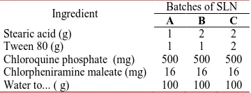
the system was kept above the melting point of the lipid by placing the beaker on a hot-plate. Subsequently, the pre-emulsion was introduced in drops into 500 ml of cold water at 2 oC. While addition ensued, stirring was provided using a magnetic stirrer assembly. This was intended to keep the particles dispersed, but not to shear further into smaller particles. Three batches of solid lipid nanoparticles (SLN A, SLN B and SLN C) were prepared according to the quantities presented in Table 1.
Table 1: Actual quantities of ingredients used in preparing the solid lipid nanoparticles
Ingredient Batches of SLN
A B C
Stearic acid (g) 1 2 2 Tween 80 (g) 1 1 2 Chloroquine phosphate (mg) 500 500 500 Chlorpheniramine maleate (mg) 16 16 16 Water to... ( g) 100 100 100
Determination of hydrodynamic size
The hydrodynamic sizes of the particle were determined after one week of preparation. The preparations were unlyophilized and 1 ml samples were first withdrawn from the bulk without shaking the dispersion, and then after shaking to redisperse any sedimented coarse particles. Measurements were done by photon correlation spectroscopy at 25 oC using a particle size analyzer (Zetasizer Nano ZS, Malvern Instruments Ltd., Worcestershire, UK) equipped with v7.03 software. The detection was performed at a scattering angle of 90o at an accumulation time of 180 s. Samples were diluted with ultrapure water (resistivity of 18.2 mΩ.cm) and data were analysed by the cumulants
method.
Determination of Zeta potential
The zeta potentials of the formulated SLN were similarly determined after one week of preparation. Each sample
was diluted with ultrapure water and at 25 ◦C and read in a Zetasizer Nano ZS (Malvern Instruments Ltd., UK) at
refractive index and pH settings of 1.33 and 7 respectively. For spherical colloidal particles of high ratio of radius to double layer thickness, the Henry function approximates 1.5 and the electrophoretic mobilities may be converted related to the zeta potential values via the Smoluchowski relation:
= / (1)
Uis the electrophoretic mobility; ε is the dielectric constant, and η the viscosity of the medium
.
Determination of entrapment efficiency
Entrapment efficiency was determined using an indirect method. This involved determination of the concentration of the free unloaded drug in the supernatant after the nanoparticles were pellet by centrifugation. Briefly, approximately 2.5 mL of the nanoparticle dispersion was centrifuged at 4000 rpm for 60 min at 37 oC in a microcentrifuge (800-B, Hunan, China). A sample of the clear supernatant was withdrawn and the amount of free chloroquine was determined by taking absorbance readings at 330 nm in a UV-Vis spectrophotometer (Jenway 6505, Bibby Scientific, Essex, UK). In addition, quantification of encapsulated chlorpheniramine was attempted using both isocratic and gradient methanol-acetonitrile systems on a Shimadzu Prominence HPLC (Shimadzu Corporation, Kyoto, Japan) equipped with a diode array detector and Phenomenex Gemini C18 column (5 µ m x 250 mm x 4.6 mm).
The entrapment efficiency (EE) was calculated using the relationship:
. . = × 100 (2)
The amount of drug entrapped was calculated by subtracting the amount of free drug from the theoretical amount of drug added to SLN.
Determination of loading efficiency
The loading efficiencies (LE) of the three formulations were determined from the entrapment efficiencies by, calculating the ratio of the entrapped amount of drug to total formulation mass.
' = (( ( × 100 (3)
In vitro release from unlyophilized nanosystems
An in vitro release study was conducted over 3 hours separately using phosphate buffered saline. For this study, 0.5 ml of the nanodispersion was placed in dialysis membrane of 10 kDa molecular weight cut off and immersed in 40 ml of release medium contained in a chamber of the dissolution testing apparatus [RC-6 dissolution]. The operating conditions were 22 rpm at a temperature of 37 °C. At predetermined time intervals, 2 ml of buffer solution outside the dialysis bag was removed and replaced with 2 ml of fresh medium. The amount of drug released was determined by taking absorbance readings against the standard calibration curves prepared previously.
Statistical analyses
Unless otherwise stated, results have been presented as mean ± standard deviation of replicate measurements (n = 3). Statistical analysis was done using Microsoft Excel 2007 and statistical significance was measured using the Welch two-sample t-test. A probability value of 0.05 or less was considered significant.
RESULTS AND DISCUSSION
[image:4.595.136.475.352.438.2]The solid lipid nanoparticle systems were translucent systems. The mean hydrodynamic diameters of the formulations are presented in Table 2. As indicated, the preparations had roughly the same size distribution, with an average polydispersity index of about 0.4 in each case. Though the dispersions remained largely stable even after one month, a light deposit was nonetheless evident after one week as some of the macro-sized particles settled under gravity. Though C and A had the same ratio of surfactant to lipid, the hydrodynamic size of C is higher than A, pointing to the influence of content of lipid on efficiency of homogenization.
Table 2: Average size and zeta potential values of chloroquine-loaded solid lipid nanoparticle systems prepared using varying quantities of stearic acid and Tween 80*
Batch Batches of SLN
A B C
Z-average size (d.nm) Without redispersion With redispersion
192.80 ± 18.1 591.30 ± 14.5
287.20 ± 6.3 498.50 ± 12.1
211.80 ± 32.90 663.80 ± 13.60 Mean polydispersity index
Without redispersion With redispersion
0.38 ± 0.08 0.40 ± 0.02
0.31 ± 0.03 0.37 ± 0.01
0.49 ± 0.13 0.38 ± 0.02 zeta potential (mV) All samples had extremely small zeta potentials values of about -0.3 mV
* Statistical differences were tested at 5 % level. Particle sizes were determined one week after preparation without lyophilisation using a small volume (1 ml) withdrawn from the bulk of the sample, first without shaking, and then after shaking to redisperse sedimented particles or
aggregates.
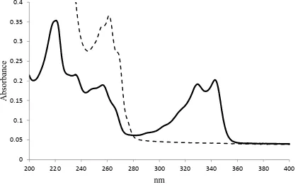
Figure 1: Uv-Vis scan of chloroquine phosphate and chlorpheniramine maleate in water
‒‒ chlorpheniramine ― chloroquine
0 0.05 0.1 0.15 0.2 0.25 0.3 0.35 0.4
200 220 240 260 280 300 320 340 360 380 400
A
bs
or
ba
nc
e
[image:4.595.87.508.474.738.2]The ultraviolet-visible spectra of the drug samples are presented in Figures 1. In view of the fact that the single extinction peak of 261 nm for chlorpheniramine significantly overlapped the 256 nm band of chloroquine, appearing roughly as a single peak in the spectrum of a mixture of the two drugs, the UV-Vis spectroscopy was incapable of reliably and accurately quantifying untrapped chlorpheniramine. In addition, HPLC-UV, which was attempted with both isocratic and gradient elution methods and varying combinations of methanol and acetonitrile as mobile phases, did not achieve significant peak resolution. In view of this, only the trapping efficiencies and loading values obtained for chloroquine are displayed (Table 3).
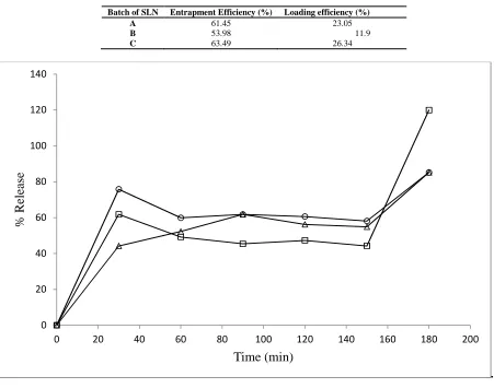
[image:5.595.74.525.223.578.2]The release curves (Figure 2) of the unlyophilized nanodispersions point to a repeat action (biphasic release) with an initial burst action due to untrapped drug, followed by rapid drug release after approximately 150 min from lipid matrices.
TABLE 3: Entrapment efficiencies of different batches of solid lipid nanoparticle system
Batch of SLN Entrapment Efficiency (%) Loading efficiency (%)
A 61.45 23.05
B 53.98 11.9
C 63.49 26.34
.
FIGURE 2: In vitro release studies of chloroquine phosphate in buffer from batches of solid lipid nanoparticles
―□― SLN A, ―○― SLN B, ―∆― SLN C. SLN A contains 1 g each of both lipid and surfactant. SLN B contains 2 g of lipid and 1 g surfactant. SLN C contains 2 g each of both lipid and surfactant.
The design of an anti-malarial formulation is influenced by the route of administration, the intravenous route being the preferred route in cerebral malaria [19]. In acute uncomplicated malaria, the oral route is preferable. The administration of drugs by the intravenous route imposes a low particle size cut off of about 5 microns, in order not to cause occlusion of small blood vessels. This generally excludes the use of coarse emulsions and most microparticle systems. The particle sizes obtained on standing the nanodispersions for one week were all less than 290 nm. On shaking and redispersion, particle sizes obtained were all lower than 640 nm. Erythrocytes are roughly 10 times bigger than this and so the particles have a size advantage over microparticle systems and may be used for targeted delivery inside erythrocytes (averaged 6 microns in size). In fact, anti-malarial drug carriers in this size range can be used in targeting the plasmodial schizonts and merozoites or their organelles, since the diameter of these blood forms (generally about1 µ m) is significantly higher than the particle size.
0 20 40 60 80 100 120 140
0 20 40 60 80 100 120 140 160 180 200
%
R
el
ea
se
However, because erythrocytes generally do not have phagocytic capacity, tertiary targeting of nanocarriers entailing delivery to intra-erythrocytic bodies is very difficult. Rather, particles which are small enough in size may pass through the new permeability pathways (NPPs) which occur on infected red blood cell membranes shortly after invasion by the parasite [20, 21, 22, 19]. For this to occur, the sizes need be less than 80 nm in size, which has been determined as the particle size cut off for passage. This may not be achievable in the present case. It is actually much more difficult to achieve such low sizes in conditions in which the drug is not dissolved, due to influences of drug crystals sizes themselves, particularly in co-formulation. To further reduce particle size of anti-malarial drug nanoformulations to attain sizes in the desired range for passage through the NPP, high pressure homogenization may be utilized. However, even after high pressure homogenization, solid lipid nanoparticles produced from Softisan® by hot pre-emulsion had particle sizes in this range (~ 550 – 700 nm) [23].
An increase in the ratio of surfactant to lipid would be expected to reduce particle size [24, 25]. From the results obtained, however, Batch A with a surfactant to lipid ratio of 1: 1 exhibited mean particle size higher than Batch B with a 1: 2 ratio, after shaking. The explanation offered is that since the preparation was unlyophilized and also size determination was done after one week of preparation, the sizes quoted represent the combined influences of homogenization efficiency and particle size growth at room temperature storage. From the viewpoint of particle size growth, low concentration (viscosity) systems such as Batch A suffer more particle size because of higher mobility, unless they are incorporation into gels or creams for increased stability. Batch C has significantly higher particle sizes (*p < 0.05) than A or B, which may result from difficulties in homogenizing the high concentration of ingredients. The particle sizes obtained before shaking were less different between the three batches: statistical difference existed only between batches A and B (*p = 0.006786). The polydispersity indices were not statistically different in either case, suggesting that the method of preparation used achieved very narrow particle size distribution. The inference drawn from particle size analysis is that particle size depends not only on surfactant to lipid ratio, but on total concentration of particles, which would affect homogenization efficiency.
For the most part, nanotechniques employed in malaria research do not seek to traverse the red cell membrane (generally because of lack of phagocytotic activity as previously mentioned), but to keep the nanoparticle in contact with the red blood cells for long enough to achieve suitable local concentrations of the entrapped drug in the neighbourhood of the red cell (19). Whereas lipid-based systems are quickly removed by the reticuloendoethelial system unless suitable hydrophilic coatings are applied, common “hydrophilization” methods like PEGylation seriously limit absorption through the lymphatic system and could predispose the particles to portal absorption and first pass hepatic metabolism. This would be a disadvantage with chlorpheniramine which undergoes significant first pass hepatic metabolism [26]. This explains why the nanoparticles were not PEGylated. Rather than PEGylate, cytoadherence should be sought.
There were no differences in the zeta potential values. The low negative values obtained would be expected from the poor ionization of stearic acid. In the absence of steric and entropic stabilization effects, fairly high values of zeta potential (± 30 mV) are needed to keep the particle apart in a colloid of this nature [27]. This low potential value is incapable of preventing close particle-particle approach, and may partly explain why the mean one-week particle sizes after agitation of the container differed significantly from the value obtained without agitation (sampling was done in the middle of the container). This suggests that multi-particle aggregates had started forming, which settled out under the influence of gravity, redispersed only on shaking. As mentioned before, these systems can be significantly stabilized by introducing viscosity enhancers, embedding into creams or gels, or by lyophilization in the presence of a cryoprotectant. Nevertheless, because surfactant-stabilized lipid systems of this nature, due to the content of viscosity-modifying lipid, can still be considerably more stable than aqueous suspensions of comparable zeta potential values.
High loading capacities were achieved in the formulations having equal ratios of lipid and surfactant, as previously reported [30]. The extent of drug loading seen in Batch C means that to prepare chloroquine phosphate injection containing 200 mg/5ml, only 760 mg of lyophilized Batch C particles would be needed per 5 ml ampoule. This is fairly high for a drug which does not dissolve in the lipid melt [28]. Loading is very important for scale up.
All release profiles show an initial a diffusion-driven period due to untrapped drug. As expected, the relative peaks attained at this point (30 minutes) inversely correspond to the trapping efficiencies, with Batch C (with highest drug trapping) having the least magnitude. However, the total percentage drug release at this point (30 minutes) far exceeds the untrapped drug content, and explanation offered is that a significant fraction of the trapped drug resides on the surface of the nanoparticles, wherein it behaves like untrapped drug and is released fairly rapidly. From 150 minutes, erosion of the non-swelling matrix is initiated leading to second phase of drug release. Batch B, having a smaller particle size (higher surface area), would be expected to exhibitthe fastest release rate (31). This is not the case, and we infer that the viscosity of the system (lowest in A due to lower concentration of ingredients) must have been a factor in the higher rate of drug diffusion across, as against release.
In future, treatment can be considerably influenced by this pattern of biphasic drug release achieved by using a mixture of trapped and untrapped drugs. Due to the high crystallinity of the lipid matrix, drug release from the system is predictably different from nanoemulsions of equivalent concentration. Because of its lipid nature, the lyophilized and reconstituted dispersion may be used in intramuscular depot medication. However, for this to happen, it may be necessary to ensure that the surfactant does not remain in sufficiently high levels after recovery of the nanoparticles and lyophilisation.
CONCLUSION
This work is based on clinical evidence of the superiority of chloroquine-chlorpheniramine co-administration in overcoming drug resistance, due possibly to effects on P-gp mediated drug efflux. We conclude that solid lipid nanoparticles co-encapsulating chloroquine and chlorpheniramine can be prepared to particle size specifications smaller than the diameter of both human capillary systems and red blood cells and merozoites, by the hot homogenization- dilution method. Equal lipid-surfactant ratios ensure high trapping and loading, but particle size is dictated by both this ratio and total system concentration, as this would affect viscosity and homogenization efficiency. In vitro release studies conducted with the unlyophilized dispersion exhibited a two-phase drug release due to untrapped and trapped drugs. Due to the limited assay methods available, the data on trapping and release of chlorpheniramine are not reported, since its spectral peak overlaps significantly with that of chloroquine. Further studies will be devoted to overcoming this problem, and also in vivo testing in both resistant and sensitive strains of Plasmodium falciparum. Studies will also be conducted to establish the toxicity profile of the formulation.
REFERENCES
[1]JF Trape; G Pison; MP Preziosi; C Enel; A Desgrees du Lou; V Delaunay; B Samb; E Lagarde; JF Molez; F Simondon, C. R. Acad. Sci. III, 1998, 321(8), 689-697.
[2]RL Hallett; CJ Sutherland; N Alexander; R Ord; M Jawara; CJ Drakeley; M Pinder; G Walraven; GA Targett; A
Alloueche, Antimicrob. Agents Chemother., 2004, 48(10), 3940-3943.
[3]EC Spoelstra; H Dekker; GJ Schuurhuis; HJ Broxterman; J Lankelma, Biochem. Pharmacol., 1991, 41(3),
349-359.
[4]J Wang; B Goh; W Lu; Q Zhang; A Chang; XY Liu; TM Tan; H Lee, Biol. Pharm. Bull., 2005, 28(5), 822-828.
[5]DJ Krogstad; IY Gluzman; DE Kyle; AM Oduola; SK Martin; WK Milhous; PH Schlesinger, Science, 1987, 238(4831), 1283-1285.
[6]DA van Schalkwyk; JC Walden; PJ Smith, Antimicrob. Agents Chemother., 2001, 45, 3171-3174.
[7]M Kalkanidis; N Klonis; L Tilley; LW Deady, Biochem. Pharmacol. 2002, 63, 833-842.
[8]AK Bhattacharjee; DE Kyle; JL Vennerstrom, Antimicrob. Agents Chemother., 2001, 45, 2655-2657.
[9]A Miki; K Tanabe; T Nakayama; C Kiryon; K Ohsawa, Exp. Parasitol. 1992, 74, 134-142.
[10]DA van Schalkwyk; TJ Egan, Drug Resist. Updat., 2006, 9(4-5) 211-226.
[11]A Sowunmi; AMJ Oduola; OAT Ogundahunsi; CO Falade; GO Gbotosho; LA Salako, Trans R Soc Trop Med
Hyg 1997; 91(1): 63-67.
[12]AMJ Oduola; A Sowunmi; WK Milhous; TG Brewer; DE Kyle; L Gerena; RN Rossan; LA Salako; BG
Schuster, Am. J. Trop. Med. Hyg. 1998, 58(5), 625-629.
[13]AR Thierry; D Vigė; SS Coughlin; JA Belli; A Dritschilo; A Rahman, Cancer Metastasis Rev. 1993, 7(6),
572-579.
[14]L Warren; JC Jardillier; A Malarska; MG Akeli, Cancer Res., 1992, 52(11), 3241-3245.
[16]YP Hu; N Henry-Toulmė; J Robert, Eur J Cancer, 1995; 31A(3), 389-394.
[17]MR Gasco; L Priano; GP Zara, The CNS. Prog. Brain Res., 2009, 180: 181-192.
[18]R Cavalli; E Marengo; L Rodriguez; MR Gasco, Eur. J. Pharm. Biopharm. 1996, 43 (2), 110-115.
[19]NS Santos-Magalhăes; VCF Mosqueira, Adv. Drug Deliv. Rev., 2010; 62 (4-5), 560-575
[20]GA Biagini; SA Ward; PG Bray, Trends Parasitol., 2005, 21 (7), 299-301.
[21]ID Goodyer; B Pouvelle; TG Schneider; DP Trelka; TF Taraschi, Mol. Biochem. Parasitol. 1997, 87(1), 13-28.
[22]S Charpian; JM Przyborski, Traffic 2008, 9(2), 157-165.
[23]EB Souto; RH Müller; S Gohla, J. Microencapsul., 2005, 22 (6): 581-592.
[24]RH Müller; S Maaben; H Weyhers; W Mehnert, J. Drug Target, 1996, 4 (3), 161-170.
[25]M Chakrapani; R Nair; RY Prasanna; P Dhanalakshmi, JITPS, 2012, 3 (3), 80-87.
[26]SH White. Remington: The Science and Practice of Pharmacy, 20 th Edition, Lippincott Williams and Wilkins,
Philadelphia, 2000, 1464-70.
[27]H Schott. Remington: The Science and Practice of Pharmacy. 20 th Edition, Lippincott Williams and Wilkins,
Philadelphia, 2000, 288-315.
[28]RH Müller; K Mäder; S Gohla, Eur. J. Pharm. Biopharm., 2000, 50 (1), 161–77.
[29]J Varshosaz; S Eskandari; M Tabakhian, Pharm. Dev. Technol., 2010, 15(1): 89–96.
[30]NS Hagalavadi; BP Pragnesh; GD Bapushheb; A Purnima; A Sinathambi, Acta Pharmaceutica, 2007, 57(3): 269-285.
[31]RH Müller; M Radtke; Wissing SA, Adv. Drug Deliv. Rev., 2002; 54(Suppl 1), S131–55.