Journal of Chemical and Pharmaceutical Research, 2016, 8(1):75-131
Review Article
CODEN(USA) : JCPRC5
ISSN : 0975-7384
Recent progress in biologically active xanthones
Arshdeep Singh
a, Navdeep Kaur
b, Sahil Sharma
a* and Preet Mohinder Singh Bedi
aaDepartment of Pharmaceutical Sciences, Guru Nanak Dev University, Amritsar, Punjab, India b
Department of Chemistry, Guru Nanak Dev University, Amritsar, Punjab, India
_____________________________________________________________________________________________
ABSTRACT
Xanthone is a novel and pharmacologically important oxygen containing hetrocyclic ring exhibiting variety of potent biological activities. This fused tricyclic ring is derived naturally from the plants of Bonnetiaceae and Clusiaceae family as well as synthesized by employing various synthetic protocols. Their wide range of reported therapeutic attributes include α-glucosidase inhibition, anti-cancer, anti-oxidant, anti-asthmatic, anti-convulsant, xanthine oxidase inhibition and anti-microbial. Among these attributes, anti-cancer and anti-oxidant potential are comprehensively studied as evidenced by number of research papers. Although, there is no marketed formulation containing xanthone derivatives except some herbal formulations, their medicinal properties can not be neglected at all. Promising activities revealed by these compounds chains their use and places them ahead as potential drug candidates for the future studies. Therefore, the present review article is entirely an assemblage of recent research work that has been done on xanthones and their analogs as therapeutic agents. This review article can be reasonably encouraging for those engaged in the area of developing efficient and effective, single therapeutic agent exhibiting a wide range of biological activities involving xanthone as crucial and central nucleus.
Keywords: α-glucosidase inhibition, anti-cancer, anti-oxidant, anti-asthmatic, anti-convulsant, xanthine oxidase inhibition, anti-microbial
_____________________________________________________________________________________________
INTRODUCTION
Heterocycles coins an exceedingly important class of compounds that form the basis of many pharmaceutical, agrochemical and veterinary products [1-3]. Many natural drugs such as quinine, papaverine, emetine, theophylline, atropine, procaine, codeine, morphine and reserpine are heterocyclic in nature. There are large number of natural and synthetic heterocyclic compounds containing pyrrole, pyrrolidine, furan, thiophene, piperidine, pyridine and thiazole having important applications [4]. Some of the heterocyclic drugs possessing varried biological activities are shown in Figure 1.
Xanthone is a special class of oxygenated tricyclic compounds which exhibits various interesting biological activities depending upon the nature and pattern of substitutions [15-17]. Xanthones are frequently valued as an effective pharmacophore in the field of medicinal chemistry [18]. Earlier, xanthones were introduced as insecticide, larvicide and ovicide codling moth eggs [19]. Later, it has been found in some scientific studies that xantone derivatives can prevent the development of cancer cells and also possesses anti-inflammatory and anti-oxidant properties [20].
• Xanthone Benefit Plus which is a herbal drink product [22].
• Xantho Plus capsules which is an anti-oxidant complex on the bases of Mangostin [23].
• MX3 (Mangostin Xanthone) capsules which is also a herbal product used as dietary supplement [24].
N Cl N H Clarinex [5] (Antihistamine) N N F O OH N O O Levofloxacin[6] (Antibacterial) O S N
N NH2
O HO Lamivudine [7] (Antiviral) N N S NCH3
CH2CH2N(CH3)2
Dercetin [10] (Anti-malarial)
N N
NHSO2CH3
H3CO
H Amsacrine [8] (Anti-neoplastic) N H O COOH Etodolac [9] (Anti-inflammatory) N H O N H O OMe OH Carvedilol [12] (Anti-hypertensive) OCH3 O O OH OH COCH3 OH
O HC CH2 C
H NH2 C H OH H C O CH3 Cerubidine [11] (Anti-cancer) O O OH OH CCH3 OH H O O O CH3 HO NH2 Idamycin [14] (Anti-cancer) H N O N N N O Alectinib [13] (Anti-tumor) .HCl
Figure 1. Heterocyclic based marketed drugs
The main component of these formulations is an extract from the well known fruit – Mangostin (Garcinia mangostana), which is also called the “Queen of fruits” mostly found in Malaysia and Southeast Asia. Mangostin extract contain xanthone derivatives i.e. 3-isomangostin, alpha mangostin, Gamma mangostin, Garcinone A, Garcinone B, C, D and E, maclurin, mangostenol (Figure 2). Over 200 naturally occurring xanthones which have been identified so far and approximately 40 of those are found in the mangostin.
In the early 80s, xanthone containing fruit, mangostin was used to treat diarrhea, dysentery, genitor-urinary infections, and also as a wash for aphthae of the mouth. Then in 90s xanthones were developed as insecticide. They also found use as ovicide, larvicide and in the preparation of xanthydrol, which is used in the determination of urea levels in the blood. In view of their therapeutic potential, lot of work has been done to explore the beneficial effects of this nucleus. Recent reports suggest that xanthones possess numerous medicinal properties such as, antiallergic, anti-inflammatory, antitubercular, antitumor, antiplatelet, anticonvulsant as well as β-adrenergic blocking property. These promising activities place xanthones ahead as potential drug candidates for the future studies.
activity and least side effects, which may, some day would help to bring new medicinal agents having xanthone as an active ingredient in market.
O O
O
HO OH
O OH
O O
HO
O OH
O HO
HO
O OH
O HO
O OH
OH OH
O HO
HO
O OH
OH OH
O H3CO
HO
O OH
OH OH
O HO
HO
O OH
O H3CO
HO
O OH
O OH
3-isomangostin Alpha-mangostin Gamma-mangostin
Garcinone A Garcinone C Garcinone D
Garcinone E Mangosterol
Figure 2. Xanthone derivatives present in marketed formulations
BIOLOGICAL ACTIVITIES OF XANTHONES
2-1. Xanthones as α-glucosidase inhibitors
In 2006, Yan Liu et al. synthesized a series of hydroxylxanthones and their acetoxy and alkoxy derivatives 1, 2, 3 (Figure 3) and evaluated as α-glucosidase inhibitors. The results indicated that these xanthone derivatives were capable of inhibiting in vitro α-glucosidase with moderate to good activities. Among them, polyhydroxylxanthones exhibited the highest activities and thus may be exploitable as a lead compound for the development of potent
α-glucosidase inhibitors [25].
The IC50 value was determined against yeast α-glucosidase in 50 µM phosphate buffer (pH 6.8) at 37 ºC (Figure 3).
A classical α-glucosidase inhibitor, 1-deoxynojirimycin, was adopted as a positive control with an IC50 value of 26.4
µM.
In 2007, Eun Jin Seo et al. proved that xanthones isolated from the root of C.tricuspidata possess highly potent
α-glucosidase inhibition properties. Compound 4 and 5 was identified as a new isoprenylated tetrahydroxyxanthone; 1,3,6,7-tetrahydroxy-2-(3-methylbut-2-enyl)-8-(2-methylbut-3-en-2-yl)-9H-xanth en-9-one 4. These are the first natural xanthones documented to exhibit such inhibition. The IC50 values of compounds 4, 5 inhibiting
O R4
R3
R5 R1
R2 O
1: R1 = R2 = R4 = OH, R3 = R5 = H
IC50 (µM) = 14.7
2: R1= R2 = R3 = R5 = OH, R4 = H
IC50 (µM) = 17.1
3: R1= R2 = OAc, R3 = R4 = R5 = H
IC50 (µM) = 31.9
O HO
HO
OH
OH O
O HO
HO
OH
OCH3 O
5 4
IC50 (µM) = 35.8 ± 1.7
Ki value = 31.7 IC50 (µM) = 16.2 ± 0.4
Ki value = 7.0
Figure 3. Xanthones exhibiting α-glucosidase inhibition properties
In 2007, Yan Liu et al. synthesized a series of novel xanthone derivatives with extended π-systems, that is, benzoxanthones (Figure 4), and their structurally perturbed analogs as α-glucosidase inhibitors. Their inhibitory activities toward yeast’s α-glucosidase were evaluated with the aim to enrich the structure–activity relationship. The results indicated that benzoxanthones 7–9 were capable of inhibiting in vitro yeast’s α-glucosidase 17 to 28 fold more strongly than xanthone derivative 6 that has smaller conjugated π-system. O-Methylation of 3-OH of benzoxanthone 10 and nitration at C4 position led to a large decrease in the activity. This indicates that 3-OH of benzoxanthone was crucial to the inhibitory activity, primarily as an H-bonding donor. The present results suggest that π–π stacking effect and H-bonding make substantial contributions to elicit the inhibitory activities of this general class of inhibitors [27].Determined against yeast’s α-glucosidase in 50 µM phosphate buffer (ph 6.8) containing 5% v/v DMSO at 370C.
O OH
OH O
O R2
R1
OH
OH O
6 O
OH
OH NO2
O
10 IC50 (µM) = 160.8 7: R
1 = R2 = H
IC50 (µM) = 9.3
8: R1 = H, R2 = OH
IC50 (µM) = 5.8 9: R1 = OH, R2 = H
IC50 (µM) = 8.0
IC50 (µM) = 20.1
Figure 4. Xanthone derivatives with extended π-system
In 2008, Yan Liu et al. employed multiple linear regression (MLR) method to establish QSAR models for xanthone derivatives 11-25 (Figure 5), that have diverse structures, to provide deep insight into the correlation between inhibitory activities and structures of xanthones. Among the 38 typical descriptors investigated, Hs (number of H-bond forming substituents), Nπ (number of aromatic rings), and S (softness value) can be utilized to model the inhibitory activity. The Hs, Nπ, S and IC50 (Figure 5). Thus, inhibitory activities of xanthone derivatives can be
O HO OH OH O O HO OH OH OH O O OAc OAc O O OH OH O
IC50 (µM) = 14.7 Hs = 2.5 N = 2 S = 0.124
11 12
IC50 (µM) = 17.1 Hs = 3.0 N = 2 S = 0.115
13 IC50 (µM) = 31.9 Hs = 1.0 N = 2 S = 0.112
14 IC50 (µM) = 9.3 Hs = 1.5 N = 3 S = 0.133
O HO OH OH O O OH OH O H O O OH OCH3 O 15 IC50 (µM) = 5.8 Hs = 2.5 N = 3 S = 0.141
25
IC50 (µM) = 8.0 Hs = 2.5 N = 3 S = 0.139
17
IC50 (µM) = 31.3 Hs = 1.0 N = 3 S = 0.134
O OH OH NO2 O 18
IC50 (µM) = 20.1 Hs = 1.5 N = 3 S = 0.134
O OH
OH
19
IC50 (µM) = 27.8 Hs = 2.0 N = 3 S = 0.113
O OH
OH O
20
IC50 (µM) = 39.9 Hs = 1.5 N = 3 S = 0.126
O OH
OH
21
IC50 (µM) = 34.9 Hs = 2.0 N = 3 S = 0.113
O OH NO2 OH O 22 IC50 (µM) = 5.9 Hs = 1.5 N = 3 S = 0.132
O OH NH2 OH O 23 IC50 (µM) = 6.3 Hs = 2.5 N = 3 S = 0.133
O OH OH NH2 O 24 IC50 (µM) = 8.3 Hs = 2.0 N = 3 S = 0.133 O
OH
OC3H6Br
O
16 IC50 (µM) = 29.7 Hs = 1.0 N = 3 S = 0.134
Figure 5. Xanthone derivatives and their Hs (number of h-bond forming substituents), Nπ (number of aromatic rings), and S (softness value)
In 2011, Gia Li Li et al. synthesized a series of novel xanthone derivatives 31-41 having non-coplanar and flexible structures as potent α-glucosidase inhibitors. Biological evaluation indicated that compounds 31-37 bearing one or two naphthol moieties exhibited up to 30-fold enhanced activities compared with their corresponding parent compounds 27-30, whereas compounds 38-41 bearing one dihydroxylnaphthalenyl group showed decreased activities compared with their corresponding analogs 31-34 having one naphthol group (Figure 6). Among them, compounds 32-33, 35-37 and 40 were more active than 1-deoxynojirimycin, a well-known inhibitor for
O OH
OH O
26 O
OH
OH O
R1
R2
R3
27: R1 = R2 = R3 = H
IC50 (µM) = 160.8
28: R1 = OH, R2 = R3 = H
IC50 (µM) = 41.5
29: R1 = R3 = OH, R2 = H
IC50 (µM) = 17.2 30: R1 = R3 = H, R2 = CH3
IC50 (µM) = >200
O R1
R2
R3 OH
OH HO O
31: R1 = R2 = R3 = H IC50 (µM) = 47.7
32: R1 = OH, R2 = R3 = H
IC50 (µM) = 10.5
33: R1 = R3 = OH, R2 = H
IC50 (µM) = 8.1
34: R1 = R3 = H, R2 = CH3 IC50 (µM) = 26.5
O R1
R2
R3 OH
OH O
HO
HO R1 O
R2
R3 OH
OH O
HO
OH
35: R1 = R2 = R3 = H
IC50 (µM) = 10.1
36: R1 = OH, R2 = R3= H
IC50 (µM) = 6.2
37: R1 = R3 = H, R2 = CH3
IC50 (µM) = 6.4
38: R1 = R2 = R3 = H
IC50 (µM) = 75.4
39: R1 = OH, R2 = R3 = H IC50 (µM) = 47.6
40: R1 = R3 = OH, R2 = H
IC50 (µM) = 17.4
41: R1 = R3 = OH, R2 = CH3
IC50 (µM) = 112.1
Figure 6. Xanthone derivatives having non-coplanar and flexible structures as potent α-glucosidase inhibitors
2-2. Anti-cancer xanthones
In 1991, Masahiro Aoki et al. reported Vinaxanthone 42 (Figure 7) a novel phospholipase C inhibitor, produced by Penicillium vinaceum Gilman and Abott NR6815. Its structure (MW 576, C28H16O14) has been elucidated as a
polycyclic xanthone with poly acidic functional groups based on various NMR studies including HMBC, COLOC, 2D-INADEQUATE and selective ID-INADEQUATE [30].
O
O HO
HO H
COOHO H
OC CH3
CO CH3
H
O
H OH
OH COOH
42
O
O
HO
OH O
OH
O
OH
HO
OH
OH OH
43 Figure 7. Vinaxanthone and Griffipavixanthone
In 2002, Madalena Pedro et al. evaluated xanthones of Formula I 44-49 (Table 1) on the proliferation of human T-lymphocytes. Oxygenated xanthones have been assessed for their capacity to inhibit in vitro the growth of three human cancer cell lines, MCF-7 (breast cancer), TK-10 (renal cancer) and UACC-62 (melanoma) (Table 1). Differences on their potency towards the effect on the growth of the human cancer cell lines as well as on the proliferation of human T-lymphocytes can be ascribed to the nature and positions of the substituents on the xanthonic nucleus [32].
O O
1 2 3 4 4a 4b 5 6 7
9a 8a 8
Formula I
Table 1. Effects of xanthone derivatives of Formula I on the growth of human cancer cell lines and proliferation of human lymphocytes [GI50 (µM)] [IC50 (µM)]
Compounds MCF-7b TK-10b UACC-62b Human lymphocytesc
(breast cancer) (renal cancer) (melanoma)
44 Xanthone >200 >200 >200 >200 45 1,2-Dihydroxy 38.4±2.7 65.8±5.1 14.0±0.3 73.3±2.2 46 1,7-Dihydroxy 57.1±11.0 60.1±9.6 51.3±9.6 20.2±0.2 47 2,3-Dihydroxy 40.6±1.3 61.4±4.3 31.7±3.7 31.3±1.5 48 3,4-Dihydroxy 40.5±1.5 59.2a 21.6±2.6 12.2±1.3
49 2-Hydroxy-1- 24.1±3.0 35.2±7.6 39.3±10.6 11.6±0.8
methoxy
50 5-Hydroxy-3- 66.1±12.6 30.9±2.3 37.7±6.7 111.2±8.3
methoxy
51 1,3-Dihydroxy- 21.9±0.4 34.3±3.8 20.0±0.5 84.4±12.4
2- methyl
52 2,3-Dihydroxy- 37.2d 76.6±7.0 19.8±1.6 17.4±2.7
4-methoxy
aResults are expressed as GI
50 (concentrations of compounds that cause 50% inhibition of cancer cell growth) or IC50 (concentrations that
cause50% inhibition of lymphocytes proliferation) and show means ±SEM of 3–6 independent observations performed in duplicate.
bDoxorubicin was used as positive control in cancer cell lines growth (GI
50 MCF-7=42.8±8.2 nM; GI50 TK-10=548.0±60.0 nM; GI50
UACC-62=94.0±9.4 nM).
c
Cyclosporin A was used as positive control in lymphocytes proliferation (IC50=0.34±0.04 µM). dData based on two independently run duplicate
experiments.
O
O OH
O
O
O
O OH
O
O
O
O OCOCH3
O
O
O
O
O
O OMe
O
O OH
53
1-Hydroxyxanthone
OH
OH
HO
OH
OCOCH3
H3CCOO
OCOCH3
OMe MeO
OM e
MeO OH
54
3-Hydroxyxanthone
55
1,4-Dihydroxyxanthone
56
2,6-Dihydroxyxanthone
57
1,2-Diacetoxyxanthone 2,6-Diacetoxyxanthone58 3-Methoxyxanthone59 1,3,7-Trimethoxyxanthone60
61
1,5-Dihydroxy-6-methoxyxanthone Figure 8. Xanthones bearing different functionalities
Table 2. Effect of xanthones on the TNF-α induced expression of ICAM-1 on endothelial cells
Serial Compound name Concentration % Inhibition No. (µg/mL)a of ICAM-1expression 53 1-Hydroxyxanthone 60 22.2 54 3-Hydroxyxanthone 66 13.7 55 1,4-Dihydroxyxanthone 65 86.0 56 2,6-Dihydroxyxanthone 18 40.9 57 1,2-Diacetoxyxanthone 75 42.4 58 2,6-Diacetoxyxanthone 100 23.8
59 3-Methoxyxanthone 46 0
60 1,3,7-Trimethoxyxanthone 13 0
61 1,5-Dihydroxy-6-methoxyxanthone 20 1.6 aThe concentration levels of different compounds are based on their maximum tolerable concentrations by the cells. Table 3. Effect of xanthones on NADPH-dependent inhibition of lipid peroxidation initiation SerialNo. Compound name NADPH-dependent lipid peroxidation initiation, percent of the controla 53 1-Hydroxyxanthone 28
54 3-Hydroxyxanthone 38
55 1,4-Dihydroxyxanthone 60
56 2,6-Dihydroxyxanthone 43
57 1,2-Diacetoxyxanthone 30
58 2,6-Diacetoxyxanthone – 59 3-Methoxyxanthone 2
60 1,3,7-Tri-methoxyxanthone 0 61 1,5-Dihydroxy-6-methoxyxanthone –
aControl represents the assay in the absence of the test compounds and the values represent the average of four observations within an error
range of 5.0%.
In 2004, Ying-Shu Zou et al. isolated eight new isoprenylated xanthones, cudratricusxanthones A–H (62–67), from the roots of Cudrania tricuspidata, together with ten known compounds, cudraxanthones H (64) and M (73), xanthone V1a (70),toxyloxanthone C (74), macluraxanthoneB (71), 1-hydroxy-3, 6, 7-trimethoxyxanthone (75),
cycloartocarpesin (76), artocarpesin (78), cudraflavone B (77), and kaempferol (79) (Figure 9). Their structures were characterized by spectroscopic methods. Xanthones 68, 69, 73 and 74 showed inhibitory effects on four kinds of human digestive apparatus tumor cell lines (HCT-116, SMMC-7721, SGC-7901, and BGC-823) with IC50 values
O HO
R2
O OH
R1
OH
62: R1 = , R2 = OH
63: R1 = , R2 = OH
64: R1 = CH2CH=C(CH3)2, R2 = H
O HO
HO OH
O OH
O
65
O R3
HO R2
O OH
R1
O
66: R1 = CH2CH=C(CH3)2, R2 = H, R3 = OH
67: R1 = C(CH3)2CH=CH2, R2 = OH, R3 = H
O R4
HO R3
O OH
R1
R2
68: R1 = CH2CH=C(CH3)2, R2 = R4 = OH, R3 = H
69: R1 = R3 = H, R2 = OCH2CH=C(CH3)2, R4 = OH
70: R1 = CH2CH=C(CH3)2, R2 = R3 = OH, R4 = H
71: R1 = C(CH3)2CH=CH2, R2 = R4 = OH, R3 = H
O R3
HO R2
O OH
R1 O
72: R1 = , R2 = H, R3 = OH
73: R1 = H, R2 = CH2CH=C(CH3)2, R3 = OH
74: R1 = R3 = H, R2 = OH
O H3CO
H3CO
O OH
OCH3
75
O O
OH O R
HO OH
76: R = H
77: R = CH2CH=C(CH3)2
O HO
R3
OH O R2
R1 OH
78: R1 = OH, R2 = H, R3 = CH2CH=C(CH3)2
79: R1 = R3 = H, R2 = OH
Figure 9. Xanthones extracted from the roots of Cudrania tricuspidata Table 4. IC50 values (µg/ml) of compounds against human tumor cell lines Compound HCT-116 SMMC-7721 SGC-7901 BGC-823 Xanthones
62 NDa ND ND 15.2 63 3.9 6.9 4.3 ND 64 ND 11.7 1.8 9.2 65 12.2 8.9 ND ND 66 4.1 4.2 9.8 ND 68 4.7 4.2 5.4 1.6 69 1.8 2.7 3.4 1.6 70 1.3 6.2 3.4 ND 73 3.4 5.1 9.5 2.6 74 2.8 8.8 11.8 5.2 Flavonoids
76 ND ND ND ND 77 ND ND ND 7.2 78 ND ND ND ND Vincristine 0.0089 0.034 0.0029 19
aND: not determined (IC
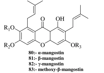
In 2005, Kenji Matsumoto et al. investigated the anti-proliferative effects of four structurally similar prenylated xanthones α-mangostin 80, β-mangostin 81, γ-mangostin 82, and methoxy-β-mangostin 83 (Figure 10), in human colon cancer DLD-1 cells. These xanthones differ in the number of hydroxyl and methoxy groups. Except for methoxy-β-mangostin, the other three xanthones strongly inhibited cell growth at 20 M and their antitumor efficacy was correlated with the number of hydroxyl groups. Hoechst 33342 nuclear staining and nucleosomal DNA-gel electrophoresis revealed that the anti-proliferative effects of α- and γ-mangostin, but not that of
β-mangostin, were associated with apoptosis. It was also shown that their anti-proliferative effects were associated with cell-cycle arrest by affecting the expression of cyclins, cdc2, and p27; G1 arrest was by α-mangostin and
β-mangostin, and S arrest by γ-mangostin. These findings provide a relevant basis for the development of xanthones as an agent for cancer prevention and combination therapy with anti-cancer drugs [35].
O R1O
R2O
O OH
OR3
80:- α-mangostin 81:- β-mangostin 82:- γ-mangostin 83:- methoxy-β-mangostin
Figure 10. Prenylated xanthones: α-mangostin, β-mangostin, γ-mangostin, and methoxy-β-mangostin
In 2006, E. M. Kithsiri Wijeratne et al. found new xanthones. Bioassay-guided fractionation of a cytotoxic EtOAc extract of the fungal strain, Chaetomium globosum, inhabiting the rhizosphere of the Christmas cactus, Opuntia leptocaulis, of the Sonoran desert afforded a new dihydroxanthenone, globosuxanthoneA 84, a new tetrahydroxanthenone, globosuxanthone B 85, two new xanthones, globosuxanthone C 86 and D 87 (Figure 11). Of the compounds encountered, 84 was found to exhibit strong cytotoxicity against a panel of seven human solid tumor cell lines, disrupt the cell cycle leading to the accumulation of cells in either G2/M or S phase, and induce classic
signs of apoptosis (Table 5) [36].
O O
OH O OH O
OH O R1
R2
OH
H
OH
H OCH3
OH OH
H
O OCH3 O OCH3
84 85
86: R1 = OH; R2 = OCH3 87: R1 = COOH; R2 = H
[image:10.595.230.401.202.322.2]Figure 11. Dihydroxanthenones, tetrahydroxanthenone and globosuxanthone
Table 5. Cytotoxicities (IC50, µM) of globosuxanthone (84) against a panel of seven human solid tumor cell lines
Cell line NCI-H460 MCF-7 SF-268 PC-3 PC-3M LNCaP DU-145
84 3.6 1.3 1.1 0.65 1.1 1.5 1.2
Dox 0.01 0.07 0.04 ND ND ND ND
Tax 0.01 0.01 0.02 ND ND ND ND
Results are expressed as IC50 values in µM; ND, not determined.
NCI-H460, non-small cell lung cancer; MCF-7, breast cancer; SF-268, CNS cancer (glioma); PC-3, hormone- (androgen) independent prostate adenocarcinoma; PC-3M, highly metastatic variant of PC-3; LNCaP, hormone-sensitive prostate cancer; DU-145, hormone-independent
prostate cancer.
Doxorubicin (Dox) and Taxol (Tax) were used as positive controls.
O R
O OH
O O
88: R = OCH3 89: R = H
O R O O O O O
90: R = H
O R O OH O X OH
[image:11.595.122.490.345.718.2]91: R = OCH3, X = Cl 92: R = OCH3, X = Br 93: R = H, X = Cl 94: R = H, X = Br
Figure 12. Some 3-(2,3-epoxypropoxy)xanthones and their epoxide ring opened halohydrin analogues Table 6. Cytotoxicities of compounds 88-90 against various human cancer cells
Cells (origin)/compound IC50a(µM)
88 89 91 92 93 94 90 Adriamycin
LnCap >100 93.1 ± 16.9 >100 >100 >100 64.4 ± 3.1 9.0 ± 0.2 4.6 ± 0.6 (prostate)
MCF-7 >100 68.4 ± 4.8 >100 97.8 ± 0.2 >100 53.5 ± 5.4 3.2 ± 0.8 4.5 ± 0.3 (breast)
HCT 116 >100 80.8 ± 3.1 31.4 ± 3.0 >100 >100 16.9 ± 0.5 10.2 ± 0.7 7.7 ± 0.2 (colon)
MDA-MB231 >100 >100 81.4 ± 3.5 99.8 ± 11.1 >100 76.3 ± 5.0 12.8 ± 0.9 17.6 ± 0.3 (breast)
Hela >100 68.7 ± 8.7 60.3 ± 3.3 >100 >100 98.1 ± 6.3 23.3 ± 1.7 3.3 ± 0.4 (cervix)
a Each value is the average of four experiments.
O OH R1 OR2 R3 O , O OH R1 OH O
95 : R1 = CH3
[GI50 (uM)] MCF-7 = 21.9 ± 0.4 NCI-H460 = 20.6 ± 0.9 SF-268 = 33.4 ± 0.2 UACC-62 = 20.0 ± 0.5
96 : R1 = H
[GI50 (uM)] MCF-7 = 50.8 ± 2.2 NCI-H460 = 37.9 ± 2.9 SF-268 = 61.4 ± 5.2 UACC-62 = 38.0 ± 1.6
97: R1 = CH3, R2 = , R3 = 98: R1 = CH3 , R2 = , R3 = H
[GI50 (µM)] [GI50 (µM)] MCF-7 = >130 MCF-7 = >160 NCI-H460 = >130 NCI-H460 = >160 SF-268 = >130 SF-268 = >160 UACC-62 = >130 UACC-62 = >160
99: R1 = H , R2 = , R3 = H 100: R1 = , R2 = , R3 = H
[GI50 (µM)] [GI50 (µM)] MCF-7 = >160 MCF-7 = 6.0 ± 0.7 NCI-H460 = >160 NCI-H460 = >130 SF-268 =>160 SF-268 =>130 UACC-62 = >160 UACC-62 = >130
101: R1 = , R2 = , R3 = H 102: R1 = H, R2 = , R3 =
[GI50 (µM)] [GI50 (µM)] MCF-7 = 9.1 ± 1.5 MCF-7 = 112.5 ± 10.1 NCI-H460 = >130 NCI-H460 = >130 SF-268 = >130 SF-268 = >130 UACC-62 = >130 UACC-62 = >130
O O
OH R1 O
103: R1 = CH3
[GI50 (µM)] MCF-7 = 18.4 ± 1.9
NCI-H460 = >160
SF-268 = >160
UACC-62 = ND
104: R1 = H
[GI50 (µM)] MCF-7 = >160
NCI-H460 = >160
SF-268 = >160
UACC-62 = ND
O O
O OH
105 [GI50 (µM)] MCF-7 = 88.6 ± 12.9 NCI-H460 = >160 SF-268 = >160 UACC-62 = ND
Figure 13. Xanthones with their anti-tumor activity against different cell lines
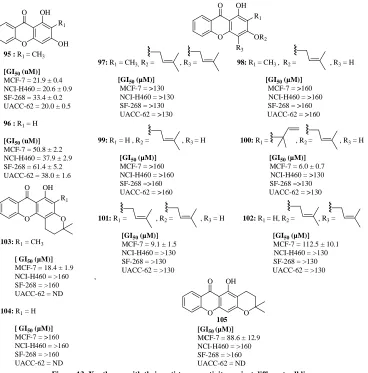
Prenylation was carried out using prenyl bromide in alkaline medium. Dihydropyranoxanthones 103–105 were obtained from compounds 98 and 99 by an oxidative ring closure. The structure of the compounds was established by IR, UV, MS, and NMR (1H, 13C, COSY, HSQC, and HMBC) techniques and for compounds 98, 100, and 105 the structure was confirmed by X-ray crystallographic analysis. The effect of the 11 xanthones on the in vitro growth of four human tumor cell lines, MCF-7 (breast adenocarcinoma), NCI-H460 (non small cell lung cancer), SF-268 (central nervous system cancer), and UACC-62 (melanoma) is also described (Figure 13) [38].
In 2008, Martine Varache-Lembe ge et al. synthesised several arylhydrazonomethyl derivatives of Formula II 106-123 (Table 7) in order to explore the antiproliferative effect associated with the xanthone framework, from various isomeric 1,3-dihydroxyxanthone carbaldehydes. Variation in the position of the aldehydic function led to three sets of compounds, bearing the hydrazonomethyl chain at positions 5, 6 or 7 on the xanthone nucleus, respectively. The anti-proliferative effect of the compounds was evaluated in vitro using the MTT colorimetric method against two human cancer cell lines (MCF-7, breast adenocarcinoma, and KB 3.1, squamous cell oral carcinoma) for two time periods (24 h and 72 h) (Table 7). Among the series, four compounds exhibited interesting growth inhibitory effects against both the cell lines, with IC50 values in the micromolar concentration range (Table
8). When compared with doxorubicin, the xanthone derivatives showed moderate cytotoxic effects. Surprisingly, unlike doxorubicin, these compounds displayed no significant time-dependent change in the concentration causing 50% inhibitory effect in proliferation. This unusual cytotoxicity profile led to the hypothesis that these molecules could be endowed with a mechanism of action distinct to that of doxorubicin [39].
O R1
R1 O
R2
R3
R4
R1 = OAc, OH
R2, R3, R4 =
X:
Y:
Z: Formula II
Table 7. Effects of studied xanthones of Formula II on the viability of MCF-7 and KB 3.1 cells
Compound R1 R2 R3 R4
MCF-7 KB 3.1 10 µM 1 µM 10 µM 1 µM
106 OAc X H H 100 100 100 100
107 OH X H H 72 100 82 100
108 OA H X H 100 100 100 100
109 OH H X H 100 100 61 100
110 OAc H H X 100 100 82 100
111 OH H H X 100 100 96 100
112 OAc Y H H 70 85 60 98
113 OH Y H H 41 58 90 95
114 OAc H Y H 100 100 87 100
115 OH H Y H 3 100 2 77
116 OAc H H Y 79 80 90 95
117 OH H H Y 12 86 3 96
118 OAc Z H H 14 64 14 100
119 OH Z H H 64 90 92 99
120 OAc H Z H 13 35 13 85
121 OH H Z H 70 73 80 93
122 OAc H H Z 48 80 66 95
123 OH H H Z 82 97 75 90
Table 8. Effect of time of incubation on anti-proliferative activity against KB 3.1 and MCF-7 cells (IC50, µM) KB 3.1 MCF-7
24 h 72 h R(24/72) 24 h 72 h R(24/72)
119 11.3 ± 1.1 4.7 ± 0.9 2.4 9.3 ± 1.3 7.0 ± 1.0 1.3 120 1.6 ± 0.3 1.8 ± 0.4 0.9 1.9 ± 0.4 2.4 ± 0.2 0.8 122 6.8 ± 0.6 6.5 ± 0.3 1.05 5.1 ± 0.6 5.6 ± 0.5 0.9 Doxorubicin 0.010 ± 0.003 0.0004 ± 0.0001 25.0 0.018 ± 0.004 0.0007 ± 0.0001 25.7
Cell viability after a further 96-h period in fresh culture medium was estimated as the compound concentration required for 50% growth inhibition. For each drug,a R(24/72) value was calculated and represents the ratio of the IC50s (µM) obtained after 24-h and 72-h treatment,
respectively. Data represent mean values (±S.D.)for three independent experiments.
In 2008, Aleksandra Isakovic et al. studied that xanthones gentiakochianin and gentiacaulein derivatives 124-131 (Figure 14) as the active principles responsible for the in vitro antiglioma action of ether and methanolic extracts of the plant Gentiana kochiana. Gentiakochianin and gentiacaulein induced cell cycle arrest in G2/M and G0/G1 phases,
respectively, in both C6 rat glioma and U251 human glioma cell lines. The more efficient anti-proliferative action of gentiakochianin was associated with its ability to induce microtubule stabilization in a cell-free assay. Both the xanthones reduced mitochondrial membrane potential and increased the production of reactive oxygen species in glioma cells, but only the effects of gentiakochianin were pronounced enough to cause caspase activation and subsequent apoptotic cell death. The assessment of structure–activity relationship in a series of structurally related xanthones from G. kochiana and Gentianella austriaca revealed dihydroxylation at positions 7, 8 of the xanthonic nucleus as the key structural feature responsible for the ability of gentiakochianin to induce microtubule-associated G2/M cell block and apoptotic cell death in glioma cells [40].
O
OH H3CO
O OH
OH
O
OH H3CO
O OCH3 OH
O
OH H3CO
O OCH3
OCH3
O
OPrim
H3CO
O OH OH
O
OH H3CO
O OH
O
OPrim H3CO
O OCH3 OH O
OGlc H3CO
O OCH3 OH
O
OH HO
O OH
OH OH
124 Gentiakochianin
125 Gentiacaulein
126 Decussatin
127
Isogentiakochianoside
128
Gentiacaulein-1-O-glucoside Gentiabavaroside129
130 Bellidifolin
131 Demethylbellidifolin
Figure 14. Xanthones gentiakochianin and gentiacaulein derivatives
In 2008, Tomohiro Itoh et al. indicated the anticancer activity induced by xanthones such as α-mangostin 132,
β-mangostin 133 and γ-mangostin 134 (Figure 15) which were major constituents of the pericarp of mangostin fruits. They examined the effect of xanthones on cell degranulation in rat basophilic leukemia RBL-2H3 cells. Antigen (Ag)-mediated stimulation of high affinity IgE receptor (FcɛRI) activates intracellular signal transductions resulting in the release of biologically active mediators such as histamine. The release of histamine and other inflammatory mediators from mast cell or basophils is the primary event in several allergic responses. These xanthones suppressed the release of histamine from IgE-sensitized RBL-2H3 cells. In order to reveal the inhibitory mechanism of degranulation by xanthones, they examined the activation of intracellular signaling molecules such as Lyn, Syk, and PLCγs. All the xanthones tested significantly suppressed the signaling involving Syk and PLCγs. In Ag-mediated activation of FcɛRI on mast cells, three major subfamilies of mitogen-activated protein kinases were activated. The xanthones decreased the level of phospho-ERKs. Furthermore, the levels of phospho-ERKs were observed to be regulated by Syk/LAT/Ras/ERK pathway rather than PKC/Raf/ERK pathway, suggesting that the inhibitory mechanism of xanthones was mainly due to suppression of the Syk/ PLCγs /PKC pathway. Although intracellular free Ca2+concentration ([Ca2+]I ) was elevated by FcɛRI activation, it was found that α- or γ-mangostin treatment
O O OH R1O
R2O OR3
132: a-Mangostin: R1=CH3, R2=R3 = H 133: b-Mangostin: R1 = R3 = CH3, R2 = H 134: g-Mangostin: R1 = R2 = R3 = H
Figure 15. Xanthones: α-mangostin, β-mangostin and γ-mangostin
In 2009, Emilia Sousa et al. synthesised bis-intercalators, a bisxanthone and a minor product, 1-(6-bromohexyloxy)-xanthone. The synthesized compounds bis-alkoxyxanthone 135, bromoalkoxyxanthone 136, dihydroxyxanthone 137, hydroxybromoalkoxyxanthone 138 and bis-bromoalkoxyxanthone 139 (Figure 16) showed no capacity to inhibit the growth of human tumor cell lines was observed for the bisxanthone, the bromoalkoxyxanthone revealed this biological activity. Inlight of these results bromoalkylation of 3,4-dihydroxyxanthone furnished two bromohexyloxyxanthones that were investigated for their effect on the in vitro growth of human tumor cell lines MCF-7 (ER+, breast), MDA-MB-231 (ER_, breast), NCI-H460 (non-small lung), and SF-268 (central nervous system) (Table 9). The X-ray structure of 1-(6-bromohexyloxy)-xanthone revealed that the xanthone skeleton remains essentially planar forming a dihedral angle of 61.3(2)0with the 6-bromohexyl side chain. These results revealed bromoalkoxyxanthones as interesting scaffolds to look for potential anticancer drugs [42].
Figure 16. Bis intercalators xanthone derivatives
Table 9. Effect of compounds on the growth of human tumor cell lines Compounds GI50 (µM)
SF-268 NCI-H460 MCF-7 MDA-MB-231 135 >100 >100 >100 >100 136 30.2 ± 3.6 30.7 ± 3.2 22.7 ± 1.3 >100 137 22.6 11.4 52.4 ± 6.3 22.8 ± 4.8 138 >100 >100 >100 >100 139 >100 >100 20.5 ± 1.9 56.8 ± 11.2
Results are given in concentrations that were able to cause 50% of cell growth inhibition (GI50) after a continuous exposure of 48 h and represent means ±SEM of3–5 independent experiments performed in duplicate and carried out independently.
Doxorubicin was used as positive control, GI50: MCF-7 = 42.8 ±8.2 nM; MDA-MB-231 = 10.86±1.28 nM; SF-268
= 94.0 ± 7.0 nM; NCI-H460 = 94.0 ± 8.7 nM. ND = not determinate.
suggested that there were intercalative interactions of the complexes with DNA. The binding affinity of complex 141 was higher than that of 140. In addition, the cytotoxic effects of both complexes were evaluated with lung adenocarcinoma (GLC-82), esophagus squamous cancer (ECA109) and human gastric cancer (SGC7901) cells using MTT assay (Table 10). Both were potent exhibiting significant cytotoxicity in vitro [43].
O O
N
O O M O O
O O N
H2O OH2
Figure 17. Metal complex of xanthone Table 10. IC50 values for the cell growth inhibition Compound IC50 value (µM)
GLC-82 ECA109 SGC7901
140 22.08 >50 19.98 141 16.20 21.04 15.40
In 2011, Jen-Hao Cheng et al. prepared xanthones to develop novel antioxidant as anticancer agents. In vitro screening, the synthetic xanthones revealed significant inhibitory effects on xanthine oxidase and ABTS radical-cation scavenging activity. The selective compounds 142 and 143 (Figure 18) induced an accumulation of NTUB1 cells in the G1 phase arrest and cellular apoptosis by the increase of ROS level. The combination of
[image:15.595.222.390.128.257.2]cisplatin and 141 significantly enhanced the cell death in NTUB1 cells. Compounds 142 and 143 did not show cytotoxic activity in selected concentrations against SV-HUC1 cells. The present results suggested that antioxidants 142 and 143 may be used as anticancer agent for enhancing the therapeutic efficacy of anticancer agents and to reduce their side effect [44].
Figure 18. Novel xanthones antioxidant as anticancer agents
In 2011, Kyu-Yeon Jun et al. synthesized Epoxide ring-opened xanthone derivatives144, 145, 146, 147 (Figure 19) and tested for their topoisomerase inhibitory activity and cytotoxicity. These compounds showed no activity against human topo I at the same concentration, where camptothecin inhibited 69% human topo I mediated relaxation of supercoiled pBR322 (Table 11). Most of the compounds showed topo IIα specific inhibitory activity. To clarify the mechanism of action of these compounds, the most potent compound (compound 145) of the synthesized analogues was further studied by testing its ATPase inhibitory activity and through molecular docking experiments. The results showed that the topo IIα inhibitory activity of compound 145 was inversely proportional to ATP concentration. In the ATPase inhibitory test, ATP hydrolysis was reduced less efficiently by compound 145 (28.5 ± 4.6%) than novobiocin (60.4 ± 8.1%). Molecular docking study revealed compound 145 to have a stable binding pattern to the ATP-binding domain of human topo II [45].
activities (IC50) are presented as the micromolar concentrations of the compounds, as listed in (Table 12).
Figure 19. Epoxide ring-opened xanthone derivatives Table 11. Topo I and II inhibitory activities of compounds
Compounds Topo I (% inhibition) Topo II (% inhibition)
Concentrations 20 µM 10 µM 20 µM 100 µM Camptothecin 69.15
Etoposide 26.04 39.11 77.07 144 0.00 26.32 49.09 63.75 145 0.00 41.23 70.01 100.00 146 0.00 23.43 43.68 89.25 147 0.00 15.80 22.24 45.33
Table 12. Cytotoxicities of compounds against the five cancer cell lines
Compd/cells IC50a(µM)
HeLa HCT116 DU145 MDA-MB231 HL60 Adriamycin 1.32 ± 0.15 6.81 ± 1.43 2.47 ± 1.03 0.84 ± 0.08 0.32 ± 0.07 Etoposide 2.36 ± 0.47 9.50 ± 1.35 2.19 ± 0.79 1.23 ± 0.38 1.28 ± 0.07 Camptothecin 1.08 ± 0.07 3.97 ± 0.76 0.31 ± 0.03 0.97 ± 0.07 0.065 ± 0.004 144 9.15 ± 2.69 23.94 ± 4.58 3.47 ± 1.80 5.13 ± 1.65 19.86 ± 2.48 145 11.62 ± 0.11 23.71 ± 1.77 0.76 ± 0.39 4.11 ± 0.57 4.15 ± 2.09 146 15.38 ± 1.04 46.70 ± 5.98 15.55 ± 2.15 10.41 ± 2.59 16.58 ± 0.64 147 4.64 ± 2.03 14.80 ± 0.26 0.004 ± 0.001 1.32 ± 0.07 3.41 ± 1.30
a Each data point represents the mean ± SD of three different experiments performed in triplicate. The cell lines used were HeLa, human cervix
tumor cell line; HCT116,human colorectal carcinoma cell line; DU145, human prostate tumor cell line; MDA-MB231, human breast adenocarcinoma cell line; HL60, human myelogenous leukemia cell line.
In 2012, Chiao-Ting Yen et al. synthesized a series of prenyl- and pyrano-xanthones 148, 149, 150 (Figure 20) derived from 1,3,6-trihydroxy-9H-xanthen-9-one, a basic backbone of gambogic acid (GA) and evaluated for in vitro cytotoxic effects against four human cancer cell lines (KB, KBvin, A549, and DU-145) and anti-inflammatory activity toward superoxide anion generation and elastase release by human neutrophils in response to fMLP/CB. Among them, prenylxanthones were generally less active than pyranoxanthones in both anticancer and anti-inflammatory assays. Two angular 3,3-dimethypyranoxanthones (148 and 150) showed the greatest and selective activity against the KBv in multidrug resistant (MDR) cell line with IC50 values of 0.9 and 0.8 lg/mL,
respectively. An angular 3-methyl-3-prenylpyranoxanthone (149) selectively inhibited elastase release with 200 times more potency than phenylmethylsulfonyl fluoride (PMSF), the positive control. The inhibitory effect of the test drugs on cell viability was measured by the MTT colorimetric method [46].
O HO
R OH O
148 R= CH3
149 R= CH2CH2CH=C(CH3)2
O HO
OH O
HO OH 150
Figure 20. Prenyl- and pyrano-xanthones
(Polygalaceae), collected at Democratic Republic of Congo, killed PANC-1 human pancreatic cancer cells preferentially in nutrient-deprived medium (NDM). Phytochemical investigation on the CHCl3 extract led to the
isolation of compounds including five new polymethoxylated xanthones [1,6,8-trihydroxy-2,3,4,5-tetramethoxyxanthone (151), 1,6-dihydroxy-2,3,4,5,8-pentamethoxyxanthone (152), 8-hydroxy-1,4,5,6-tetramethoxy-2,3-methylenedioxyxanthone (153), 4,6,8-trihydroxy-1,2,3,5-tetra methoxyxanthone (154), 4,8-dihydroxy-1,2,3,5,6 pentamethoxyxanthone (155)] and a new benzyl benzoate [benzyl 3-hydroxy-2-methoxybenzoate (156)] (Figure 21). Among them, 1,6,8-trihydroxy-2,3,4,5-tetramethoxyxanthone (151) and 1,6-dihydroxy-2,3,4,5,8-pentamethoxyxanthone (152) displayed the potent preferential cytotoxicity with PC50 of 22.8 and 17.4 µM, respectively (Table 13). They triggered apoptosis-like PANC-1 cell death in NDM with a
glucose-sensitive mode [47].
Figure 21.Polymethoxylated xanthones
Table 13. Preferential cytotoxicity of compounds on human pancreatic cancer PANC-1cells in NDM
Compound PC50 (µM)
151 22.7 152 17.4 Others >100 Arctigenina 0.5 Taxolb >100
a,b Positive and negative controls, respectively.
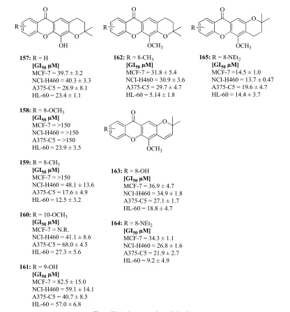
In 2013, Carlos M. G. Azevedo et al. optimized antitumor xanthone derivatives with their GI50 values 157-165
(Figure 22) following a multidimensional approach that involved the synthesis of analogues, the study of their lipophilicity and solubility, and the evaluation of their growth inhibitory activity on four human tumor cell lines. A new synthetic route for the hit xanthone derivative was also developed and applied for the synthesis of its analogues. Among the used cell lines, the HL-60 showed to be in general more sensitive to the compounds tested, with the most potent compound 157 having a GI50 of 5.1 µM, lower than the hit compound. Lipophilicity was evaluated by the
partition coefficient (Kp) of a solute between buffer and two membrane models, namely liposomes and micelles. The
Partition coefficients in liposomes-buffer and micelle-buffer for synthesized compounds are also shown (Table 14) [48].
Table 14. Partition coefficients in liposomes-buffer and micelle-buffer for compounds synthesized
Compounds LogKp liposomes LogKp micelles
157 158 159 160 161 162 163 165
3.35 ± 0.02 3.25 ± 0.08 3.86 ± 0.08 3.53 ± 0.04 3.32 ± 0.10 4.02 ± 0.03
— 4.62 ± 0.02
O O O
OH R
157:R = H [GI50µM]
MCF-7 = 39.7 ± 3.2 NCI-H460 = 40.3 ± 3.3 A375-C5 = 28.9 ± 8.1 HL-60 = 23.4 ± 1.1
158:R = 8-OCH3 [GI50µM] MCF-7 = >150 NCI-H460 = >150 A375-C5 = >150 HL-60 = 23.9 ± 3.5
159:R = 8-CH3 [GI50µM] MCF-7 = >150
NCI-H460 = 48.1 ± 13.6 A375-C5 = 17.6 ± 4.9 HL-60 = 12.5 ± 3.2
160:R = 10-OCH3
[GI50µM] MCF-7 = N.R. NCI-H460 = 41.1 ± 8.6 A375-C5 = 68.0 ± 4.5 HL-60 = 27.3 ± 5.6
161:R = 9-OH [GI50µM]
MCF-7 = 82.5 ± 15.0 NCI-H460 = 59.1 ± 14.1 A375-C5 = 40.7 ± 8.5 HL-60 = 57.0 ± 6.8
O O
O
R
OCH3 162:R = 8-CH3
[GI50µM]
MCF-7 = 31.8 ± 5.4 NCI-H460 = 30.9 ± 3.6 A375-C5 = 29.7 ± 4.7 HL-60 = 5.14 ± 1.8
O
O R
O
OCH3
163:R = 8-OH [GI50µM] MCF-7 = 36.9 ± 4.7 NCI-H460 = 34.9 ± 1.8 A375-C5 = 27.1 ± 1.7 HL-60 = 18.8 ± 4.7
164:R = 8-NEt2 [GI50µM] MCF-7 = 34.3 ± 1.1 NCI-H460 = 26.8 ± 1.6 A375-C5 = 21.9 ± 2.7 HL-60 = 9.2 ± 4.9
O
O R
O
OCH3
165:R = 8-NEt2
[GI50µM] MCF-7 =14.5 ± 1.0 NCI-H460 = 13.7 ± 0.47 A375-C5 = 19.6 ± 4.7 HL-60 = 14.4 ± 3.7
Figure 22. Antitumor xanthone derivatives
[image:18.595.99.515.65.513.2]In 2013, Carlos M.G. Azevedo et al. synthesized new pyranoxanthones 165-176 (Figure 23). The benzopyran and dihydrobenzopyran moieties can be considered as “privileged motifs” in drug discovery being good platforms for the search of new bioactive compounds. Accordingly, with the aim of rationalizing the importance of the fused ring orientation and oxygenation pattern in pyranoxanthones, this study describes the synthesis of new pyranoxanthones and evaluation of their cell growth inhibitory activity in four human tumor cell lines as well as their lipophilicity (Table 15 and Table 16) [49].
Table 15. Cell growth inhibitory activity in four human tumor cell lines
Compound GI50 (µM)
MCF-7 NCI-H460 A375-C5 HL-60
166 168 169 170 171 172 173 174 175 176
Figure 23. Pyranoxanthones
Table 16. Partition coefficients in liposomes-buffer and micelle-buffer for a chemical library of Pyranoxanthones
Compound
Log Kp liposomes Log Kp micelles
166 167 168 169 170 171 172 173 174 175 176
3.42 ± 0.02 3.50 ± 0.12 4.17 ± 0.06 4.15 ± 0.08 3.92 ± 0.04 3.88 ± 0.02 - 4.32 ± 0.04 4.14 ± 0.08 4.10 ± 0.04 3.60 ± 0.08 3.59 ± 0.06 3.06 ± 0.16 3.29 ± 0.06 3.32 ± 0.12 3.33 ± 0.02 3.35 ± 0.02 3.28 ± 0.02 3.09 ± 0.18 3.58 ± 0.08 3.54 ± 0.01 3.76 ± 0.03
O O O
H2N O NH2
O O O
H2N O NH2
178 (X2S, n = 6) 6 6
177 (X2S)
O O O
H2N
O
NH2
O O O
N O N
179 (X2S -1-Me) 180 (X2S -N,N-diMe)
S O O
H2N
O
NH2
S O O
N O N
182 (X2S -N,N-diMe) 181 (X2SS)
In 2013, Asako Murata et al. demonstrated that an aminoalkoxy-substituted thioxanthone derivatives 177-182 (Figure 24) interferes Dicer-mediated processing of pre-miRNA. Various biological processes have been found to be regulated by miRNA-mediated gene silencing. A small molecule that modulates the miRNA pathway will provide the biological tool for elucidating mechanisms of miRNA-mediated gene regulation, and can be the drug lead for miRNA related diseases [50].
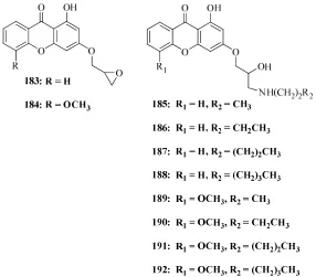
In 2013, So-Eun Park et al.prepared several alkylamine (n= 3-6) 183-192 (Figure 25) and evaluated for the pharmacological activity and mode of action. In the topoisomerase IIα (topo IIα) inhibition test, compound 186 showed strongest inhibitory activity among the compounds at 10 µM. Inhibitory activities (topo I and topo II) of the compounds are in the order of 186 (n = 4) >177 (n = 3) >>187 (n = 5) 188 (n = 6); 190 (n = 4) >>189 (n = 3)
191 (n = 5) 192 (n = 6) where n is the number of carbon in the aliphatic side chain in ring C (Table 17) and
compounds 189-192 have additional methoxy group in ring A compared to compounds 185, 186-188. Compound
186 showed efficient cytotoxicities against T47D (IC50: 0.93 ± 0.04 µM) and HCT15 (IC50: 0.78 ± 0.01 µM) cells,
which are higher than etoposide (Table 18). Compound 186 was also an ATP-competitive human topo IIα catalytic inhibitor with partially blocking human topo IIα-catalyzed ATP hydrolysis and intercalating into DNA. Compound 186 induced much less DNA damage than etoposide in HCT15 human colorectal carcinoma cells. Overall, compound 186 can be a potential anticancer agent acting as topo IIα catalytic inhibitor with low DNA damage [51].
[image:20.595.163.449.284.535.2]Figure 25. Alkylamine xanthone derivatives Table 17. Topo I and IIa inhibitory activities of compounds
Compounds Topo I(% inhibition) Topo II (% inhibition) Concentration 20 µM 10 µM 20 µM 100 µM IC50 (µM)
Camptothecin 26.1
Table 18. Cytotoxicities of compounds 1, 4-10 against cancer cell lines
Compd/cells IC50a(µM)
T47D HCT15 DU145 HEK293 Adriamycin 0.41 ± 0.09 1.38 ± 0.39 2.68 ± 0.51 3.07 ± 1.67 Etoposide 3.42 ± 2.26 5.12 ± 3.64 9.29 ± 0.71 0.72 ± 0.11 Camptothecin 0.3 ± 0.09 0.42 ± 0.10 0.39 ± 0.34 0.78 ± 0.87 185 6.49 ± 0.09 5.49 ± 0.13 1.98 ± 0.12 4.28 ± 0.03 186 0.93 ± 0.04 0.78 ± 0.01 1.98 ± 0.01 3.44 ± 0.08 187 1.62 ± 0.02 0.71 ± 0.004 1.13 ± 0.11 2.65 ± 0.04 188 1.28 ± 0.03 1.53 ± 0.11 1.15 ± 0.02 1.45 ± 0.01 189 3.33 ± 0.14 5.39 ± 0.08 1.48 ± 0.08 3.62 ± 0.66 190 2.97 ± 0.04 9.11 ± 0.9 3.31 ± 0.06 1.74 ± 0.03 191 9.80 ± 0.07 1.00 ± 0.004 0.91 ± 0.02 7.22 ± 0.97 192 6.17 ± 0.02 6.32 ± 0.03 1.79 ± 0.11 4.53 ± 0.87
a Each data point represents the mean ± SD of three different experiments performed in triplicate.
The cell lines used were T47D, human ductal breast epithelial tumor cell line; HCT15, human colorectal carcinoma cell line; DU145, human prostate tumor cell line; HEK293, human embryonic kidney 293 cell line.
In 2013, Ming Dai et al.synthesized a series of novel derivatives of phenyl substituted tetramethoxy xanthone based on Formula III 193-196 and evaluated for their in vitro cytotoxicity against human hepatocellular carcinoma (HCC) and non-tumor hepatic cells (Table 19). Among these derivatives, compound 193 was more potent than positive control 5-fluorouracil (5-Fu) on QGY-7703 and SMMC-7721 cells with IC50 values of 6.27 µM, 7.50 µM and 15.56
µM, 14.55 µM, respectively (Table 20). Furthermore, compounds 193, 194, 195, and 196 exhibited much better selectivity toward the normal hepatic cell line QSG-7701 than 5-Fu. Additionally, compound 193 significantly induced cell apoptosis in QGY-7703 cells [52].
O O
O
O O
O
[image:21.595.243.368.354.461.2]R Formula III
Table 19. Inhibition rates of compounds of Formula III against the QGY-7703 cell line at 10 M
Compound R Yield Inhibitory ratio (%) 193 H 91% 66.45 194 4′-CH3(CH)CH3 88% 52.67
195 4′-C(CH3)3 88% 56.31
196 3′-CH3 82% 51.36
Table 20. Effects of the target compounds on proliferation of human HCC cells and normal hepatic cells
Compd
IC50 (µM)
QGY-7703 HepG-2 SMMC-7721 QSG-7701 193 6.27 19.18 7.50 >100 194 26.14 32.46 20.11 >100 195 33.13 39.21 18.15 >100 196 13.26 5.00 14.82 >100 5-Fu 15.66 12.48 14.55 0.60
In 2014, Zheng-Min Yang et al. synthesized a series of novel 1-hydroxyl-3-aminoalkoxy xanthone derivatives and evaluated for in-vitro anticancer activity against four selected human cancer cell lines (nasopharyngealneoplasm CNE, liver cancer BEL-7402, gastric cancer MGC-803, lung adenocarcinoma A549). Compound 197 (Figure 26) shows excellent broad spectrum anticancer activity with IC50 values ranging from 3.57 to 20.07 µM. The in vitro
Figure 26. 1-hydroxyl-3-aminoalkoxy xanthone derivative and anticancer activity in-vitro
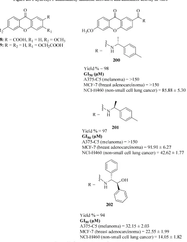
[image:22.595.127.497.267.747.2]In 2014, Carla Fernandes et al.developed a highly efficient and practical methodology for synthesis of new chiral derivatives of xanthones (CDXs) 198-202 (Figure 27) in enantiomerically pure form. According to this approach, CDXs were synthesized by coupling a carboxyxanthone 198 and a carboxymethoxyxanthone 199 with both enantiomers of commercially available chiral building blocks, namely six amino alcohols, one amine and one amino ester. The synthesized CDXs were evaluated for their effect on the in vitro growth of three human tumor cell lines, namely A375-C5 (melanoma), MCF-7 (breast adenocarcinoma), and NCI-H460 (non-small cell lung cancer). The most active compound was CDX 5 being active in all human tumor cell lines. The growth inhibitory effects, in some cases, demonstrated to be depending on the stereo chemistry of the CDXs. An interesting example was observed with the enantiomers 200 and 201, which demonstrated high enantioselectivity for MCF-7 and NCI-H460 cell lines [54].
In 2014, Xiang Fei et al. synthesized novel xanthone derivatives 204-208 based on α-mangostin 203 and evaluated as anti-cancer agents by cytotoxicity activity screening using 5 human cancer cell lines (Figure 28). A xanthone-derived natural product, α-mangostin is isolated from various parts of the mangostin, Garcinia mangostana L. (Clusiaceae), a well-known tropical fruit. The structure–activity relationship studies revealed that phenol groups on C3 and C6 are critical to anti-proliferative activity and C4 modification is capable to improve both
anti-cancer activity and drug-like properties [55].
O R1O
OH O
OR2
OH 1
3
7
6
a-mangostin R1 = H, R2 = CH3 b-mangostin R1 = CH3, R2 = CH3 g-mangostin R1 = H, R2 = H
203
O HO
OH O
OH O
204 IC50 (µM) NCI-H460 = 3.23
SW-620 = 2.97
AsPC-1 = 4.02
MDA-MB-231 = 3.04
B16F10 = 3.23
O HO
OH
H O
O
OH
205 IC50 (µM) NCI-H460 = 4.17
SW-620 = 6.42
AsPC-1 = 4.64
MDA-MB-231 = 4.91
B16F10 = 6.54 O
HO OH
NH2
O
O
OH
206 IC50 (µM) NCI-H460 = 15.1
SW-620 = 6.24
AsPC-1 = 12.96
MDA-MB-231 = 17.57
B16F10 = 12.34
O HO
Cl
OH O
OH O
207 IC50 (µM) NCI-H460 = 9.96
SW-620 = 9.59
AsPC-1 = 19.26
MDA-MB-231 = 10.11
B16F10 = 12.44
O HO
OH O
OH OH
208 IC50 (µM) NCI-H460 = 4.48
SW-620 = 5.69
AsPC-1 = 5.92
MDA-MB-231 = 5.87
Figure28. Xanthone derivatives based on α-mangostin
In 2014, Tsung-Chih Chen et al. synthesized a series of tetracyclic heterocyclic azathioxanthones 209-213 and evaluated for cell proliferations, topoisomerase inhibitions, and NCI-60 cell panel assay, respectively. The IC50
value of each target compound was measured by MTT assay against PC-3 and DU-145 cells, respectively. The values of IC50 for five compounds 209, 210, 211, 212, and 213 were less than 10 µM against PC-3 and/or DU-145
Camptothecin, Etoposide and Doxorubicin were used as standard derivatives for MTT assay.
S N
Cl
O O
S N
Cl
N O
N H
209 210
IC50 (µM)
DU-145 = 10.84 ± 6.55
PC- 3 = 3.89 ± 0.54
IC50 (µM)
DU- 145 = 5.01 ± 1.68
PC- 3 = 2.84 ± 0.64
S N
Cl
N O
N
S N
Cl
N O
OH
211 212
IC50 (µM)
DU- 145 = 12.94 ± 0.26 PC- 3 = 7.18 ± 2.45
IC50 (µM)
DU- 145 = 11.12 ± 4.18 PC- 3 = 9.55 ± 2.42
S N
Cl
N O
N
NH 213
IC50 (µM)
DU- 145 = 9.02 ± 1.20 PC- 3 = 6.36 ± 0.17
Figure 29. Tetracyclic heterocyclic azathioxanthones
Compounds 209, 210, 211, 212 and 213 were selected for primary topo I and II activity assays at 25 and/or 50 µM, respectively. Compounds 210, 212, and 213 showed various inhibitory effect against topo I at 50 µM. Compounds 209 and 210 exerted slight inhibitory effect against topo II at 50 µM. Compound 210 was chosen for further testing against topo I and II in a concentration-dependent manner doses at 1, 5, 10, 25, 50 µM. Compound 7 not only exhibited more potent inhibitory activity than compound 209, 212, 213 and CPT, but also completely blocked topos-mediated DNA relaxation at 25 µM. Compound 210-treated PC-3 cells were examined to see the effect of compound 210 on apoptosis and related protein, PARP, and procaspase-3. Compound 210 induced apoptosis in PC-3 cells followed by increasing the DNA fragmentation via cleavage of PARP and decreasing procaspase-3 [56]. In 2014, Somayeh Motavallizadeh et al. prepared several novel N-(9-oxo-9H-xanthen-4-yl)benzene sulfonamide derivatives as anti-proliferative agents. The synthesized compounds was investigated for in vitro anti-proliferative activity against a panel of tumor cell lines including breast cancer cell lines (MDA-MB-231, T-47D) and neuroblastoma cell line (SK-N-MC) using MTT colorimetric assay.4-methoxy-N-(9-oxo-9Hxanthen- 4-yl)benzenesulfonamide 214 (Figure 30) showed the highest antiproliferative activity against MDA-MB- 231, T-47D, and SK-N-MC cells. Etoposide was used as a positive standard drug.
Substituted benzenesulfonamide derivatives was also evaluated against human leukemia (CCRF-CEM), breast adenocarcinoma (MDA-MB-468), and colorectal carcinoma (HCT-116) cell lines at the concentration of 50 µM. pentafluoro derivatives 215 and 216 (Figure 30) exhibited higher antiproliferative activity than doxorubicin against human leukemia cell line (CCRF-CEM) and breast adenocarcinoma (MDA-MB-468) cells [57].
O H
O
HN
S O
O
H3CO 214
IC50(µM)
SK-N-MC = 25.2 ± 26.5
MDA-MB-231 = 54.4 ± 21
T-47D = 19.7 ± 0.18
O H
O
HN S O
O
F F
F F F
O Cl
O
HN S O
O
F F
F F F
[image:24.595.145.468.92.320.2]215 216
2-3. Anti-convulsant xanthones
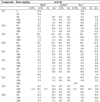
In 2008, Henryk Marona et al. prepared a series of appropriate alkanolamine and amide derivatives related to Formula IV 217-240 of xanthone (Table 21) and evaluated for anticonvulsant activity using maximal electroshock (MES) and subcutaneous pentylenetetrazole (scMet) induced seizures, and for neurotoxicity (TOX) using the roto rod test on mice and rats. A modification of the chemical structure of the most active compound, 218 and 219 as well as (R,S)-2-N-(6-chloro-2-xanthonemethyl)- amino-1-propanol 237 by the introduction of a tertiary amine instead of secondary one, and the replacement of the hydroxy group in the relevant compounds mentioned above by a chloride did not increase or eliminate anti-MES activity (compounds 228 and 230 are active at 100 mg/kg; compound 229 is inactive), and greatly increased the neurotoxicity (neurotoxic at 100 mg/kg) (Table 22).The Anti-MES activity determined in rats treated with 30 mg/kg (po) of the compounds under investigation is summarized in (Table 23).
O R
O
XZ
Formula IV
Table 21. Alkanolamine and amide derivatives Formula IV of xanthone Compounds R X Z
217 Cl CH2
HN OH
218 Cl CH2 NH
OH
219 Cl CH2 NH OH
220 Cl CH2 NH
OH
221 Cl CH2 NH
OH
222 Cl CH2 NH
OH
223 Cl CH2 N
OH
x HCl
224 Cl CH2 N
OH
225 Cl CH2 NH
OH
x HCl
226 Cl CH2 NH
OH
227 Cl CH2 N
H OH
228 Cl CH2 N
OH
229 Cl CH2 NH Cl
x HCl
230 Cl CH2 NH Cl
231 Cl CH2
N Cl
x HCl
232 Cl CH
2
N Cl
x HCl
233 Cl CH
2 NH
OH
234 Cl CH
2 NH
OH
235 H CO
NH O O
236 H CO NH
NH2
O
237 H CO HO HN
D,L-trans
238 H CO NH
OH
239 H CO NH
OH
240 Cl CO NH OH
[image:26.595.146.466.438.763.2]a(R) isomer. b(S) isomer
Table 22. Anticonvulsant screening project (ASP), phase I: test results in mice after intraperitoneal injection Compound Dose (mg/kg) Activity
MESa ScMetb Toxc
0.25h 0.5h 1h 4h 0.5h 4h 0.25h 0.5h 1h 4h 217 3 0/4 0/4
10 0/4 0/4
30 1/1 0/1 0/1 0/1 2/4 0/2 100 3/3 3/3 0/1 0/1 6/8 1/4 300 1/1 1/1 0/1 0/1 4/4 2/2 218 30 0/1 0/1 0/1 0/1 0/4 0/2 100 2/3 0/3 0/1 0/1 1/8 0/4 300 1/1 1/1 0/1 0/1 2/4 0/2 219 30 0/1 0/1 0/1 0/1 0/4 0/2
100 2/3 2/3 2/5 0/1 0/8 0/4 300 1/1 1/1 0/1 0/1 2/4 0/2 220 30 0/1 0/1 0/1 0/1 0/4 0/2
100 2/3 0/3 0/1 0/1 5/8 1/4 300 1/1 1/1 0/1 0/1 4/4 2/2 221 30 0/1 0/1 0/1 0/1 0/4 0/2
100 3/3 0/3 0/1 0/1 2/8 1/4 300 1/1 1/1 0/1 0/1 4/4 2/2 222 30 0/1 0/1 0/1 0/1 1/4 0/2 100 1/3 0/3 0/1 0/1 1/8 0/4 300 1/1 1/1 0/1 0/1 3/4 0/2 223 30 0/1 0/1 0/1 0/1 0/4 0/2 100 1/3 0/3 0/1 0/1 6/8 0/4 300 0/4 4/4 1/1 224 30 1/2 1/3 0/1 0/1 0/4 0/4 100 1/1 1/1 0/1 0/1 1/8 2/2 300 4/4