Lee, Hyunah and Patschull, Anathe and Bagneris, Claire and Ryan, H. and
Sanderson, C.M. and Ebrahimi, B. and Nobeli, Irene and Barrett, Tracey
E. (2017) KSHV SOX mediated host shutoff: the molecular mechanism
underlying mRNA transcript processing. Nucleic Acids Research 45 (8), pp.
4756-4767. ISSN 0305-1048.
Downloaded from:
Usage Guidelines:
Please refer to usage guidelines at
or alternatively
KSHV SOX mediated host shutoff: the molecular
mechanism underlying mRNA transcript processing
Hyunah Lee
1,2,†, Anathe O.M. Patschull
1,†, Claire Bagn ´eris
1, Hannah Ryan
3, Christopher
M. Sanderson
4, Bahram Ebrahimi
5, Irene Nobeli
1and Tracey E. Barrett
1,*1Institute for Structural and Molecular Biology, Department of Biological Sciences, Birkbeck College, Malet Street,
London WC1E 7HX, UK,2Department of Structural and Molecular Biology, University College London, London, WC1E 6BT, UK,3Liverpool School of Tropical Medicine, Pembroke Place, Liverpool, L3 5QA, UK,4Department of Cellular and Molecular Physiology, Institute of Translational Medicine, University of Liverpool, Crown Street,
Liverpool, L69 3BX, UK and5Department of Functional and Comparative Genomics, Institute of Integrative Biology, University of Liverpool, Crown Street, Liverpool L69 7ZB, UK
Received January 31, 2016; Revised December 7, 2016; Editorial Decision December 21, 2016; Accepted December 22, 2016
ABSTRACT
Onset of the lytic phase in the KSHV life cycle is ac-companied by the rapid, global degradation of host (and viral) mRNA transcripts in a process termed host shutoff. Key to this destruction is the virally encoded alkaline exonuclease SOX. While SOX has been shown to possess an intrinsic RNase activity and a potential consensus sequence for endonucle-olytic cleavage identified, the structures of the RNA substrates targeted remained unclear. Based on an analysis of three reported target transcripts, we were able to identify common structures and confirm that these are indeed degraded by SOXin vitroas well as predict the presence of such elements in the KSHV pre-microRNA transcript K12-2. From these studies, we were able to determine the crystal structure of SOX productively bound to a 31 nucleotide K12-2 fragment. This complex not only reveals the struc-tural determinants required for RNA recognition and degradation but, together with biochemical and bio-physical studies, reveals distinct roles for residues implicated in host shutoff. Our results further con-firm that SOX and the host exoribonuclease Xrn1 act in concert to elicit the rapid degradation of mRNA substrates observed in vivo, and that the activities of the two ribonucleases are co-ordinated.
INTRODUCTION
KSHV predominantly targets immunocompromised indi-viduals and has been directly linked to Kaposi’s sarcoma (KS), the most common form of AIDS-related cancer to-gether with lymphoproliferative disorders that include
mul-ticentric Castleman’s disease and primary effusion
lym-phoma (PEL) (1). In common with members of the ␥
-herpesviridiae, KSHV has a biphasic life cycle and follow-ing an as-yet-unknown stimulus, can transition from the la-tent to lytic phase. This is concomitant with the large-scale overexpression of viral genes required for genomic repli-cation and the ultimate production of progeny. Transition to the lytic phase coincides with the rapid degradation of
∼80% of all host mRNA transcripts in a process termed
host shutoff (HSO) that is thought to promote immune eva-sion and enable viral co-option of the host replicative
ma-chinery (2). In the␥-herpesviridiae, HSO is initiated by the
bifunctional alkaline exonuclease SOX that has been shown
to possess exonucleolytic DNase and RNase activities (3,4).
Owing to the presence of a PD-(D/E)XK sequence
span-ning two of seven conserved motifs shown to be essential for DNA turnover, KSHV SOX has been classified as a member of the type II restriction endonuclease-like
super-family (5) confirmed by the crystal structures of the the
re-lated Epstein–Barr virus alkaline exonuclease BGLF5 (3)
and apo form (6). In addition, a bridge or arch structure
linking the N and C-terminal lobes of SOX is also present (similar to that observed in bacteriophage lambda exonu-clease) that has been shown to function in substrate recog-nition in flap endonucleases and suggested to have a
simi-lar role by mutagenesis studies performed in BGLF5 (3,6–
8). The crystal structure of SOX bound to a DNA duplex
has been reported which revealed that the degradation of DNA substrates is likely to proceed via a bi-metal nuclease mechanism involving the catalytic carboxylate groups D221 and E244. Although it has been shown that the processing of DNA and RNA substrates requires the same catalytic
center (4,9), a number of residues have been identified that
attenuate HSO while having no impact on DNA process-ing. The exact manner in which they participate, however, is
*To whom correspondence should be addressed. Tel: +44 0207 631 6822; Fax: +44 0207 631 6803; Email: [email protected] †These authors contributed equally to the paper as first authors.
C
The Author(s) 2017. Published by Oxford University Press on behalf of Nucleic Acids Research.
unclear given their disparate nature, broad spatial distribu-tions and the fact that none were observed to impair SOX’
5-3exonucleolytic RNase activity (10).
Despite the in vitro detection of a 5-3 exonucleolytic
RNase activity against unstructured oligonucleotides, it
was unclear whether they were the only targets (4). More
recentin vivostudies, however, indicate that SOX-mediated
HSO is instead initiated by endonucleolytic ‘nicking’ of tar-get transcripts followed by exonucleolytic degradation by the co-opted host scavenger exoribonuclease Xrn1
remi-niscent of nonsense-mediated decay (9). It has also been
reported that a pre-requisite for processing is a degener-ate sequence containing two or more unpaired adenine
nu-cleotides located 5 to the incision site within the context
of a stem loop structure (11). Furthermore, cleavage has
been cited to preferentially occur directly 5to pyrimidines
or adenine where guanine is highly under-represented. De-spite these advances, it has yet to be established whether spe-cific elements or more general characteristics are being rec-ognized for endonucleolytic mRNA processing given that a number of the targets lacked some or all of these features. In this study, common stem loops and bulges were identified in
a detailedin silicoanalysis of three distinct mRNA reporter
transcripts shown to be degraded by SOXin vivo(9). Based
on our findings, we were able to both predict and verify the presence of similar elements in the KSHV pre-microRNA (pre-mirna) K12-2 (K2), an important factor in the
sup-pression of anoikis (12). A 31mer K12-2 fragment was
sub-sequently co-crystallized with a catalytically inactive SOX mutant to yield a productive complex. This first structure of a viral pre-miRNA fragment bound to a viral nuclease
in conjunction with biochemical/biophysical studies reveals
that despite targeting stem loop elements and bulges, SOX-mediated turnover appears not to require recognition of a particular consensus sequence and that there are no obvious
restrictions on the size of the loop/bulge elements incised.
Through re-evaluating five non-catalytic HSO mutants in terms of their ability to process RNA substrates endonu-cleolytically, our results reveal that while some function di-rectly in RNA degradation, a subset have roles downstream of initial endonucleolytic processing. We also demonstrate that the activities of SOX and Xrn1 are coupled for the ef-ficient degradation of host mRNA transcripts.
MATERIALS AND METHODS
Protein expression and purification
The procedures used for SOX overexpression and
purifica-tion (together with mutants) are described in Bagn´eriset al.
(4). The 6His-tagged K.lactisXrn1 plasmid construct was
a kind gift from L. Tong, Columbia University. Details for protein production and purification are described in Chang
et al.(13).
Oligonucleotides
Oligonucleotides used in the various studies are shown in
Table1.
All oligonucleotides were purchased from Eurogentec™. Those requiring further purification were subjected to gel
extraction and processing using a ZR small-RNA™PAGE
recovery kit following the manufacturer’s instructions in-house.
RNase cleavage assay
The assay used to assess RNA degradation was based on
that reported in Bagn´eriset al.(4) which was adapted from
the original protocol cited by Buissonet al.(3). While these
conditions (1.35 M SOX, 0.2 M RNA, 25 mM Tris–
HCl pH 9.0, 200 mM NaCl, 10 mM MgCl2 and 5 mM
-mercaptoethanol) supported efficient exonucleolytic
pro-cessing of the unstructured 51mer (UN51) used by Buis-sonet al., poor endonucleolytic cleavage was observed for GFP51 (Supplementary Figure S1A). It was therefore nec-essary to screen for more optimal conditions. To achieve
this, 0.2 M RNA was incubated with 1.35M SOX in
25 mM Tris-HCl (at pH intervals of 0.5 between 7.5 and
9.0), NaCl (varied between 50 and 200 mM), 10 mM MgCl2
and 5 mM-mercaptoethanol (see Supplementary Figure
S1B and C). Optimal cleavage was found to occur at pH 9.0 and 50 mM NaCl prompting all assays involving wild-type (WT) SOX alone (or mutants) to be performed in the original buffer reported for exonucleolytic processing, but with the NaCl concentration adjusted to 50 mM. For those involving Xrn1, that is inactive under these conditions, 1.35
M WT SOX and/or 1.35M Xrn1 was incubated with 0.2
M RNA in a buffer comprising 25 mM Tris–HCl pH 8.0,
100 mM NaCl, 1 mM DTT (dithiothreitol), 10 mM MgCl2,
100g/l bovine serum albumin (BSA). In this buffer,
how-ever, SOX is less active owing to the reduced pH and in-creased NaCl concentration (Supplementary Figure S1B and C). To compare the rates of turnover between oligonu-cleotides more quantitatively, time course assays were also conducted where samples were taken at 20-min intervals for
1 h. All other reactions were incubated for 1 h at 37◦C and
7.5l of each mixture combined with 7.5l of Novex TBE
(Tris-borate, EDTA)-urea sample buffer (Invitrogen) prior to loading onto 15% TBE-urea gels (Invitrogen or in-house)
that were subsequently run in 1×TBE. The gels were then
visualized using an FLA3000 transilluminator (FujiFilms) at excitation and emission wavelengths of 473 and 520 nm, respectively.
Crystallization
E244S KSHV SOX at a concentration of 2 mg/ml was
in-cubated at 4◦C for 2–3 h with K2-31 using a protein:RNA
ratio of 1:1.2 in a buffer comprising 32 mM Tris pH 8.3,
189 mM NaCl, 1.6g/l BSA and 10 mM DTT. The
com-plex was then concentrated to 6 mg/ml (KSHV SOX) in a
0.5 ml MilliporeT3 KDa cut-off centrifugal concentrator.
A MosquitoT robot was used to set up 200–300 nl drops
in vapour diffusion, sitting drop crystallization trials using several commercially available screens. Both 1:1 and 1:2 ra-tios of complex to mother liquor solution were trialed. The best crystals were obtained using a 1:2 ratio after several
days at 20◦C from the condition 0.1 M magnesium acetate,
0.1 M MES, pH 6.5, 10% w/v PEG 10 000. These were
Table 1. RNA and DNA sequences
Oligonucleotide Sequence
GFP51 5-UACGGCAAGCUGACCCUGAAGUUCAUCUGCACCACCGGCAAGCUGCCCGUG-3FAM
HBB58 5-AGGUGAAGGCUCAUGGCAAGAAAGUGCUCGGUGCCUUUAGUGAUGGCCUGGCUCACCU-3FAM
GFP51-UCUCU 5-UACGGCAAGCUGACCCUCUCUUUCAUCUGCACCACCGGCAAGCUGCCCGUG-3FAM
GFP51-UGCAC 5-UACGGCAAGCUGACCCUGCACUUCAUCUGGUGCACCGGCAAGCUGCCCGUG-3FAM
K2-31 5-GAUCUGAGCCAUUGAAGCAAGCUUCCAGAUC-3FAM
K2-31A4 5-GAUCUGAGCCAUUGAAGCAAAAAGCUUCCAGAUC-3FAM
K2-31A9 5-GAUCUGAGCCAUUGAAGCAAAAAAAAAAAGCUUCCAGAUC-3FAM
UN51 5-GGCCAUCCUGUUUUUUUCCCUUUUUUUUUUUCUUUUUUUUUUUUUUUUUUU-3FAM
dsUN51 5-GGCCAUCCUGUUUUUUUCCCUUUUUUUUUUUCUUUUUUUUUUUUUUUUUUU-3FAM
3-CCGGUAGGUCAAAAAAAGGGAAAAAAAAAAAGAAAAAAAAAAAAAAAAAAAAAA-5
dsDNA5P 5-pGGGGATCCTCCCAGTCGACC-3
3FAM-CCCCTAGGAGGATCAGCTGG-5
Data were collected on beamline I04 at DIAMOND on a
single flash-frozen crystal to 3.3 ˚A. Images were processed
and scaled using XDS and AIMLESS from the CCP4 suite
(14). The structure was solved by Molecular Replacement
using PHASER (15), manually re-built in COOT (16) and
refined using AUTOBUSTER (17) incorporating cycles of
TLS refinement. The final model comprises a single protein
monomer, 18 nucleotides, 9 water molecules and has anRfree
of 26.32%/Rwork20.77% (see Table2). All stereochemical
parameters are within the expected ranges for a structure at this resolution. The co-ordinates and structure factors have been deposited in the protein data bank under the accession code 5HSW.
Fluorescence polarization anisotropy (FPA)
FPA was performed using 3FAM labeled oligonucleotides
(see Table1and Results section). Details of the
experimen-tal setup and procedures used are as reported in Bagn´eris
et al.(4) with the exception that the data were processed in Graphpad PRISM6 and fitted using a single site binding equation.
Yeast two hybrid screening
A previously validated library of human genes associated with mRNA degradation pathways was used to identify potential SOX partners where screens were performed as
previously described (19). In brief, the SOX open reading
frame was PCR (polymerase chain reaction) amplified from
cDNA using the primers 5-ATG GAG GCC ACC CCC
ACA CCC GCG GAC TTG-3 (forward), 5- GAT TTC
TCC TAT CTA TCT GCA AAC GTC CCT CAC AGC
CCG TAG-3(reverse). The PCR product was then
ampli-fied using the primers 5- GAA TTC ACA AGT TTG TAC
AAA AAA GCA GGC TGG ACC-3(forward), 5- CGG
GCT GTG AGG GAC GTT TGC AGA TAG ATA GGA
GAA ATC CCA TTT GAT ATA TGG A-3(reverse) for
gap repair cloning into the pGBAD-B and pGACTBD-B vectors. These were then transformed into the yeast strain pJ69-4A and the red colonies picked from the resultant plate subjected to PCR to confirm that SOX had been suc-cessfully inserted as bait and prey plasmids. Autoreactive clones were removed from further mating. Positive clones were subsequently inoculated into the selective media SD-WHL+AT and SD-WAL for both bait and prey and
incu-bated at 30◦C for a minimum of 48 h. All positive colonies
were further checked by plasmid sequencing and a
func-tional-galactosidase assay as described in Lehneret al.
(19).
RESULTS
SOX-mediated endonucleolytic cleavage in vitro requires stem loop or bulge motifs
In studies monitoring SOX-mediated turnover of GFP (green fluorescent protein), DsRed2 (red fluorescent
pro-tein) and -globin (HBB) reporter mRNA in 293T cells
(9), it was shown that although endonucleolytic cleavage
yielded fragments of different lengths, the dominant sites all
mapped to regions 3to a UGAAG motif. More recently, a
transcriptome-wide analysis has indicated that the sequence recognized is more likely to be degenerate, favoring di or tri-adenine within 6 nucleotides of the cleavage site, and that particular structures (i.e. stem loops) are the targets for
mRNA degradation (11). Interestingly, analysis of the
orig-inal GFP, DsRed2 and HBB transcripts revealed that ad-ditional cleavage sites (though less predominant) were also
reported in regions devoid of any consensus sequence ((9)
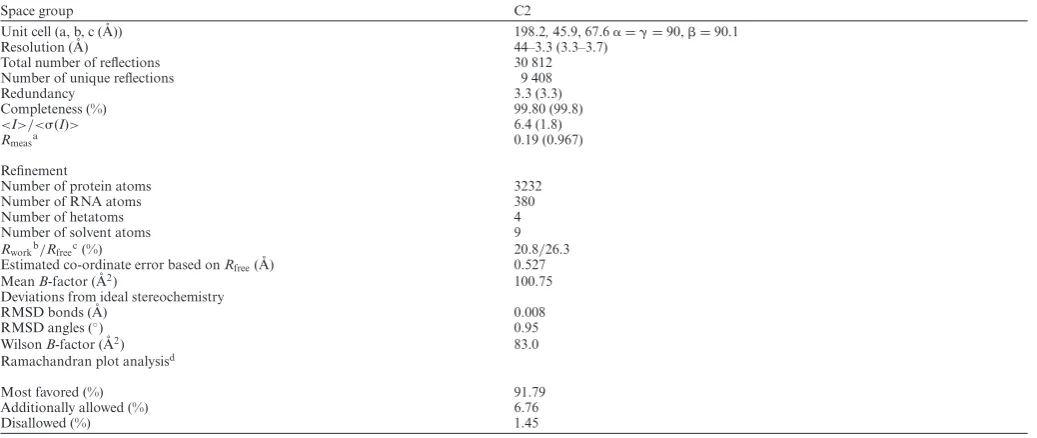
and Figure1A). These observations prompted us to
fur-ther investigate the three transcripts computationally. To facilitate this, mRNA sequences for the full-length GFP,
DsRed2 and HBB transcripts were first subjected toin silico
folding using default parameters in the program MFOLD
(20). For GFP, 13 unique structures were obtained, 10 of
which shared the same secondary structure in the vicinity of the UGAAG motif. These 10 structures corresponded to the lowest energy folds out of the 13 identified. Sim-ilar results were obtained with HBB, the only exception was that 14 solutions were obtained, 10 of which shared the same fold in the target UGAAG region. In contrast, DsRed2 yielded 39 structures where only 5 were found to be equivalent. Following the identification of stable substruc-tures, the sequences were reduced to 201 nucleotides based on the lengths of transcripts reported to support cleavage
(9). In all three, the substructures encompassing the
tar-geted UGAAG sites remained unchanged. Cycles ofin
sil-icofolding were then iteratively performed on sequences of
incrementally reduced length until the minimal number of nucleotides could be identified which maintained the orig-inal substructures. These corresponded to 51 nucleotides for GFP (GFP51, nucleotides 117 to 168), 58 nucleotides for HBB (HBB58, nucleotides 179–237) and 61 nucleotides for DsRed2 (DsRed61, nucleotides 489–550). The resulting
structures were also analyzed using the MC-Fold|MC-Sym
Table 2. Crystallographic parameters
Space group C2
Unit cell (a, b, c ( ˚A)) 198.2,45.9, 67.6␣=␥=90,=90.1
Resolution ( ˚A) 44–3.3 (3.3–3.7)
Total number of reflections 30 812
Number of unique reflections 9 408
Redundancy 3.3 (3.3)
Completeness (%) 99.80 (99.8)
<I>/<(I)> 6.4 (1.8)
Rmeasa 0.19 (0.967)
Refinement
Number of protein atoms 3232
Number of RNA atoms 380
Number of hetatoms 4
Number of solvent atoms 9
Rworkb/Rfreec(%) 20.8/26.3
Estimated co-ordinate error based onRfree( ˚A) 0.527
MeanB-factor ( ˚A2) 100.75
Deviations from ideal stereochemistry
RMSD bonds ( ˚A) 0.008
RMSD angles (◦) 0.95
WilsonB-factor ( ˚A2) 83.0
Ramachandran plot analysisd
Most favored (%) 91.79
Additionally allowed (%) 6.76
Disallowed (%) 1.45
Values in parentheses are for the highest resolution shell (3.3–3.7 ˚A).
Rmeas=((N/N-1))1/2(|Ii–<I>|)/(<I>)), where the sum is calculated over all observations of a measured reflection (Ii),<I>is the mean intensity of all the measured
observations (Ii) and N the total number of observations for each reflection.
Rwork=(|Fobs– Fcalc|)/(Fobs), Fobsare the observed structure factor amplitudes, and Fcalcthose calculated from the model.
Rfreeis equivalent to Rworkbut where 5% of the measured reflections have been excluded from refinement and set aside for cross-validation purposes.
d Ramachandran plot analysis was from molprobity (18).
which non-canonical base pairs are included in energy cal-culations (the lowest energy predictions are shown in Figure
1A). Despite each being distinct, all form simple (GFP51)
or more complex (HBB58 and DsRed61) structures consist-ing of one or more stem loops, where the cleavage sites are located within the loops themselves or adjacent to regions containing unpaired nucleotides. All have pyrimidines
di-rectly 3 to the major cleavage sites consistent with the
re-cent studies of Gagliaet al.(11).
We next assessed whether the elements identified could be
processedin vitro. To achieve this, the GFP51 and HBB58
sequences were synthesized as 3FAM substituted
oligonu-cleotides (see Materials and Methods and Table1) and
ana-lyzed using RNase assays (Figure1B). Initial attempts using
reaction buffers designed for unstructured substrates that are cleaved exonucleolytically (UN51, Supplementary
Fig-ure S1A and shorter oligonucleotides cited in Bagn´eriset al.
(11)) resulted in poor levels of cleavage. This necessitated
buffer re-screening to establish the optimum conditions for degradation (see Materials and Methods together with Sup-plementary Figures S1A–C). Under these conditions, both GFP51 and HBB58 were found to be susceptible to
degra-dation, in particular GFP51 (Figure1B and Supplementary
Figure S1D). Although discrete products resulting from en-donucleolytic cleavage appear absent, this is largely due to rapid exonucleolytic processing following the initial incision given that fragments were frequently observed in assays of mutants defective in cleavage activity (see ‘Results’ section below).
To investigate whether the loops/bulges identified in
GFP51 and HBB58 rendered substrates more readily pro-cessed than Watson-Crick base paired double-stranded RNA lacking these elements, assays were also performed
with a double-stranded 51mer comprising UN51 and its
complementary sequence (dsUN51, Table1). dsUN51 was
largely resistant to degradation (as shown in Figure 1B
and the corresponding time course assays in Supplemen-tary Figures S1D and E) in all buffers (data are only shown for those performed in the optimal endonucleolytic buffer) indicating that SOX-induced endonucleolytic processing is entirely dependent on the presence of loop or bulge ele-ments within duplex regions of RNA substrates.
Crystal structure of SOX bound to the KSHV pre-miRNA stem loop fragment K2-31
Attempts to co-crystallize SOX with either GFP51 or HBB58 were unsuccessful therefore, based on our computa-tional studies, we sought to identify shorter RNA sequences more amenable to crystallographic studies. Since SOX de-grades KSHV mRNA transcripts in addition to those of
the host for the purposes of self-regulation (22), we focused
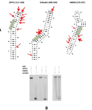
on the KSHV transcriptome and in particular pre-miRNA’s given their simple stem loop configurations. We were able to identify KSHV pre-miRNA K12-2 (K2), that also con-tains a similarly positioned UGAAG motif upstream of a bulge, as a likely target and using the same strategy to that outlined above, deduce that the structure could be
main-tained with only 31 nucleotides (K2-31, Figure2A). This
sequence was synthesized (incorporating a 3FAM group)
and found to be degraded by SOX in RNase assays
(Fig-ure 2A and Supplementary Figure S1E). K2-31 was
Figure 1. (A) The lowest energy secondary structure predictions for GFP51, DsRed61 and HBB58 generated by the MC-Fold|MC-Sym pipeline. Red arrows indicate cleavage sites reported by Covarrubiaset al.(9) and green boxes highlight the UGAAG sequences. Thicker arrows represent sites that were targeted at higher frequency. Pink boxes highlight loops capped by two unpaired nucleotides reported to be cleavage sites. (B) RNase assays performed with GFP51, HBB58 (left) and dsUN51 (right).
Supplementary Figure S2) mediated by symmetry-related molecules although only poor density could be observed for those involved in intermonomer contacts (nucleotides 1–3 and 29–31). These nucleotides were subsequently omitted from the deposited co-ordinates.
Similar to the SOX-DNA structure (3POV), K2-31 is ac-commodated in the ‘canyon’ between the N and C-terminal
lobes of SOX (Figure 2B) where the apex of the loop
projects directly into the active site. Closer inspection re-vealed that K2-31 has a near continuous stem loop config-uration, interrupted by the presence of a 7 nucleotide bulge
(disordered in our structure) immediately 5to the UGAAG
motif. No density could be observed for A19 (Figure2C).
The overall K2-31 conformation is therefore congruent with the lowest energy secondary and tertiary structure MC-Fold
|MC-sym predictions (Figure2A) in which two nucleotides
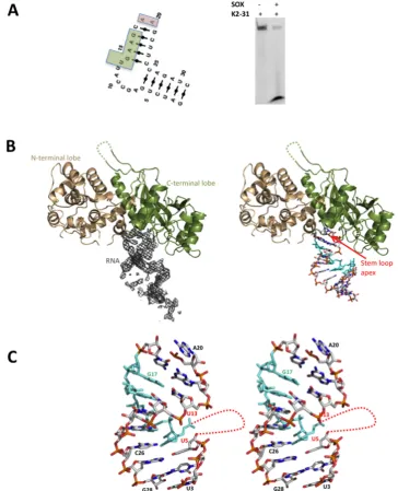
(A19 and A20) forming the loop apex are unpaired. The only deviation is that G6 (absent in our structure) appears not to form a Watson Crick base pair with C26 which al-ternatively stacks opposite U13 within the body of the stem duplex. Interestingly, the protein-RNA contacts stabilising the complex are narrowly distributed with most restricted
to the catalytic region and the K2-31 loop (Figure3A). In
our structure, A20 and G21 at the apex are recognized by a series of hydrogen bonds involving Y373, R248 and F249
(Figure3B and Supplementary Figure S3). While N⑀1 of
R248 donates a hydrogen bond to O4of A20, the OH group
of Y373 donates hydrogen bonds to both O4and O1P. Also
evident is a hydrogen bond donated by the 2OH group of
A20 to the carbonyl oxygen of C247 as well as Van der Waals interactions contributed by the side chain of K246. Additionally, the peptide NH group of F249 donates a
hy-drogen bond to O1P of G21 (Figure3B).
Further stabilization of K2-31 is derived from a stacking interaction between the adenine base of A20 and the
aro-matic side chain of F179 located in the bridge (Figure3C).
In order to achieve this, F179 moves∼5 ˚A (based on C␣-C␣
distances) relative to its position in the DNA complex. Sur-prisingly, this alternative configuration of F179 and the N-terminal end of the bridge is stabilized by a disulphide bond between the sulphydryl groups of C183 and C247
(Fig-ure 3C). These interactions, absent in the reported
sub-Figure 2. (A) Left. The lowest energy MC-Fold|MC-Sym secondary structure prediction for K2-31 (the UGAAG sequence is highlighted by a green box). Right. RNase assay performed with K2-31. (B) Cartoon representations of SOX (N and C-terminal lobes coloured wheat and dark green, respectively) together with (Left) the associated 2mFo-DFc omit map density (contoured at 1) for K2-31 bound in the ‘canyon’ and (right) with the RNA from the final model represented as sticks. The UGAAG sequence (U13-G17) is highlighted in cyan. Missing loops are included as dashed lines. (C) Stereoview showing the overall conformation of K2-31 (nucleotides U3-G28). Nucleotides 3to U5 and 5to U13 (red dotted line) constitute an internal 7 nucleotide loop that could not be modeled owing to disorder.
strates. To investigate this, F179A and C247S mutants were constructed and their ability to incise GFP51 assessed. Al-though C247S was only slightly impaired in cleavage activ-ity compared to apo SOX, F179A was highly defective il-lustrating its important role in endonucleolytic processing
(Figure3D). These defects are RNA specific since both the
C247S and F179A mutants readily degrade a DNA duplex
substituted with a 5phosphate (dsDNA5P).
Analysis of the SOX-K2-31 structure revealed that no specific interactions are observed between SOX and
nu-cleotides 5to A19. This apparent confinement of
protein-RNA contacts to the K2-31 loop results in an overall bind-ing configuration which contrasts markedly with that of the DNA duplex in 3POV, where there are additional contacts with the putative nuclear localization motif as well as those
involving R248 and F249 detailed above (4), (Figure3E).
Consistent with this, the RNA stem loop and DNA du-plex are mostly non-overlapping beyond the active site
re-gion, where an∼90◦rotation would be required to
[image:7.612.115.480.71.521.2]con-Figure 3. (A) Schematic diagram of the observed protein-RNA contacts in the SOX-K2-31 complex. Hydrogen bonding and-stacking interactions are shown in grey and red, respectively. (B) The protein RNA contacts that function in loop recognition, central to which are those mediated by A20, G21 and residues K246, R248, F249 and Y373 within the catalytic region. (C) The protein–RNA interactions involving the bridge and the conformational re-arrangements observed in transitioning from the SOX–DNA complex, 3POV, (light blue) to SOX-K2-31 (dark green). The disulphide bond between C183 and C247, absent in 3POV, is shown in magenta. (D) RNase assays performed using the C247S and F179A mutants illustrating that while the C247S substitution is mildly defective, F179A is significantly impaired. (E) Superposition of 3POV and SOX-K2-31. Although the protein moieties in both complexes are near identical, an∼90◦rotation would be required to map the DNA co-ordinates onto those of K2-31 despite both interacting with the same catalytic residues. This is owing to K2-31 being stabilized exclusively in the loop region while additional contacts are evident in the DNA complex, notably, those involving the nuclear localization motif (NLS, red in 3POV).
stitute 240 and 480 ˚A2for the SOX–K2-31 and SOX–DNA
complexes, respectively.
Despite use of the inactivated E244S SOX mutant, the ab-sence of density for A19 and lack of a phosphate group in close proximity to the essential catalytic carboxylate D221, is indicative of K2-31 having undergone cleavage over the duration of the crystallization experiment to produce a product complex. Thus in order to investigate the most likely cleavage geometry for a stem loop substrate, an
‘in-tact loop’ model was generated for K2-31 by combining the co-ordinates for loop residues 18–22 from the lowest energy
structure prediction of K12-2 (MC-Fold|Mc-Sym pipeline)
with the remaining stem nucleotides from the crystal
struc-ture (Figure4A). This model was subsequently docked onto
the K2-31-SOX crystal structure. Although a configuration was observed in which the scissile phosphate could form a ligand with the non-canonical magnesium ion in 3POV, K2-31 would be distant from S144 that has also been shown to
[image:8.612.136.491.72.521.2]Figure 4. (A) Potential cleavage geometry of K2-31 based on an alignment with 3SM4. The most favorable configuration for catalysis is one in which the scissile phosphate (magenta) is located between magnesium ions A and B (MgAand MgB, blue) where MgBis in the canonical site observed in 3SM4 and
other type II restriction endonuclease-like enzymes but not 3POV. In this position, K2-31 is not only able to contact residues 247–249 (green) but also S144 reported to be essential for cleavage which forms a cluster with S145 and S146 that is conserved in a number of type II restriction-like enzymes (yellow). (B) RNase assays using the GFP51 template in which the UGAAG motif was substituted for UCUCU and UGCAC. (C) FPA assays in which WT SOX was titrated against GFP51, HBB58, K2-31 and dsUN51.
form a cluster that is conserved in other type II
restriction-like enzymes where it has been shown to function in 5
phos-phate stabilization in the bacteriophage lambda DNA sub-strate complex (3SM4). Alternatively, aligning the scissile phosphate with that observed in 3SM4, which is
approx-imately equidistant from MgA and MgB in their
canoni-cal positions, produces a configuration consistent with in-line attack based on the relative juxtapositions of the
cat-alytic residues E244, D221 and K246 (Figure4A). In this
position, the phosphate group of A19 can be stabilized by S144 and important contacts between nucleotides 20–22 and residues 247–249 (though distinct to those in the prod-uct complex) still maintained. These findings point toward a
geometry consistent with a canonical SN2 bi-metal nuclease
mechanism for endonucleolytic cleavage.
RNA turnover by SOX is consensus sequence independent
Inspection of the SOX-K2-31 structure reveals a com-plete absence of interactions between SOX and either the phophodiester backbone or bases of the UGAAG motif. To ascertain whether this might be a feature of the crystal structure or specific to K2-31, the role of the motif in cleav-age was investigated by substituting the UGAAG sequence in GFP51 for UGCAC and UCUCU (GFP-UGCAC and
GFP-UCUCU in Table 1). UGCAC was chosen to
dis-rupt the AAG/A sequence reported to potentially
influ-ence targeting (11) while maintaining Watson Crick base
MC-Fold with those corresponding to the lowest energies re-vealing that the overall morphology of GFP51 remained largely unchanged (Supplementary Figure S4), although an additional two nucleotide bulge was observed for the
UG-CAC substitution three nucleotides 5 to the first major
cleavage site. RNase assays revealed that substitution of the UGAAG motif for either UGCAC or UCUCU failed to appreciably impair SOX-mediated degradation suggesting
that cleavagein vitrois not significantly dependent on
con-servation of a consensus sequence (Figure4B). Although it
could be argued that this apparent lack of impairment could be attributable to the incubation times and quantities of SOX used, a time course experiment was also conducted in-volving GFP51 and GFP51-UCUCU (Supplementary Fig-ure S5). This revealed that turnover of GFP51-UCUCU by SOX is as efficient as that observed for GFP51 confirming that the UGAAG motif is not the key driver for transcript processing.
Given that endonucleolytic processing appears to be
in-dependent of a consensus sequence while loop/bulge
ele-ments are required for cleavage, we next investigated the affinity of SOX for GFP51, HBB58, K2-31 and dsUN51 to establish whether such features are preferentially bound
(Figure4C). Of the oligonucleotides tested, SOX had the
highest affinity for GFP51 with a Kd of 5M compared
to 42 and 49M for HBB58 and K2-31, respectively. The
highestKdwas observed for dsUN51 at∼73M. These
re-sults demonstrate a clear binding preference for simple stem loops over bulges. Additionally, expanding the loop in K2-31 to incorporate three and nine additional adenines failed
to negatively impact on turnover (Figure5A) indicating that
although stem loops are required for optimal endonucle-olytic processing, there are no limitations on size. This lack of apparent specificity would account for the disparate
na-ture of the RNA targets identifiedin vivoreported by Gaglia
et al.(11).
The HSO mutants have distinct roles in RNA processing
Having probed the nature of the SOX RNA targets, we next deduced whether the apparent failure of the HSO mu-tants to impair unstructured single-stranded RNA process-ing reported, could be attributed to the absence of stem loops and bulges in such oligonucleotides. They were there-fore re-evaluated in terms of their capacity to degrade GFP51. The mutants tested constitute A61T (within the N-terminal lobe), P176S (located in the bridge region), V369I (at the center of the C-terminal lobe), D474N and Y477Stop (Y477*) at the C-terminus of SOX (Supplemen-tary Figure S6). T24I (an additional HSO mutant) could not be assessed owing to insolubility as previously reported
(4)). RNase assays revealed that the mutants can be largely
subdivided into two categories: those that are profoundly defective in GFP51 cleavage activity and those that have
a minor-to-moderate defect (Figure 5B). The mutations
P176S and V369I almost completely abolish processing in
agreement within vivostudies where they virtually abrogate
HSO (10). Given that P176 is located within the ‘bridge’,
it can be envisaged that the P176S mutation has the poten-tial to disrupt the conformational re-arrangements required for F179 to engage with RNA targets in order to confer
substrate/product stabilization based on our earlier results.
Similarly, V369 is located in a small hydrophobic recess, where its replacement with isoleucine would result in the positioning of residues directly adjacent to the catalytic re-gion, in particular those in close proximity to A20. By con-trast, D474N and Y477* have a moderate to minor effect on RNase activity. Their lower impact can be explained by
their locations at the C-terminus of SOX (>30 ˚A from the
catalytic region). Similarly, the A61T mutant, located∼29
˚
A from the active site, appears to have wild-type activity. These results suggest that the far C-terminus of SOX and A61 do not have a role in the initial endonucleotlytic cleav-age of mRNA transcripts.
The RNase activities of SOX and Xrn1 are coupled
Although it has been shown that Xrn1 is essential for HSO
(9), it was unclear whether the products of SOX cleavage
generate Xrn1 substrates. To assess this, RNase assays in-volving SOX using optimized conditions for Xrn1 activity (sub-optimal for SOX cleavage, see Materials and
Meth-ods), were conducted in the presence and absence of K.
lac-tisXrn1 that has 54% sequence identity to the human
ho-molog in the catalytic domain. While Xrn1 in isolation is
in-capable of degrading GFP51 (Figure5C), enhanced
degra-dation is observed when it is combined with SOX. Our re-sults therefore confirm that the products of SOX cleavage are substrates for Xrn1 and that additional factors are un-likely to be essential at these initial stages of target process-ing. Moreover, the increased rate of turnover indicates that the activities of SOX and Xrn1 act in concert to facilitate rapid processing. In agreement with this, yeast two hybrid studies involving SOX as both bait and prey using a library composed of proteins exclusively involved in human mRNA degradation pathways reveal that Xrn1 and SOX physically
interact (Figure5D–F).
DISCUSSION
Pivotal to a detailed understanding of KSHV mediated HSO is knowledge of the targets degraded and in partic-ular, the nature of their interaction with the HSO nuclease
SOX. Although recentin vivostudies have identified RNA
stem loops as substrates and a potential requirement for se-quence dependency, little could be ascertained about their structure. This problem derived from the seemingly diverse nature of the elements targeted given not only the variations in their sequences, but also the fact that incision sites have been identified in both loop and stem segments of struc-tured mRNA transcripts. Despite this, cleavage appears to be restricted to regions within, or flanked by, unpaired
nu-cleotides (11). In efforts to address these important issues,
we first generatedin silicomodels of three transcripts shown
to be SOX targets and were able to identify the minimal se-quences required to maintain the folds of these elements.
We were able to showin vitrothat they are incised by SOX
while Watson Crick base paired double-stranded RNA of a comparable length is resistant to processing.
Analysis of the identified elements revealed that all three
transcripts incorporate single stem loops or bulge/loop
Figure 5. (A) RNase assays performed using the K2-31 variants K2-31A4 and K2-31A9 where three and nine adenines were introduced to the K2-31 loop apex, respectively. (B) RNase assays of WT SOX and the HSO mutants A61T, P176S, V369I, D474N and Y477 stop using GFP51. The D221S and E244S catalytic mutants were included as negative controls. (C) RNase assays performed using WT SOX, GFP51 and Xrn1 illustrating enhanced cleavage when SOX and Xrn1 are combined. (D) A yeast two hybrid screen using a selective library involving human proteins exclusively involved in mRNA degradation illustrating an interaction between SOX and Xrn1. Cells containing SOX as bait (SOX-pGBAD-B) and the library proteins as prey (Lib-pGACTBD-B) were grown in the selective media SD-WHL + AT (top) and SD-WAL (bottom), respectively. (E) As for (D), but where the library was used as bait (Lib-pGBAD-B) and SOX prey (SOX- pGACTBD-B). Colonies were only observed for those containing SOX and Xrn1 that were verified by sequencing. (F)
-galactosidase assay of clones identified as positive and negative for SOX and Xrn1. The most intense blue plaque corresponds to those positive for SOX and Xrn1.
Based on these common features, we were able to both identify and verify KSHV pre-miRNA K12-2 (K2) as a potential target using a 31mer fragment (K2-31). K2, an RNA polymerase II transcript that is expressed from la-tency, has been reported to have an important role in in-fluencing cytoskeletal organization through suppressing the expression of high molecular weight tropomyosin 1 splice
variants (HMW-TPM1) (12). Although how K2 functions
remains unclear, the suppression of HMW-TPM1 has been directly linked to downregulated anoikis. This has been as-sociated with KSHV infection as well as metastasis in sev-eral non-viral cancers where their expression is near abol-ished. An important contributor to viral trafficking could
thus be the overproduction of K2 and it is interesting to speculate that SOX may have a role in the maturation of its pre-miRNA. In support of this, it has been established that SOX-mediated degradation of the KSHV transcrip-tome has an important role in the production of viable
progeny (22) and that pre-miRNAs containing short loops
are often poor substrates for dicer, the host nuclease that is required for miRNA maturation via the canonical pathway
(23) (although other mechanisms have been identified). It
has also been reported that KSHV miRNA’s 1 to K12-8 are highly expressed during the lytic phase in the PELs
cell line BC3 (24). Intriguingly, the 5-3degradation of
cleav-age at the loop apex, would result only in destruction of the passenger strand (Supplementary Figure S7) leaving the guide strand intact. This could potentially be passed to the RISC complex for engagement with its target. Our findings thus present the possibility that KSHV has the potential to not only subvert the host RNA degradation machinery but also machineries involved in miRNA targeting.
In addition to identifying K2 as a targetin vitro, we were
able to successfully crystallize a product complex involv-ing a K2 31mer fragment. This structure has established the structural basis for mRNA transcript recognition and pro-cessing. Key to its formation is loop recognition in which one of the two unpaired adenines at the apex, together with
the following 3guanine, contact residues that collectively
form part of the catalytic region (namely K246, R248 and F249). As well as contacts involving residues key to cataly-sis, F179 further stabilizes the SOX–K2-31 complex follow-ing re-modelfollow-ing of the bridge that appeared to involve for-mation of a disulphide bond between the sulphydryl groups of C183 and C247. Our mutagenesis studies have shown that while C247 has a minor role, F179 is essential for RNA but not DNA processing confirming the bridge as an
im-portant factor in substrate/product recognition where it
directly functions in RNA binding as previously
hypothe-sized (7). Interestingly, comparison of the bridge regions in
BGLF5 and SOX reveals several differences, the most sig-nificant of which are re-arrangements in the P158 region (equivalent to P176 in KSHV SOX) in response to the dele-tion of two upstream amino acid residues and the substi-tution of A174 for glycine. These differences may alter how RNA substrates are bound and processed potentially result-ing in distinct preferential cleavage sites to those observed
for KSHV SOX as reported in Covarrubiaset al.(25).
Despite K2-31 in our structure having undergone pro-cessing, we have been able to model a potential substrate complex arising from alignments involving a ‘loop intact’ model and the co-ordinates from 3SM4 in keeping with a bi-metal nuclease mechanism. Based on the positions of the conserved magnesium ions and sites known to be essential for binding (i.e. S144 and residues 247–249) and cleavage, the scissile phosphate is more likely to interact with MgB in its canonical position with respect to other type II restric-tion endonuclease-like enzymes.
Surprisingly, the SOX–K2-31 complex showed no evi-dence of protein–RNA contacts beyond the catalytic region including the UGAAG motif. This apparent lack of depen-dency on a consensus sequence for transcript recognition was supported by our RNase assays in which its substitu-tion for UCUCU or UGCAC in GFP51 failed to attenu-ate cleavage. Also noteworthy is the absence of this motif in
the vicinity of the loop structure of DsRed61 (Figure1A)
reported as an incision target (9). Our studies additionally
show that SOX appears to have a binding preference for simple stem loops over substrates comprising those that are more complex or bulges, consistent with the higher rates of turnover associated with GFP51 and UCUCU. There also appear to be no restrictions on loop size based on assays involving the K2-31 loop variants.
Although our results point to architecture and not se-quence being the major pre-requisite for cleavage, the pref-erence for an adenine rich degenerate consensus sequence
5 to cleavage sites in vivo was reported by Gaglia et al.
(11). This could be explained by the propensity of
ade-nine to induce bends/bulges into RNA duplexes when
un-paired. In keeping with this, it has been shown that the inser-tion of unpaired adenine stretches has a greater capacity to
distort RNA duplexes than pyrimidines (26,27). Although
purines have been shown to be under-represented directly
3to cleavage sites, the origins of this preference remain
un-clear based on our current data. This poor representation may arise from the potential of adenine and in particular guanine to adopt conformations that inhibit substrate bind-ing or product dissociation in this position as a result of interactions with non-catalytic residues. These hypotheses, however, have yet to be investigated.
The identification of substrates susceptible to endonucle-olytic cleavage led us to re-evaluate the HSO mutants to as-certain whether their activities might be impaired relative to the unstructured substrates originally trialed. Interestingly, a range of results were obtained in RNase assays involving
GFP51 that largely mirrored those reportedin vivo.Perhaps
not surprisingly, the P176S and V369I mutants are highly defective in RNase activity most likely as a consequence of
their roles in substrate/product stabilization and/or
prox-imity to key residues forming the active site. In contrast, D474N showed attenuation while A61T and Y477* had near wild-type activity. Based on the crystal structure, these residues are distant from the catalytic site and are thus un-likely to have a direct impact on RNA recognition. We were also able to ascertain that the products of SOX cleavage are indeed substrates for Xrn1 and that the enhanced degra-dation of transcripts evident when both are combined is suggestive of their exoribonuclease activities being coupled, most likely through a physical interaction as suggested by the results of yeast two hybrid analysis. On aggregate, our results are consistent with a dynamic system for KSHV
me-diated mRNA degradation in line within vivostudies.
SUPPLEMENTARY DATA
Supplementary Data are available at NAR Online.
ACKNOWLEDGEMENTS
We thank Dr Ambrose Cole (X-ray Lab, Birkbeck College) for his assistance with data collection and also Dr Tina Daviter (Biophysics Centre, Birkbeck College) for her help
with the biophysical/biochemical assays. We would also like
to thank Dr Mark Williams for many useful discussions.
FUNDING
Wellcome Trust [WT090147MA to A.P. and T.B.]. Funding for open access charge: Wellcome Trust [WT090147MA].
Conflict of interest statement.None declared.
REFERENCES
1. Kaplan,L.D. (2013) Human herpesvirus-8: Kaposi sarcoma, multicentric Castleman disease, and primary effusion lymphoma.
Hematol. Am. Soc. Hematol. Educ. Program,2013, 103–108.
3. Buisson,M., Geoui,T., Flot,D., Tarbouriech,N., Ressing,M.E., Wiertz,E.J. and Burmeister,W.P. (2009) A bridge crosses the active-site canyon of the Epstein-Barr virus nuclease with DNase and RNase activities.J. Mol. Biol.,391, 717–728.
4. Bagneris,C., Briggs,L.C., Savva,R., Ebrahimi,B. and Barrett,T.E. (2011) Crystal structure of a KSHV-SOX-DNA complex: insights into the molecular mechanisms underlying DNase activity and host shutoff.Nucleic Acids Res.,39, 5744–5756.
5. Bujnicki,J.M. and Rychlewski,L. (2001) The herpesvirus alkaline exonuclease belongs to the restriction endonuclease PD-(D/E)XK superfamily: insight from molecular modeling and phylogenetic analysis.Virus Genes,22, 219–230.
6. Dahlroth,S.L., Gurmu,D., Schmitzberger,F., Engman,H., Haas,J., Erlandsen,H. and Nordlund,P., (2009) Crystal structure of the shutoff and exonuclease protein from the oncogenic Kaposi’s sarcoma-associated herpesvirus.FEBS J.,276, 6636–6645. 7. Horst,D., Burmeister,W.P., Boer,I.G., van Leeuwen,D., Buisson,M.,
Gorbalenya,A.E., Wiertz,E.J. and Ressing,M.E., (2012) The ‘Bridge’ in the Epstein-Barr virus alkaline exonuclease protein BGLF5 contributes to shutoff activity during productive infection.J. Virol., 86, 9175–9187.
8. AlMalki,F.A., Flemming,C.S., Zhang,J., Feng,M., Sedelnikova,S.E., Ceska,T., Rafferty,J.B., Sayers,J.R. and Artymiuk,P.J. (2016) Direct observation of DNA threading in flap endonuclease complexes.Nat. Struct. Mol. Biol.,23, 640–646.
9. Covarrubias,S., Gaglia,M.M., Kumar,G.R., Wong,W., Jackson,A.O. and Glaunsinger,B.A. (2011) Coordinated destruction of cellular messages in translation complexes by the gammaherpesvirus host shutoff factor and the mammalian exonuclease Xrn1.PLoS Pathog., 7, e1002339.
10. Glaunsinger,B., Chavez,L. and Ganem,D. (2005) The exonuclease and host shutoff functions of the SOX protein of Kaposi’s
sarcoma-associated herpesvirus are genetically separable.J. Virol.,79, 7396–7401.
11. Gaglia,M.M., Rycroft,C.H. and Glaunsinger,B.A. (2015) Transcriptome-wide cleavage site mapping on cellular mRNAs reveals features underlying sequence-specific cleavage by the viral ribonuclease SOX.PLoS Pathog.,11, e1005305.
12. Kieffer-Kwon,P., Happel,C., Uldrick,T.S., Ramalingam,D. and Ziegelbauer,J.M. (2015) KSHV MicroRNAs repress tropomyosin 1 and increase anchorage-independent growth and endothelial tube formation.PLoS One,10, e0135560.
13. Chang,J.H., Xiang,S., Xiang,K., Manley,J.L. and Tong,L., (2011) Structural and biochemical studies of the 5–>3exoribonuclease Xrn1.Nat. Struct. Mol. Biol.,18, 270–276.
14. Winn,M.D., Ballard,C.C., Cowtan,K.D., Dodson,E.J., Emsley,P., Evans,P.R., Keegan,R.M., Krissinel,E.B., Leslie,A.G., McCoy,A.
et al.(2011) Overview of the CCP4 suite and current developments.
Acta Crystallogr. D Biol. Crystallogr.,67, 235–242.
15. McCoy,A.J., Grosse-Kunstleve,R.W., Adams,P.D., Winn,M.D., Storoni,L.C. and Read,R.J. (2007) Phaser crystallographic software.
J. Appl. Crystallogr.,40, 658–674.
16. Emsley,P., Lohkamp,B., Scott,W.G. and Cowtan,K. (2010) Features and development of Coot.Acta Crystallogr. D Biol. Crystallogr.,66, 486–501.
17. Smart,O.S., Womack,T.O., Flensburg,C., Keller,P., Paciorek,W., Sharff,A., Vonrhein,C. and Bricogne,G. (2012) Exploiting structure similarity in refinement: automated NCS and target-structure restraints in BUSTER.Acta Crystallogr. D Biol. Crystallogr.,68, 368–380.
18. Chen,V.B., Arendall,W.B. 3rd, Headd,J.J., Keedy,D.A.,
Immormino,R.M., Kapral,G.J., Murray,L.W., Richardson,J.S. and Richardson,D.C. (2010) MolProbity: all-atom structure validation for macromolecular crystallography.Acta Crystallogr. D Biol.
Crystallogr.,66, 12–21.
19. Lehner,B. and Sanderson,C.M. (2004) A protein interaction framework for human mRNA degradation.Genome Res.,14, 1315–1323.
20. Zuker,M. (2003) Mfold web server for nucleic acid folding and hybridization prediction.Nucleic Acids Res.,31, 3406–3415.
21. Parisien,M. and Major,F. (2008) The MC-Fold and MC-Sym pipeline infers RNA structure from sequence data.Nature,452, 51–55. 22. Abernathy,E., Clyde,K., Yeasmin,R., Krug,L.T., Burlingame,A.,
Coscoy,L. and Glaunsinger,B. (2014) Gammaherpesviral gene expression and virion composition are broadly controlled by accelerated mRNA degradation.PLoS Pathog.,10, e1003882. 23. Feng,Y., Zhang,X., Graves,P. and Zeng,Y. (2012) A comprehensive
analysis of precursor microRNA cleavage by human Dicer.RNA,18, 2083–2092.
24. Umbach,J.L. and Cullen,B.R. (2010) In-depth analysis of Kaposi’s sarcoma-associated herpesvirus microRNA expression provides insights into the mammalian microRNA-processing machinery.J. Virol.,84, 695–703.
25. Gaglia,M.M., Covarrubias,S., Wong,W. and Glaunsinger,B.A. (2012) A common strategy for host RNA degradation by divergent viruses.
J. Virol.,86, 9527–9530.
26. Kulinski,T., Bielecki,L. and Adamiak,R.W. (2001) Structure and dynamics of adenosine loops in RNA bulge duplexes as revealed by linked application of thermodynamics, spectrofluorimetry and simulation of molecular dynamics.Nucleic Acids Res. Suppl.,1, 139–140.