RESEARCH REPORT
DEFECTIVE KERNEL 1 promotes and maintains plant epidermal
differentiation
Roberta Galletti1,*, Kim L. Johnson2, Simon Scofield3, Rita San-Bento1, Andrea M. Watt2, James A. H. Murray3 and Gwyneth C. Ingram1,*
ABSTRACT
During plant epidermal development, many cell types are generated from protodermal cells, a process requiring complex co-ordination of cell division, growth, endoreduplication and the acquisition of differentiated cellular morphologies. Here we show that theArabidopsisphytocalpain DEFECTIVE KERNEL 1 (DEK1) promotes the differentiated epidermal state. Plants with reduced DEK1 activity produce cotyledon epidermis with protodermal characteristics, despite showing normal growth and endoreduplication. Furthermore, in non-embryonic tissues (true leaves, sepals), DEK1 is required for epidermis differentiation maintenance. We show that the HD-ZIP IV family of epidermis-specific differentiation-promoting transcription factors are key, albeit indirect, targets of DEK1 activity. We propose a model in which DEK1 influences HD-ZIP IV gene expression, and thus epidermis differentiation, by promoting cell adhesion and communication in the epidermis.
KEY WORDS: DEK1, Epidermis, Differentiation maintenance, Arabidopsis thaliana
INTRODUCTION
The production of specific function-adapted organ morphologies in plants is achieved by the exquisitely complex spatial and temporal control of cell proliferation, expansion and differentiation. The importance of this co-ordination is well illustrated in the cotyledon epidermis ofArabidopsis thaliana. To perform its basic functions this cell layer must produce both mature pavement cells (PCs), which can be very large and have a complex morphology, and appropriately spaced pairs of stomatal guard cells, which are tiny and adapted to their role in gas exchange (Glover, 2000). Producing this complex mosaic demands that neighbouring cells co-ordinate their growth.
Epidermal specification and differentiation are linked to the activity of subfamily IV of the plant-specific homeodomain-leucine zipper (HD-ZIP) transcription factors (TFs) (Abe et al., 2003; Depege-Fargeix et al., 2011; Di Cristina et al., 1996; Horstman et al., 2015; Javelle et al., 2011a, 2010; Nakamura et al., 2006; Peterson et al., 2013; Roeder et al., 2012; San-Bento et al., 2014; Takada et al., 2013; Vernoud et al., 2009; Wu et al., 2011; Yang et al., 2011). Cell-cell communication influences both epidermal cell fate decisions and subsequent differentiation (reviewed by Javelle et al., 2011b;
Schiefelbein et al., 2014; Takada and Iida, 2014). For example, several receptor-like kinases (RLKs) and ligand-like peptides control patterning of the stomatal lineage (reviewed by Richardson and Torii, 2013) and the CRINKLY4 (CR4) RLK, and its Arabidopsis homologue ACR4, are required for the control of epidermal identity in maize (Becraft et al., 1996) and the expression of HD-ZIP IV genes inArabidopsis(San-Bento et al., 2014).
DEFECTIVE KERNEL 1 (DEK1) regulates growth coordination and epidermal characters in plants (Becraft et al., 2002; Johnson et al., 2005, 2008; Lid et al., 2002). Loss ofDEK1function causes early embryo lethality in both Arabidopsis and maize (Becraft et al., 2002; Johnson et al., 2005). In Petunia and Arabidopsis, strong downregulation of DEK1 is accompanied by proliferation of disorganised callus-like cells (Ahn et al., 2004; Lid et al., 2005). DEK1 is also required for multicellular development in the moss Physcomitrella patens(Demko et al., 2014; Liang et al., 2013; Perroud et al., 2014). DEK1 has a modular structure, with several predicted transmembrane domains, a linker domain and a cytoplasmically localised C-terminal domain comprising a sequence similar to animal calpains and a C2-like domain (Lid et al., 2002). The complete C-terminal domain (which we refer to as the CALPAIN domain) is sufficient to complementdek1loss-of-function alleles, suggesting that this is the active domain of the protein (Johnson et al., 2008; Liang et al., 2013). A weakDEK1allele,dek1-4, which causes a single amino acid substitution in the C2-like domain, and mutant alleles ofACR4and the HD-ZIP IV-encoding genesHDG11andATML1, were recently reported to lack sepal giant cells (GCs) (Roeder et al., 2012).
Here we aim to understand the role of DEK1 in epidermis differentiation, and particularly to clarify its relationship with the HD-ZIP IV TFs.
RESULTS AND DISCUSSION
dek1-4shows protodermal characteristics in cotyledon epidermis but no significant defects in cell expansion, ploidy or cell cycle-related gene expression
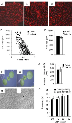
We found that the PCs in the adaxial epidermis ofdek1-4were less complex and more homogeneous than in wild-type (WT; Col-0) controls (Fig. 1). Three-day-old Col-0 cotyledon PCs included small cells with almost no lobes, intermediate cells with few lobes and larger, fully interdigitated cells (Fig. 1A). At the same stage,dek1-4 PCs ranged from showing no lobes ( polygonal shape) to showing some interdigitation, although less than in Col-0 (Fig. 1B,C). We noted an abnormally high frequency of four-way, or near four-way, junctions (usually found in meristematic cells) indek1-4(Fig. 1B,C). In thedek1-4mutant, intermediate size and large cells were rounder than WT cells of similar size (Fig. 1D). Despite their shape differences, the average final cell size of PCs in dek1-4 and WT was similar (Fig. 1E). Thus, PCs indek1-4have, on average, less complex shapes. This cellular phenotype persisted in fully expanded cotyledons (Fig. 1H,I). Neighbouring PCs in dek1-4 frequently had shared
Received 19 January 2015; Accepted 7 April 2015
1
Laboratoire de Reproduction et Développement des Plantes, Ecole Normale Supérieure de Lyon, 46 allée d’Italie, Lyon 69364, Cedex 07, France.2ARC Centre of Excellence in Plant Cell Walls, School of Botany, University of Melbourne, Royal Parade, Parkville, Victoria 3010, Australia.3Cardiff School of Biosciences, Cardiff University, Museum Avenue, Cardiff CF10 3AX, UK.
*Authors for correspondence ([email protected]; [email protected])
DEVEL
O
straight walls (Fig. 1I) suggesting that they had undergone late divisions (observed in 100% of mutant seedlings, but only twice in 40 WT seedlings analysed). No defect in blade expansion was observed (Fig. 1J), but cotyledons were frequently epinastically curled (Fig. 1G).
Leaf blade curling was described inCYCLIN D3;1(CYCD3;1 )-overexpressing plants, where it was associated with dramatic changes in ploidy distribution (Scofield et al., 2013). However, no significant differences in ploidy distribution were detected between WT anddek1-4cotyledons (Fig. 1K). Consistent with this, no differences in the expression ofCYCD3;1, or other cell cycle-related genes such asLOSS OF GIANT CELLS FROM ORGANS (LGO), SIAMESE (SIM), KIP-RELATED PROTEIN 6 (KRP6), HOBBIT(HBT) orDEL1, which encodes an endocycle inhibiting E2F-like protein, were observed between Col-0 and dek1-4
seedlings (supplementary material Fig. S1). Therefore, the phenotypes observed are not associated with ploidy defects.
DEK1 controls epidermal differentiation via a pathway involving multiple HD-ZIP IV-encoding genes
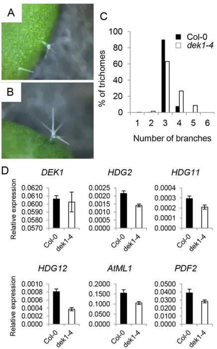
dek1-4leaves showed a significantly higher proportion of trichomes with more than three branches than WT controls (Wilcoxon rank sum test,P=1.7–20) (Fig. 2A-C). A similar phenotype was described
[image:2.612.51.346.53.557.2]for hdg11 and hdg12 hdg11 mutants (Nakamura et al., 2006). We analysed the expression ofHDG11,HDG12and of additional HD-ZIP IV genes (ATML1,PDF2,HDG2) in thedek1-4mutant and found that all genes tested were reproducibly downregulated (Fig. 2D). We then assessed the genetic relationship between DEK1, HDG11 and HDG12. hdg12-2 dek1-4 double mutants showed highly branched trichomes, as seen in thedek1-4parental Fig. 1. DEK1 promotes cell differentiation.(A-C) Confocal images of the adaxial side of 3-day-old Col-0 (A) anddek1-4
(B,C)Arabidopsiscotyledons stained with PI. Scale bars: 50μm. (D) Epidermal cell area plotted against shape factor (combined data from cells measured in four independent seedlings). Guard cells were eliminated from the analysis.
n=135 for Col-0 andn=189 fordek1-4. (E) Average area±s.e. of epidermal pavement cells (PCs). (F-I) Images and agarose imprints of the adaxial side of 8-day-old Col-0 (F,H) anddek1-4
(G,I) seedlings. Asterisks indicate four-way junctions (B,C) or neighbouring PCs with shared straight cell walls (I) in the mutant. (J) Average area±s.e. of 8-day-old cotyledons.n=19 for Col-0 andn=16 fordek1-4. (K) Nuclear DNA content/ploidy distribution profile of 10-day-old Col-0 anddek1-4cotyledons.
n=nuclei counted per sample.
DEVEL
O
line, but did not exhibit additional defects. Indek1-4 hdg11-2plants we observed no significant additivity of the trichome phenotypes of the two mutants (supplementary material Fig. S2). The sepals of hdg12-2 dek1-4and of dek1-4 hdg11-2mutants resembled those ofdek1-4mutants (supplementary material Fig. S3). Together, our results are consistent with multiple HD-ZIP IV proteins acting downstream of DEK1 to regulate epidermal differentiation.
To confirm whether the phenotype of thedek1-4allele is caused by reduced DEK1 activity, we expressed an artificial microRNA (Ossowski et al., 2008) targeted to theDEK1coding sequence (amiRDEK1) under the CaMV 35S promoter and under a dexamethasone (DEX)-inducible promoter (Craft et al., 2005; Samalova et al., 2005). T135S:amiRDEK1 plants showed similar phenotypes to those reported forAtDEK1RNAi lines (Johnson et al., 2005) and thedek1-4mutant (Roeder et al., 2012). Some seedlings had fused cotyledons, discontinuous epidermis and died. Surviving seedlings had PCs with shallow lobes, true leaves with multi-branched trichomes, twisted rosette leaves with elongated petioles and sepals devoid of GCs (supplementary material Fig. S4). Three independent homozygous lines showing reduced expression levels ofDEK1(Fig. 3A) were analysed. The expression levels of all HD-ZIP IV genes previously analysed were not only reduced, but also correlated tightly with the levels ofDEK1transcript (Fig. 3A) and with
the severity of PC phenotypes (Fig. 3B) and trichome branching defects (Wilcoxon rank sum test:P=4.24–23for line C,P=3.03–23for
line L) (Fig. 3C). Aborted trichomes were observed (Fig. 3D) but not included in the statistical analysis.
To study the effects of overexpressing the active form of DEK1 (the CALPAIN domain) on the expression of HD-ZIP IV genes, we used transgenic seedlings harbouring apRPS5A:CALPAIN-6XHIS construct (OEX [WT]). Endogenous levels ofDEK1were similar to that of WT plants, but CALPAIN transcript levels (transgene+ endogenous) were several times higher than in Col-0 seedlings (supplementary material Fig. S5). No consistent changes in the expression of HD-ZIP IV genes were observed in these lines compared with WT seedlings (supplementary material Fig. S5), suggesting that HD-ZIP IV gene regulation by DEK1 is either indirect or requires additional factors.
Fine-tuned silencing ofDEK1expression leads to the appearance of dividing GC-like structures in sepals
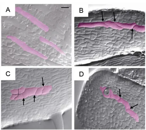
Two independent homozygous lines in whichamiRDEK1expression was triggered only upon DEX application were selected. Induction of amiRDEK1 expression loweredDEK1mRNA levels to about half those detected in control plants (supplementary material Fig. S4J). Upon flowering, control sepals showed a classic epidermal structure with GCs surrounded by smaller cells (Fig. 4A). In DEX-grown plants, we observed sepals with few or no GCs, as well as GC-like structures that had undergone late divisions after the onset of differentiation (Fig. 4B-D). Thus, reduced levels of DEK1 affect the maintenance of GC differentiation.
Cell shape defects indek1-4are not caused by changes in microtubule behaviour
PC shape is affected in mutants with altered cortical microtubule (CMT) dynamics (Ivakov and Persson, 2013). Thedek1-4allele was introgressed into a line harbouring a GFP-TUA6 expression construct (Ueda et al., 1999) and the resulting plants analysed. In WT seedlings, small polygonal PCs have highly oriented CMTs arranged transversely with respect to their long axis, while in more lobed PCs CMTs usually orient more randomly and bundle densely in neck regions. Indek1-4, CMTs are mainly oriented perpendicularly to the long axis of the cell (supplementary material Fig. S6B,C). Quantification confirmed that CMTs in mutant PCs are more highly oriented than in WT PCs (supplementary material Fig. S6D).
To test whether their abnormal arrangement in the mutant was due to an inability of CMTs to rearrange their distribution, or was a consequence of cell shape defects, we studied CMT reorganisation upon ablation in cotyledons (Sampathkumar et al., 2014). CMTs in both mutant and WT cotyledons reoriented in cell files surrounding the ablation site (supplementary material Fig. S6E-H), suggesting that CMT dynamics are not disrupted indek1-4. This observation supports the hypothesis that the CMT arrangement in the mutant is a consequence of altered cell shape.
[image:3.612.66.284.57.408.2]DEK1 activity may define epidermal cell-cell contact zones A recent study has provided strong evidence that the expression of HD-ZIP IV-encoding genes is maintained by intercellular signalling mediated by ACR4 (San-Bento et al., 2014). We measured the height of epidermal cell contact zones and found that they are less uniform in height indek1-4 than in WT seedlings (supplementary material Fig. S7). Consistent with a defect in epidermal cell adhesion (Singh et al., 2005), we also observed examples of cell separation in the cotyledons of 35S:amiRDEK1 lines and the abnormal accumulation of callose at epidermal cell boundaries in Fig. 2.dek1-4mutants show reduced expression of
differentiation-promoting HD-ZIP IV TF-encoding genes.(A,B) Ten-day-old Col-0 (A) and
dek1-4(B) seedlings. (C) Distribution of branch point numbers in the first and second true leaves of 15-day-old plants. Number of trichomes:n=500 for Col-0 andn=400 fordek1-4.(D) Gene expression levels in 6-day-old Col-0 anddek1-4
seedlings were quantified by qRT-PCR and normalised using the expression of
EIF4A. Shown is the average of three independent biological replicates±s.d.
DEVEL
O
the expanding cotyledons of dek1-4 seedlings (supplementary material Fig. S7).
Unlike animal calpains, which exist in many isoforms,DEK1is a single-copy gene unique to land plants (Liang et al., 2013; Lid et al., 2002). Here we show that defects in DEK1 perturb epidermal cell differentiation via a mechanism affecting the transcription of genes encoding HD-ZIP IV family TFs. The ancestral HD-ZIP IV protein, like DEK1, is thought to have arisen in the common algal ancestor of land plants, where it might have been crucial in the evolution of a specialised outer cell layer with the characteristics necessary for life on land, such as cuticle formation and stomatal development (Zalewski et al., 2013). Apoplastic signalling mediated by ACR4, like DEK1 function, is necessary for the maintenance of HD-ZIP IV gene expression (San-Bento et al., 2014). However, genetic analysis
carried out inArabidopsisand maize (Becraft et al., 2002; Roeder et al., 2012) suggests that DEK1 and ACR4 act in parallel.
[image:4.612.46.411.51.560.2]A key requirement of the epidermis is that it should be continuous, implying the maintenance of a highly regulated zone of circumferential contact between neighbouring cells. A common feature of phenotypes associated with loss of DEK1 activity is epidermal cell separation, suggesting that DEK1 might regulate this zone of cell-cell contact (Ahn et al., 2004; Johnson et al., 2005; Lid et al., 2005). Here we show that althoughdek1-4mutants produce a continuous epidermis, the zone of cell-cell contact between epidermal cells is perturbed, further supporting this hypothesis. We propose that DEK1 influences HD-ZIP IV gene expression, and thus epidermal differentiation, by regulating epidermal cell adhesion, a function that might have been crucial in the successful move of plants onto dry land. Fig. 3.DEK1transcript levels correlate with HD-ZIP IV gene expression and with epidermal phenotypes.(A) Gene expression levels in the aerial parts of 6-day-old Col-0 and
35S:amiRDEK1seedlings were quantified by qRT-PCR and normalised using the expression ofEIF4A. Shown is the average of three independent biological replicates±s.d. (B) Confocal images of the adaxial side of 4-day-old cotyledons stained with PI. Scale bars: 50μm. (C) Distribution of branch point numbers in the first and second true leaves of 15-day-old plants. Number of trichomes:
n=507 for Col-0,n=515 for line E,n=498 for line C andn=143 for line L. Red bar, aborted trichomes. (D) Scanning electron micrographs of true leaves from 15-day-old plants. Scale bars: 50μm.
DEVEL
O
MATERIALS AND METHODS Plant material and growth conditions
Seeds were obtained from the Nottingham Arabidopsis Stock Centre unless
otherwise stated. The dek1-4 allele was donated by Adrienne Roeder
(Cornell University, USA) and introgressed into the Col-0 background.
dek1-4genotyping involved amplifying genomic DNA using the primers shown in supplementary material Table S1 and sequencing the resulting product with primer RG15 (supplementary material Table S1, Fig. S2). T-DNA insertion lines were genotyped using the primers described in
supplementary material Table S1. ADEK1-specific artificial microRNA
was designed using MicroRNA Designer (WMD, http://wmd3.weigelworld. org) and generated using the PCR primers in supplementary material
Table S1 according to the protocol from WMD. The pre-amiRNAwas
recombined into pDONR221 (Invitrogen) and then into the Gateway-compatible binary vector pART27 (Lee and Gelvin, 2008) under control of a CaMV 35S promoter, or into the pOPON2.1 vector (a gift from Ian Moore,
University of Oxford, UK). Plants expressingpRPS5A:CALPAIN-6XHIS
were generated as described forpRPS5A:CALPAIN-GFP(Johnson et al.,
2008), but with the insertion of an in-frame two times 6×His tag with a
STOP codon in place of theGFPsequence.
Soil-grown plants were placed at 20°C and 55% relative humidity under a
16-h light/8-h dark cycle. Forin vitrocultures, seeds were surface sterilised with
chlorine gas, stratified on media for 2-3 days in the dark and germinated under a
16-h light/8-h dark cycle at 21°C. To induceDEK1silencing, germinated
seedlings were transferred after 5 days onto medium supplemented with 0.4% DMSO (as control) or 25 µm dexamethasone (DEX).
Imaging and image analysis
Scanning electron microscopy samples were mounted on graphite paste (Electron Microscopy Sciences) and visualised using an SH-3000 table-top
scanning electron microscope (HIROX) at −30°C or −50°C with an
accelerating voltage of 5 kV.
Cotyledon PC cell walls were stained with 1 mg/ml propidium iodide (PI) for 5 min, briefly rinsed, and imaged at a point midway between the midrib and margin and halfway along the blade. Cell areas and perimeters were
measured using ImageJ (NIH). A‘shape factor’(SF=4πarea/perimeter2)
was calculated for each cell (Dewitte et al., 2007). CMT alignments were quantified using the ImageJ plug-in FibrilTool (Boudaoud et al., 2014). All confocal images were acquired using an LSM700 confocal microscope (Carl Zeiss).
For tissue imprints, we used an agarose-based method (Mathur and Koncz, 1997). Light micrographs were taken with an Axio Imager.M2 microscope (Carl Zeiss) using DIC optics.
RNA extraction and quantitative gene expression analysis Seedlings were grown vertically for 6 days on square plates and aerial parts harvested. Three independent biological replicates of 15-20 seedlings were used. RNA extraction, DNase treatment and qPCR analysis were carried out as described previously (Xing et al., 2013). Expression levels of each gene,
relative toEIF4A, were determined as previously described (Ferrari et al.,
2006). Primers used for qRT-PCR analysis are shown in supplementary material Table S1.
Ploidy analysis
Cotyledons were harvested with forceps, chopped with a razor blade in nuclear lysis buffer (Dewitte et al., 2007), filtered and stained with DAPI. The nuclei-containing solution was analysed with a Partec ploidy analyser. Three biological replicates (20 cotyledons from ten independent plants for each replicate) were performed.
Acknowledgements
We thank Adrienne Roeder (Cornell University, USA) for providing the originaldek1-4 mutant line and for helpful discussions; Ian Moore (University of Oxford, UK) for providing the binary vector pOPON2.1; and Jeremy Just (RDP, ENS de Lyon, France) for helping with statistical analysis.
Competing interests
The authors declare no competing or financial interests.
Author contributions
R.G. and G.C.I. designed the experiments and wrote the paper; R.G., G.C.I., S.S. and A.M.W. performed the experiments; K.L.J. and R.S.-B. generated materials; J.A.H.M. contributed analytic tools.
Funding
R.G. was supported by an Agence Nationale de la Recherche (France) Chaire D’Excellence [ANR-10-CHEX-0011 - 01 to G.C.I.], R.S.-B. by the Marie Curie ITN SIREN, and A.M.W. by a Victorian Government postdoctoral training fellowship.
Supplementary material
Supplementary material available online at
http://dev.biologists.org/lookup/suppl/doi:10.1242/dev.122325/-/DC1
References
Abe, M., Katsumata, H., Komeda, Y. and Takahashi, T.(2003). Regulation of shoot epidermal cell differentiation by a pair of homeodomain proteins in Arabidopsis.Development130, 635-643.
Ahn, J.-W., Kim, M., Lim, J. H., Kim, G.-T. and Pai, H.-S.(2004). Phytocalpain controls the proliferation and differentiation fates of cells in plant organ development.Plant J.38, 969-981.
Becraft, P. W., Stinard, P. S. and McCarty, D. R.(1996). CRINKLY4: a TNFR-like receptor kinase involved in maize epidermal differentiation. Science 273, 1406-1409.
Becraft, P. W., Li, K., Dey, N. and Asuncion-Crabb, Y.(2002). The maize dek1 gene functions in embryonic pattern formation and cell fate specification.
Development129, 5217-5225.
Boudaoud, A., Burian, A., Borowska-Wykręt, D., Uyttewaal, M., Wrzalik, R., Kwiatkowska, D. and Hamant, O.(2014). FibrilTool, an ImageJ plug-in to quantify fibrillar structures in raw microscopy images.Nat. Protoc.9, 457-463.
Craft, J., Samalova, M., Baroux, C., Townley, H., Martinez, A., Jepson, I., Tsiantis, M. and Moore, I. (2005). New pOp/LhG4 vectors for stringent glucocorticoid-dependent transgene expression in Arabidopsis.Plant J. 41, 899-918.
Demko, V., Perroud, P.-F., Johansen, W., Delwiche, C. F., Cooper, E. D., Remme, P., Ako, A. E., Kugler, K. G., Mayer, K. F. X., Quatrano, R. et al.(2014). Genetic analysis of DEFECTIVE KERNEL1 loop function in three-dimensional body patterning in Physcomitrella patens.Plant Physiol.166, 903-919.
Depege-Fargeix, N., Javelle, M., Chambrier, P., Frangne, N., Gerentes, D., Perez, P., Rogowsky, P. M. and Vernoud, V.(2011). Functional characterization of the HD-ZIP IV transcription factor OCL1 from maize.J. Exp. Bot.62, 293-305.
[image:5.612.54.294.54.268.2]Dewitte, W., Scofield, S., Alcasabas, A. A., Maughan, S. C., Menges, M., Braun, N., Collins, C., Nieuwland, J., Prinsen, E., Sundaresan, V. et al.(2007). Arabidopsis CYCD3 D-type cyclins link cell proliferation and endocycles and Fig. 4. Silencing of theDEK1gene leads to the appearance of dividing
GCs in sepals.Agarose imprints of sepal epidermis from DEX-grown Col-0 (A) andamiRDEK1line 2 (B,C) and 3 (D) plants. GCs are false coloured in pink. Arrows indicate divisions that have occurred in GCs; dotted lines indicate common division planes in a group of cells. Scale bar: 50μm.
DEVEL
O
are rate-limiting for cytokinin responses. Proc. Natl. Acad. Sci. USA 104, 14537-14542.
Di Cristina, M., Sessa, G., Dolan, L., Linstead, P., Baima, S., Ruberti, I. and Morelli, G.(1996). The Arabidopsis Athb-10 (GLABRA2) is an HD-Zip protein required for regulation of root hair development.Plant J.10, 393-402.
Ferrari, S., Galletti, R., Vairo, D., Cervone, F. and De Lorenzo, G.(2006). Antisense expression of the Arabidopsis thaliana AtPGIP1 gene reduces polygalacturonase-inhibiting protein accumulation and enhances susceptibility to Botrytis cinerea.Mol. Plant Microbe Interact.19, 931-936.
Galletti, R., Denoux, C., Gambetta, S., Dewdney, J., Ausubel, F. M., De Lorenzo, G. and Ferrari, S.(2008). The AtrbohD-mediated oxidative burst elicited by oligogalacturonides in Arabidopsis is dispensable for the activation of defense responses effective against Botrytis cinerea.Plant Physiol.148, 1695-1706.
Glover, B. J. (2000). Differentiation in plant epidermal cells.J. Exp. Bot. 51, 497-505.
Horstman, A., Fukuoka, H., Muino, J. M., Nitsch, L., Guo, C., Passarinho, P., Sanchez-Perez, G., Immink, R., Angenent, G. and Boutilier, K.(2015). AIL and HDG proteins act antagonistically to control cell proliferation.Development142, 454-464.
Ivakov, A. and Persson, S.(2013). Plant cell shape: modulators and measurements.
Front. Plant Sci.4, 439.
Javelle, M., Vernoud, V., Depege-Fargeix, N., Arnould, C., Oursel, D., Domergue, F., Sarda, X. and Rogowsky, P. M.(2010). Overexpression of the epidermis-specific homeodomain-leucine zipper IV transcription factor Outer Cell Layer1 in maize identifies target genes involved in lipid metabolism and cuticle biosynthesis.Plant Physiol.154, 273-286.
Javelle, M., Klein-Cosson, C., Vernoud, V., Boltz, V., Maher, C., Timmermans, M., Depege-Fargeix, N. and Rogowsky, P. M. (2011a). Genome-wide characterization of the HD-ZIP IV transcription factor family in maize: preferential expression in the epidermis.Plant Physiol.157, 790-803.
Javelle, M., Vernoud, V., Rogowsky, P. M. and Ingram, G. C.(2011b). Epidermis: the formation and functions of a fundamental plant tissue.New Phytol.189, 17-39.
Johnson, K. L., Degnan, K. A., Ross Walker, J. and Ingram, G. C.(2005). AtDEK1 is essential for specification of embryonic epidermal cell fate.Plant J.44, 114-127.
Johnson, K. L., Faulkner, C., Jeffree, C. E. and Ingram, G. C.(2008). The phytocalpain defective kernel 1 is a novel Arabidopsis growth regulator whose activity is regulated by proteolytic processing.Plant Cell20, 2619-2630.
Lee, L.-Y. and Gelvin, S. B.(2008). T-DNA binary vectors and systems.Plant
Physiol.146, 325-332.
Liang, Z., Demko, V., Wilson, R. C., Johnson, K. A., Ahmad, R., Perroud, P.-F., Quatrano, R., Zhao, S., Shalchian-Tabrizi, K., Otegui, M. S. et al.(2013). The catalytic domain CysPc of the DEK1 calpain is functionally conserved in land plants.Plant J.75, 742-754.
Lid, S. E., Gruis, D., Jung, R., Lorentzen, J. A., Ananiev, E., Chamberlin, M., Niu, X., Meeley, R., Nichols, S. and Olsen, O.-A.(2002). Thedefective kernel 1 (dek1) gene required for aleurone cell development in the endosperm of maize grains encodes a membrane protein of the calpain gene superfamily.Proc. Natl.
Acad. Sci. USA99, 5460-5465.
Lid, S. E., Olsen, L., Nestestog, R., Aukerman, M., Brown, R. C., Lemmon, B., Mucha, M., Opsahl-Sorteberg, H.-G. and Olsen, O.-A.(2005). Mutation in the Arabidopisis thaliana DEK1 calpain gene perturbs endosperm and embryo development while over-expression affects organ development globally.Planta
221, 339-351.
Mathur, J. and Koncz, C.(1997). Method for preparation of epidermal imprints using agarose.Biotechniques22, 280-282.
Nakamura, M., Katsumata, H., Abe, M., Yabe, N., Komeda, Y., Yamamoto, K. T. and Takahashi, T.(2006). Characterization of the class IV homeodomain-leucine zipper gene family in Arabidopsis.Plant Physiol.141, 1363-1375.
Ossowski, S., Schwab, R. and Weigel, D.(2008). Gene silencing in plants using artificial microRNAs and other small RNAs.Plant J.53, 674-690.
Perroud, P.-F., Demko, V., Johansen, W., Wilson, R. C., Olsen, O.-A. and Quatrano, R. S. (2014). Defective Kernel 1 (DEK1) is required for three-dimensional growth in Physcomitrella patens.New Phytol.203, 794-804.
Peterson, K. M., Shyu, C., Burr, C. A., Horst, R. J., Kanaoka, M. M., Omae, M., Sato, Y. and Torii, K. U.(2013). Arabidopsis homeodomain-leucine zipper IV proteins promote stomatal development and ectopically induce stomata beyond the epidermis.Development140, 1924-1935.
Richardson, L. G. L. and Torii, K. U.(2013). Take a deep breath: peptide signalling in stomatal patterning and differentiation.J. Exp. Bot.64, 5243-5251.
Roeder, A. H. K., Cunha, A., Ohno, C. K. and Meyerowitz, E. M.(2012). Cell cycle regulates cell type in the Arabidopsis sepal.Development139, 4416-4427.
Samalova, M., Brzobohaty, B. and Moore, I.(2005). pOp6/LhGR: a stringently regulated and highly responsive dexamethasone-inducible gene expression system for tobacco.Plant J.41, 919-935.
Sampathkumar, A., Krupinski, P., Wightman, R., Milani, P., Berquand, A., Boudaoud, A., Hamant, O., Jönsson, H. and Meyerowitz, E. M. (2014). Subcellular and supracellular mechanical stress prescribes cytoskeleton behavior in Arabidopsis cotyledon pavement cells.Elife3, e01967.
San-Bento, R., Farcot, E., Galletti, R., Creff, A. and Ingram, G.(2014). Epidermal identity is maintained by cell-cell communication via a universally active feedback loop in Arabidopsis thaliana.Plant J.77, 46-58.
Schiefelbein, J., Huang, L. and Zheng, X.(2014). Regulation of epidermal cell fate in Arabidopsis roots: the importance of multiple feedback loops.Front. Plant Sci.
5, 47.
Scofield, S., Dewitte, W., Nieuwland, J. and Murray, J. A. H. (2013). The Arabidopsis homeobox gene SHOOT MERISTEMLESS has cellular and meristem-organisational roles with differential requirements for cytokinin and CYCD3 activity.Plant J.75, 53-66.
Singh, S. K., Eland, C., Harholt, J., Scheller, H. V. and Marchant, A.(2005). Cell adhesion in Arabidopsis thaliana is mediated by ECTOPICALLY PARTING CELLS 1–a glycosyltransferase (GT64) related to the animal exostosins.Plant J.
43, 384-397.
Takada, S. and Iida, H.(2014). Specification of epidermal cell fate in plant shoots.
Front. Plant Sci.5, 49.
Takada, S., Takada, N. and Yoshida, A.(2013). ATML1 promotes epidermal cell differentiation in Arabidopsis shoots.Development140, 1919-1923.
Ueda, K., Matsuyama, T. and Hashimoto, T.(1999). Visualization of microtubules in living cells of transgenic Arabidopsis thaliana.Protoplasma206, 201-206.
Vernoud, V., Laigle, G., Rozier, F., Meeley, R. B., Perez, P. and Rogowsky, P. M.
(2009). The HD-ZIP IV transcription factor OCL4 is necessary for trichome patterning and anther development in maize.Plant J.59, 883-894.
Wu, R., Li, S., He, S., Wassmann, F., Yu, C., Qin, G., Schreiber, L., Qu, L.-J. and Gu, H.(2011). CFL1, a WW domain protein, regulates cuticle development by modulating the function of HDG1, a class IV homeodomain transcription factor, in rice and Arabidopsis.Plant Cell23, 3392-3411.
Xing, Q., Creff, A., Waters, A., Tanaka, H., Goodrich, J. and Ingram, G. C.(2013). ZHOUPI controls embryonic cuticle formation via a signalling pathway involving the subtilisin protease ABNORMAL LEAF-SHAPE1 and the receptor kinases GASSHO1 and GASSHO2.Development140, 770-779.
Yang, C., Li, H., Zhang, J., Luo, Z., Gong, P., Zhang, C., Li, J., Wang, T., Zhang, Y., Lu, Y. et al.(2011). A regulatory gene induces trichome formation and embryo lethality in tomato.Proc. Natl. Acad. Sci. USA108, 11836-11841.
Zalewski, C. S., Floyd, S. K., Furumizu, C., Sakakibara, K., Stevenson, D. W. and Bowman, J. L. (2013). Evolution of the class IV HD-zip gene family in streptophytes.Mol. Biol. Evol.30, 2347-2365.