Cell cycling through
development
Edward M. Levine
Department of Ophthalmology and Visual Sciences,
Department of Neurobiology and Anatomy, University of Utah, Salt Lake City, UT 84112, USA
E-mail: [email protected]
Development 131, 2241-2246
Published by The Company of Biologists 2004 doi:10.1242/dev.01180
Summary
Regardless of the species, the development of a multicellular organism requires the precise execution of essential developmental processes including patterning, growth, proliferation and differentiation. The cell cycle, in addition to its role as coordinator of DNA replication and mitosis, is also a coordinator of developmental processes, and is a target of developmental signaling pathways. Perhaps because of its central role during development, the cell cycle mechanism, its regulation and its effects on developing tissues is remarkably complex. It was in this light that the Keystone meeting on the cell cycle and development at Snowbird, Utah in January 2004 was held.
The meeting covered many topics and addressed several crucial questions regarding the cell cycle and development. How is cell number controlled during organogenesis, and what is the relationship between cell size and the cell cycle? How is cell division coordinated during metazoan development of multicellular organisms, and how are developmental cues integrated with the cell cycle? How do cells know when to start and stop dividing? While I attempt to cover these topics here, one message that became clear from this meeting was that the cell cycle, despite its conserved nature, can be modified in
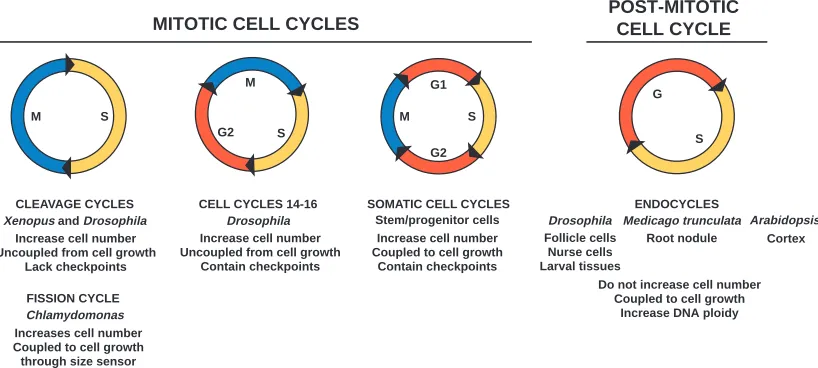
diverse and novel ways to adapt to the demands of a growing cell, tissue or embryo (Fig. 1).
Size matters
In most proliferating somatic tissues, growth is tightly coupled to the cell cycle, which makes the increase in cell number a reliable indicator of tissue growth and size during development. There are several ways in which cell numbers in a tissue can be modulated. These include alterations in patterning (which determines the initial size of the stem or founder cell population), the rate and mode of proliferation of founder and progenitor cells, cell death and cell migration. In plants, cell proliferation and growth primarily contribute to organ size; the removal of overproliferated cells and cell migration does not occur. How do plants coordinate these mechanisms to produce an organ of the appropriate size and cell number? Keiko Torri (University of Washington, Seattle, WA, USA) showed that the leucine-rich receptor-like kinases ERECTA, ERL1 and ERL2 together are crucial for the normal proliferation of cells that form the above-ground organs in Arabidopsis. It is not known whether these proteins directly regulate the cell cycle, but the expression of some cell cycle regulator genes was reduced in erecta, erl1, erl2 triple mutants. Interestingly, these mutants had much larger cells than wild-type plants, suggesting that cell growth compensated to some extent for the reduced proliferation (Shpak et al., 2004).
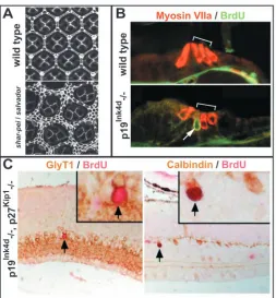
In Drosophila, the coordination of cell proliferation and cell death is essential for determining the correct numbers of cells in a tissue. Georg Halder’s lab (M.D. Anderson Cancer Center, Houston, TX, USA) performed a genetic screen to find mutants with enlarged tissues. They found that flies with mutations in hippo, salvador and warts have larger tissues than normal due to a combination of ectopic proliferation and reduced cell death (Fig 2A). Warts and Hippo are kinases, and Salvador is an adaptor protein. These three proteins form a complex and negatively regulate the G1/S phase cyclin Cyclin E (CycE), and the anti-apoptotic protein DIAP1 (Drosophila Inhibitor of Apoptosis 2241
Meeting review
G
S
Drosophila Follicle cells
Nurse cells Larval tissues
Medicago trunculata Root nodule
Arabidopsis Cortex ENDOCYCLES
Do not increase cell number Coupled to cell growth
Increase DNA ploidy M
S G2
CELL CYCLES 14-16 Drosophila
G1
S
G2 M
SOMATIC CELL CYCLES S
M
CLEAVAGE CYCLES Xenopus and Drosophila
Increase cell number Uncoupled from cell growth
Lack checkpoints
FISSION CYCLE Chlamydomonas
MITOTIC CELL CYCLES POST-MITOTICCELL CYCLE
Increase cell number Uncoupled from cell growth
Contain checkpoints
Increase cell number Coupled to cell growth
Contain checkpoints
Increases cell number Coupled to cell growth through size sensor
[image:1.612.104.513.510.694.2]Stem/progenitor cells
Protein 1), to control proliferation and cell death, respectively (Udan et al., 2003). Halder suggested that this complex acts to coordinate proliferation and apoptosis.
In mammals, the contributions of proliferation, migration and death to the regulation of cell number vary greatly between tissues. One mammalian tissue in which proliferation has a significant effect on cell number during development is the retina. Ed Levine (University of Utah, Salt Lake City, UT, USA) described data showing that Chx10, a homeobox gene expressed in retinal progenitors, regulates retinal cell proliferation rate during development. In the Chx10 mutant, the overall progression of neurogenesis is relatively normal, but the tissue has a major deficit in cell number due to a slowing down of the cell cycle. This correlates with an increase in the percentage of cells expressing the cyclin-dependent kinase inhibitor (CKI) protein p27Kip1, and removal of p27Kip1in the
Chx10 mutant significantly alleviated the cell number defect (Green et al., 2003). Interestingly, Cyclin D1 (CycD1) – an activator of the G1 cyclin-dependent kinases Cdk4 and Cdk6 (Cdk4/6) – is a likely mediator of the interaction between Chx10 and p27Kip1, but this pathway on its own is insufficient
to fully account for Chx10 function in regulating proliferation. Although the late embryonic retinal phenotypes of the Chx10 null mouse and Cycd1 KO mouse are similar, in that both exhibit hypocellularity, the Chx10 phenotype is much more severe. Further work is being carried out to determine whether Chx10 also regulates other components of the cell cycle. If so, Chx10 might act to tailor the cell cycle such that it meets the proliferative demands of the developing retina, and this may be an example of a more general model in which tissue-specific factors control the cell cycle during tissue formation.
Cell growth is tightly coupled to cell cycle progression. However, how this coupling occurs is not clear, and several speakers presented diverse models that addressed this question. Bruce Edgar (Fred Hutchinson Cancer Center, Seattle, WA, USA) discussed how cell growth in polyploid tissues of the Drosophila larvae is tightly coupled to nutritional availability. His lab has previously shown that overexpression of the insulin receptor, other insulin pathway components, or the transcription factor Dmyc (Dm – FlyBase), drives cell growth in larval tissues (Britton et al., 2002; Saucedo et al., 2003; Pierce et al., 2004). However, although the insulin pathway acts as a nutrient sensor, the effects on cell growth by Dmyc are not dependent on nutrient availability or the insulin pathway, indicating that Dmyc drives cell growth by an independent mechanism. Furthermore, they found that Dmyc selectively promotes the expression of genes involved with protein synthesis and RNA transcription (Orian et al., 2003). Another question raised by Edgar was, how do growth regulators like Dmyc and the insulin pathway promote endocycles (a modified cell cycle associated with increased cell size in which growth and DNA replication are uncoupled from mitosis, resulting in polyploidy; Fig. 1, and see below)? Edgar proposed a relatively simple oscillatory mechanism, in which CycE levels accumulate in phase with DNA replication and dE2F (E2f – FlyBase) activity increases out of phase with DNA replication, and these two proteins regulate each other through feedback mechanisms. Growth cues such as the insulin pathway and Dmyc feed into this oscillation by increasing the levels of CycE protein, thereby promoting the G to S phase transition in endocycling tissues (Edgar and Orr-Weaver, 2001).
[image:2.612.311.564.69.342.2]Martin Raff (University College, London, UK) addressed how cell growth and proliferation are coordinated in mitotic mammalian cells, and specifically whether mammalian cells have cell-size checkpoints that are analogous to those in yeast (checkpoints arrest the cell cycle if proper cell-cycle events are not completed). Experiments in which the cell-cycle and growth rate of rat Schwann cells were modulated in culture by extracellular factors indicate that these cells may not have such checkpoints (Conlon and Raff, 2003). Instead, cell size at cell division appears to depend on how fast the cells progress through the cell cycle and how fast they grow, which in turn depends on extracellular signals that control cell cycle progression, cell growth, or both. By contrast, Jim Umen (Salk Institute, La Jolla, CA, USA) has found that, in the unicellular green alga Chlamydomonas reinhardtii, the activity of the retinoblastoma-like protein Mat3/Rb is sensitive to a minimum
cell size and acts as a sensor of size threshold. Cells grow during their gap (G) phases of the cell cycle, and in Chlamydomonas, the G1 phase varies in length; cells can grow to be very large and of varying size. Following the initial entry into S phase, the cell then goes through multiple rounds of S to M (S-M) cycles, termed fission cycles (Fig. 1), during which all daughters ultimately attain a uniform size. The coupling of cell size to the number of fission cycles depends on Mat3/Rb activity (Umen and Goodenough, 2001), which might (1) prevent premature entry into fission cycles and (2) restrict the number of fission cycles according to cell size. Interestingly, Umen described how this mechanism of cell size regulation is coupled to developmental programming in multicellular relatives of Chlamydomonas, like Volvox carteri, and how Volvox may use the Mat3/Rb pathway to regulate asymmetric cell division and germ/soma differentiation.
Early embryonic cell cycles
Metazoans undergo a period of rapid cell cycles, termed cleavage cycles, immediately following fertilization (Fig. 1). In Drosophila, these divisions produce the syncitial blastoderm and in Xenopus, the blastula, but these cycles are remarkably similar in these two species in that the size or mass of the embryo does not increase compared with the fertilized eggs from which they arise. In each case, the cell cycles are similar in design; they have S-M oscillations without intervening gap phases and they function independently of transcription. In Drosophila, the early cleavage cycles (cycles 2-9) are remarkably fast; they take approximately 8.5-9 minutes and S phase possibly lasts ~3.5 minutes. How then, is the entire genome replicated in this short time? This is an especially important question because, during S phase, heterochromatin replicates later than euchromatin, and the entire genome must be precisely and completely duplicated during each cycle. Does heterochromatin exist during the early cleavage cycles? According to Patrick O’Farrell (UC San Francisco, CA, USA), heterochromatin is present but its replication is not delayed, suggesting that DNA replication through heterochromatin is regulated differently in cleavage cell cycles compared to later cell cycles. O’Farrell suggested that zygotic gene products are responsible for the appearance of late-replicating heterochromatic DNA and the lengthening of S-phase, because its later replication does not occur until the maternal-zygotic transition (MZT).
Another important question regarding the Drosophila cleavage cycles is how the activity of the mitotic cyclin and cyclin-dependent kinase (CDK) complex, CycB/Cdk1, is regulated to promote mitosis. High CycB/Cdk1 activity prevents DNA re-replication and promotes entry into the mitotic program, and low CycB/Cdk1 activity causes cells to exit mitosis. This typically occurs via the anaphase promoting complex (APC)-dependent degradation of CycB. During the cleavage cycles, however, CycB/Cdk1 activity does not oscillate, except for highly localized Cdk1 inactivation due to CycB degradation at mitotic spindles in late metaphase; this appears to be sufficient for mitotic exit during these cycles (Huang and Raff, 1999; Su et al., 1998). Are high CycB levels the default during the cleavage cycles? Evidence suggests that high CycB levels are actively regulated prior to the MZT by proteins encoded by pan gu (png), plutonium (plu) and giant nuclei (gnu). png putatively encodes a serine/threonine kinase,
and forms a complex with PLU and GNU that is required for kinase activity, as discussed by Terri Orr-Weaver (MIT, Cambridge, MA, USA). However, she also showed that CycB is not a direct substrate of this kinase, so work is under way to identify substrates that regulate CycB (Lee et al., 2003).
The cell cycle and the switch from maternal to zygotic control
The MZT in Drosophila and the mid-blastula transition (MBT) in Xenopus are both characterized by a shift in developmental control from maternally provided proteins to those produced by the embryo. This shift also coincides with a switch from cleavage cycles to cell cycles that have gap phases, which is partly due to an acquired dependence on the nascent transcription of cell cycle genes, including histones. Prior to the MZT in Drosophila, histone mRNA is maternally supplied, but once zygotic transcription begins, histone RNA levels begin to oscillate during the cell cycle and accumulate to high levels only in S-phase, when DNA synthesis occurs. In cell cycles that have a G1 phase, histone transcription is upregulated at the G1/S boundary and depends on CycE/Cdk2 activity. However, in Drosophila, the cell cycles immediately following the MZT (cycles 14-16) have a G2 phase, but lack a G1 phase (Fig. 1). What then triggers the synthesis of histone RNA during these cycles? Bob Duronio (University of North Carolina, Chapel Hill, NC, USA) described certain components of this regulation. He showed that zygotic histone transcription depends on the G2 phosphatase Stringcdc25a, which is limiting in these cycles, and
that new histone mRNA synthesis occurs after the histone mRNA from the previous S-phase is degraded. Furthermore, the oscillation in histone mRNA levels during different phases of the cell cycle depends on blocking polyadenylation, which is mediated by Stem Loop Binding Protein (SLBP) binding to a stem-loop in the 3′ untranslated region (UTR) of histone mRNA. In slbp mutants, histone mRNAs are inappropriately polyadenylated at the MZT, resulting in the aberrant accumulation and loss of their cell cycle oscillation (Lanzotti et al., 2002; Sullivan et al., 2001). Similarly, in Xenopus, a change in the regulation of adenylation of maternal cell cycle transcripts occurs at the MBT. Rebecca Hartley (University of New Mexico, Albequerque, NM, USA) showed that the deadenylation of CycA1 and CycB2 mRNA is required for the timed downregulation of CycA1 and B2 proteins, which is necessary for the slowing of S phase and introduction of the G2 phase (Audic et al., 2001). As in Drosophila, this deadenylation depends on RNA-binding proteins interacting with the 3′UTRs of the cyclin transcripts.
Patterning and morphogenesis
regulated gene activation (Fisher and Mechali, 2003). In contrast to Fischer’s work, which exemplifies the importance of how promoting cell cycle activity influences patterning, Paul Mueller (University of Chicago, IL, USA) described how restricting cell cycle activity influences morphogenesis. Following the MBT, the Xenopus embryo undergoes convergent extension, during which the cell cycle arrests transiently in the paraxial mesoderm. Preventing this block disrupts the positioning and segmentation of the paraxial mesoderm and convergent extension for reasons unknown. However, Mueller showed that this cell cycle arrest requires the G2 kinase Wee2, a zygotically transcribed gene that is activated at the MBT and expressed in the paraxial mesoderm. Morpholino knockdown of Wee2 expression levels relieved the cell cycle block and disrupted convergent extension (Leise and Mueller, 2004).
Switching from mitotic cell cycles to endocycles
Mechanisms have evolved to ensure that the entire genome is replicated once and precisely each mitotic cell cycle to maintain genomic stability. However, endocycles can also occur (Fig. 1), which have alternating G to S phases (G-S), thus these cycles do not cause an increase in cell number. Endocycles are important for cell growth and for several developmental processes in plants and animals, and as such considerable attention is being given to understanding their regulation. As may be expected, endocycles use the cell cycle machinery to block mitosis.
One model system being used to study endocycle regulation is the Drosophila oocyte cyst. The cyst consists of 16 clonally related cells, the oocyte and 15 nurse cells, which are cytoplasmically coupled to each other by ring canals. By stage 9, the oocyte is arrested in prophase of Meiosis I, but the nurse cells are highly polyploid due to endocycles. Enveloping the cyst are follicle cells, which have several important functions, including establishing dorsoventral (DV) polarity. Once
enough follicle cells are produced by cell division, they undergo endocycles during formation of the oocyte cyst. Two important questions raised at the meeting were: (1) how does the oocyte stay arrested in meiosis while the nurse cells undergo endocycles, especially considering their cytoplasmic linkage; and (2) how does the mitotic-to-endocycle transition occur in the follicle cells? Mary Lilly (NIH, Bethesda, MD, USA) addressed the first question by presenting evidence that expression levels of the CKI Dacapo (Dap) are highly locally regulated within the cyst (Fig. 3A). High levels of Dap in the oocyte block CycE/Cdk2 activity and low levels of Dap around the nurse cell nuclei activates CycE/Cdk2, allowing nurse cells to progress into S phase (Hong et al., 2003). Hannele Ruhola-Baker (University of Washington, Seattle, WA, USA) addressed the second question and showed that Notch signaling is required for the transition from mitotic cycles to endocycles (Fig. 3A). They found that in response to Notch, Dap and Stringcdc25aexpression is repressed and Fizzy-related (FzrCdh1)
expression is promoted (Shcherbata et al., 2004). One model is that Notch activity coordinates the regulation of important checkpoints during this transition: FzrCdh1 promotes APC
activity by keeping CDK activity low, which is important for progression through early G1; low levels of Dap allows the transition from G1 to S by raising CycE/Cdk2 activity; and low Stringcdc25ablocks mitosis by keeping CycB/Cdk1 activity low.
It became apparent at the meeting that in plants, similar molecules and mechanisms to those found in animals may regulate the transition from a mitotic cell cycle to an endocycle (Fig. 3B). Eva Kondorosi (CNRS, Gif sur Yvette, France) described data showing that Ccs52b, the FzrCdh1component of
the APC in the legume Medicago trunculata, is necessary for polyploidy to occur in the large nitrogen-fixing root nodule cells (Vinardell et al., 2003). Jim Murray (University of Cambridge, UK) and Dirk Inze (Ghent University, Belgium) described independent data indicating that levels of both
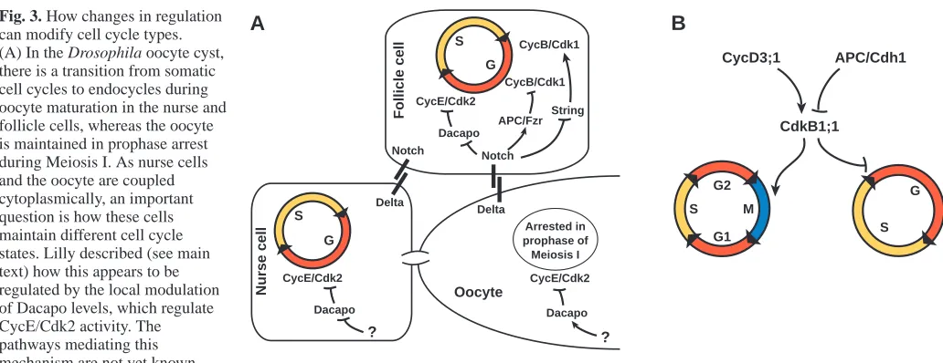
D-Fig. 3. How changes in regulation can modify cell cycle types. (A) In the Drosophila oocyte cyst, there is a transition from somatic cell cycles to endocycles during oocyte maturation in the nurse and follicle cells, whereas the oocyte is maintained in prophase arrest during Meiosis I. As nurse cells and the oocyte are coupled cytoplasmically, an important question is how these cells maintain different cell cycle states. Lilly described (see main text) how this appears to be regulated by the local modulation of Dacapo levels, which regulate CycE/Cdk2 activity. The pathways mediating this mechanism are not yet known.
Ruholla-Baker described how the transition to endocycles in the follicle cells is mediated by activation of the Notch pathway through Delta ligand expressed by the oocyte and nurse cells (see main text). Notch signaling may regulate the cell cycle in at least three ways: repression of CycB/Cdk1 activity by downregulation of Stringcdc25; upregulation of Fzr/Cdh1-dependent APC activity; and activation of CycE/Cdk2 activity by downregulation of Dacapo. (B) In Arabidopsis and Medicago trunculata, the transition from a somatic cell cycle to an endocycle during differentiation appears to depend on the activity of the CDK CdkB1;1. This may involve Fzr/Cdh1-dependent APC activity and downregulation of CycD3;1, although the specific nature of the interactions of these proteins with CdkB1;1 is not yet known.
APC/Cdh1
CdkB1;1 CycD3;1
G
S G1
S G2
M
Delta Notch
APC/Fzr CycB/Cdk1
String
Dacapo CycE/Cdk2
CycB/Cdk1
Dacapo CycE/Cdk2 Oocyte
N
u
rs
e
c
ell
F
o
ll
ic
le
c
ell
G S
CycE/Cdk2
? ?
G S
Dacapo Delta
Notch
B
A
[image:4.612.43.559.69.268.2]cyclin and of a plant-specific S-G2 regulated CDK, known as Cdkb1;1, need to be downregulated for endocycles to occur. One possible model is that reduced D-cyclin expression combined with increased FzrCdh1-dependent APC activity
downregulates mitotic cyclins leading to reduced activity of Cdkb1;1 and the activation of endocycles. Plants apparently lack a cyclin E equivalent, and certain plant D-type cyclins may play an equivalent role in providing a limiting activity for S-phase entry (Dewitte et al., 2003).
Consistent with the role of FzrCdh1 in plant endocycles,
Yukiko Mizukami (UC Berkeley, CA, USA) overexpressed FzrCdh1 in Arabidopsis to alter the timing of transition from
mitotic cycles to endocycles in the leaf epidermal lineage to study the relationship of cell fate and patterning, and cell cycle regulation. Interestingly, modifying the timing of endocycle transition caused changes in patterning but not cell fate within the leaf epidermis. Mizukami suggested that endocycles do not drive differentiation per se, but rather are necessary for the correct patterning of the leaf epidermis.
Exiting the cell cycle
Once metazoan embryos begin organogenesis, most somatic cells are undergoing the prototypical mitotic cell cycle (Fig. 1). In these somatic cell cycles, the G1 phase provides an opportunity for external inputs to influence the autonomous mechanisms of the cell cycle. This is an especially critical phase for most developing tissues, as it is often when cell cycle exit is coordinated with terminal differentiation. This has been a strong focus of investigation in the vertebrate central nervous system (CNS), and Martine Roussel (St. Jude Children’s Research Hospital, Memphis, TN, USA) described how areas of the brain utilize combinations of two CKI classes, the Ink4 and Cip/Kip proteins, to initiate cell cycle exit at the onset of neuronal differentiation and to maintain neurons in a postmitotic state. Roussel and Neil Segil showed that p19Ink4dloss in the mouse
induces deafness due to inappropriate cell cycle re-entry in differentiated, sensory hair cells, which is followed by apoptosis (Fig. 2B) (Chen et al., 2003). Roussel also demonstrated that many CNS neurons require both p19Ink4d and p27Kip1 to
maintain cell cycle exit in their differentiated state (Cunningham et al., 2002; Zindy et al., 1999). In p19Ink4dand p27Kip1double
knockout mice, postmitotic, differentiated neurons re-entered the cell cycle in many parts of the brain and retina (Fig. 2C), a phenotype not observed in the single knockouts. These findings indicate that the cell cycle arrest normally maintained in terminally differentiated cells is dependent on CKI activity beyond the initial event of cell cycle exit.
How CKIs are regulated to promote cell cycle arrest is a question with no single answer. Examples abound that suggest that CKIs are regulated at many levels, from transcription to post-translational modification. Two distinct examples were described at the meeting by Joan Seoane (Sloan Kettering, New York, NY, USA) and Ludger Hengst (Max Planck Institute, Martinsreid, Germany). Seoane described work with Joan Massague in which TGFβ-induced cell cycle arrest depends not on TGFβ-dependent SMAD transcription factors, but rather on the direct interactions of SMAD proteins with other transcription factors, such as FOXO (Seoane et al., 2004), ATF3 (Kang et al., 2003) and E2F4/5 (Chen et al., 2002), which are downstream of signals other than TGFβ. This indicates that the cell cycle arrest mediated through the CKIs that are the
transcriptional targets of TGFβ (such as p21Cip1) is probably
due to combined extracellular inputs. Hengst showed that the RNA-binding Hu proteins regulate the translational efficiency of p27Kip1. p27Kip1contains a small open reading frame (µORF)
at the extreme 5′end of the mRNA and an internal ribosome entry site (IRES) between the µORF and the p27Kip1ORF. The
HuR protein binds in the IRES region and may interfere with the access of the IRES to ribosomes, thereby reducing the translational efficiency of the p27Kip1ORF (Gopfert et al., 2003;
Kullmann et al., 2002). As HuR is expressed in proliferating cells, this mode of p27Kip1 regulation explains how p27Kip1
protein levels might be kept in check in proliferating cells that express high levels of p27Kip1mRNA.
G1 regulators in development: critical…or not?
Surprisingly, several labs have found that many cell cycle components predicted to be essential for cell cycle regulation in embryonic mouse tissues are largely dispensable during development. Knockouts of D-cyclins, E-cyclins, Cdk2, Cdk4 and all G1 regulators do not directly or globally disrupt embryogenesis. For example, Peter Sicinski (Dana Farber Cancer Institute, Boston, MA, USA) pointed out that many embryonic tissues still developed fairly well when D-cyclins were not detected in mice with double knockout combinations (Ciemerych et al., 2002), and Philip Kaldis (NIH, Bethesda, MD, USA) showed that in the absence of Cdk2, mice are viable but sterile due to a requirement for Cdk2 in both the male and female germlines (Berthet et al., 2003; Ortega et al., 2003). Although the lack of global phenotypes resulting from knockouts of these essential cell cycle regulators could be explained by the compensatory actions of other proteins, these studies suggest that the embryonic somatic cell cycle has a high degree of plasticity that is more sophisticated than simple redundancy. An important goal is to identify how the cell cycle adapts in the absence of these key proteins.
basis for genetic identification of parallel acting genes and for genes that act in the temporal control of cell division, several of which regulate levels of the CKI Cki-1.
Concluding remarks
These were just some of the excellent talks that were presented at the meeting. While many of the talks reveal just how versatile the cell cycle can be during different developmental events, it is also clear that we have much more to learn about the basic workings of the cell cycle within the context of developing tissues, and how it is that the cell cycle can be modified to fit into a developmental program, and yet can be so adaptable as to function ‘normally’ in the absence of supposed key components. Fortunately, these questions will not, and should not, be answered in isolation because this meeting revealed just how much common ground exists between the diverse model systems being investigated. In many ways, this meeting launched the study of cell cycle and development as a global, rather than as an organism-specific, field.
I wish to thank the speakers who were kind enough to answer my inquiries, and I also apologize to those whose work I was unable to highlight.
References
Audic, Y., Anderson, C., Bhatty, R. and Hartley, R. S. (2001). Zygotic
regulation of maternal cyclin A1 and B2 mRNAs. Mol. Cell. Biol. 21, 1662-1671.
Berthet, C., Aleem, E., Coppola, V., Tessarollo, L. and Kaldis, P. (2003).
Cdk2 knockout mice are viable. Curr. Biol. 13, 1775-1785.
Britton, J. S., Lockwood, W. K., Li, L., Cohen, S. M. and Edgar, B. A.
(2002). Drosophila’s insulin/PI3-kinase pathway coordinates cellular metabolism with nutritional conditions. Dev. Cell 2, 239-249.
Brumby, A. M. and Richardson, H. E. (2003). scribble mutants cooperate
with oncogenic Ras or Notch to cause neoplastic overgrowth in Drosophila.
EMBO J. 22, 5769-5779.
Brumby, A. M., Zraly, C. B., Horsfield, J. A., Secombe, J., Saint, R., Dingwall, A. K. and Richardson, H. (2002). Drosophila cyclin E interacts
with components of the Brahma complex. EMBO J. 21, 3377-3389.
Chen, C. R., Kang, Y., Siegel, P. M. and Massague, J. (2002). E2F4/5 and
p107 as Smad cofactors linking the TGFbeta receptor to c-myc repression.
Cell 110, 19-32.
Chen, P., Zindy, F., Abdala, C., Liu, F., Li, X., Roussel, M. F. and Segil, N.
(2003). Progressive hearing loss in mice lacking the cyclin-dependent kinase inhibitor Ink4d. Nat. Cell Biol. 5, 422-426.
Ciemerych, M. A., Kenney, A. M., Sicinska, E., Kalaszczynska, I., Bronson, R. T., Rowitch, D. H., Gardner, H. and Sicinski, P. (2002). Development
of mice expressing a single D-type cyclin. Genes Dev. 16, 3277-3289.
Conlon, I. and Raff, M. (2003). Differences in the way a mammalian cell and
yeast cells coordinate cell growth and cell-cycle progression. J. Biol. 2, 7.
Cunningham, J. J., Levine, E. M., Zindy, F., Goloubeva, O., Roussel, M. F. and Smeyne, R. J. (2002). The cyclin-dependent kinase inhibitors
p19(Ink4d) and p27(Kip1) are coexpressed in select retinal cells and act cooperatively to control cell cycle exit. Mol. Cell. Neurosci. 19, 359-374.
Dewitte, W., Riou-Khamlichi, C., Scofield, S., Healy, J. M., Jacqmard, A., Kilby, N. J. and Murray, J. A. (2003). Altered cell cycle distribution,
hyperplasia, and inhibited differentiation in Arabidopsis caused by the D-type cyclin CYCD3. Plant Cell 15, 79-92.
Duman-Scheel, M., Weng, L., Xin, S. and Du, W. (2002). Hedgehog
regulates cell growth and proliferation by inducing Cyclin D and Cyclin E.
Nature 417, 299-304.
Edgar, B. A. and Orr-Weaver, T. L. (2001). Endoreplication cell cycles: more
for less. Cell 105, 297-306.
Fisher, D. and Mechali, M. (2003). Vertebrate HoxB gene expression requires
DNA replication. EMBO J. 22, 3737-3748.
Gopfert, U., Kullmann, M. and Hengst, L. (2003). Cell cycle-dependent
translation of p27 involves a responsive element in its 5′-UTR that overlaps with a uORF. Hum. Mol. Genet. 12, 1767-1779.
Green, E. S., Stubbs, J. L. and Levine, E. M. (2003). Genetic rescue of cell
number in a mouse model of microphthalmia: interactions between Chx10 and G1-phase cell cycle regulators. Development 130, 539-552.
Hong, A., Lee-Kong, S., Iida, T., Sugimura, I. and Lilly, M. A. (2003). The
p27cip/kip ortholog dacapo maintains the Drosophila oocyte in prophase of meiosis I. Development 130, 1235-1242.
Huang, J. and Raff, J. W. (1999). The disappearance of cyclin B at the end
of mitosis is regulated spatially in Drosophila cells. EMBO J. 18, 2184-2195.
Kang, Y., Chen, C. R. and Massague, J. (2003). A self-enabling TGFbeta
response coupled to stress signaling: Smad engages stress response factor ATF3 for Id1 repression in epithelial cells. Mol. Cell 11, 915-926.
Kango-Singh, M., Nolo, R., Tao, C., Verstreken, P., Hiesinger, P.R., Bellen, H. J., and Halder, G. (2002). Shar-pei mediates cell proliferation arrest
during imaginal disc growth in Drosophila. Development 129, 5719-5730.
Kullmann, M., Gopfert, U., Siewe, B. and Hengst, L. (2002). ELAV/Hu
proteins inhibit p27 translation via an IRES element in the p27 5’UTR.
Genes Dev. 16, 3087-3099.
Lanzotti, D. J., Kaygun, H., Yang, X., Duronio, R. J. and Marzluff, W. F.
(2002). Developmental control of histone mRNA and dSLBP synthesis during Drosophila embryogenesis and the role of dSLBP in histone mRNA 3′end processing in vivo. Mol. Cell. Biol. 22, 2267-2282.
Lee, L. A., Van Hoewyk, D. and Orr-Weaver, T. L. (2003). The Drosophila
cell cycle kinase PAN GU forms an active complex with PLUTONIUM and GNU to regulate embryonic divisions. Genes Dev. 17, 2979-2791.
Leise, W. F., III and Mueller, P. R. (2004). Inhibition of the cell cycle is
required for convergent extension of the paraxial mesoderm during Xenopus neurulation. Development 131, 1703-1715.
Orian, A., van Steensel, B., Delrow, J., Bussemaker, H. J., Li, L., Sawado, T., Williams, E., Loo, L. W., Cowley, S. M., Yost, C. et al. (2003).
Genomic binding by the Drosophila Myc, Max, Mad/Mnt transcription factor network. Genes Dev. 17, 1101-1114.
Ortega, S., Prieto, I., Odajima, J., Martin, A., Dubus, P., Sotillo, R., Barbero, J. L., Malumbres, M. and Barbacid, M. (2003).
Cyclin-dependent kinase 2 is essential for meiosis but not for mitotic cell division in mice. Nat. Genet. 35, 25-31.
Pierce, S. B., Yost, C., Britton, J. S., Loo, L. W. M., Flynn, E. M., Edgar, B. A and Eisenman, R. N. (2004). dMyc is required for larval growth and
endoreplication in Drosophila. Development 131, 2317-2327.
Saucedo, L. J., Gao, X., Chiarelli, D. A., Li, L., Pan, D. and Edgar, B. A.
(2003). Rheb promotes cell growth as a component of the insulin/TOR signalling network. Nat. Cell Biol. 5, 566-571.
Seoane, J., Le, H.-V., Shen, L., Anderson, S. A. and Massagué, J. (2004).
Integration of Smad and Forkhead pathways in the control of neuroepithelial and glioblastoma cell proliferation. Cell (in press).
Shcherbata, H., Althauser, C., Schaeffer, V., Findley, S. and Ruohola-Baker, H. (2004). The mitotic-to-endocycle switch in Drosophila follicle
cells is executed by Notch dependent regulation of G1/S-, G2/M- and M/G1-cell cycle transitions. Development (in press).
Shpak, E. D., Berthiaume, C. T., Hill, E. J. and Torri, K. U. (2004).
Synergistic interaction of three ERECTA-family receptor-like kinases controls Arabidopsis organ growth and flower development by promoting cell proliferation. Development 131, 1491-1501.
Su, T. T., Sprenger, F., DiGregorio, P. J., Campbell, S. D. and O’Farrell, P. H. (1998). Exit from mitosis in Drosophila syncytial embryos requires
proteolysis and cyclin degradation, and is associated with localized dephosphorylation. Genes Dev. 12, 1495-1503.
Sullivan, E., Santiago, C., Parker, E. D., Dominski, Z., Yang, X., Lanzotti, D. J., Ingledue, T. C., Marzluff, W. F. and Duronio, R. J. (2001).
Drosophila stem loop binding protein coordinates accumulation of mature histone mRNA with cell cycle progression. Genes Dev. 15, 173-187.
Udan, R. S., Kango-Singh, M., Nolo, R., Tao, C. and Halder, G. (2003).
Hippo promotes proliferation arrest and apoptosis in the Salvador/Warts pathway. Nat. Cell Biol. 5, 914-920.
Umen, J. G. and Goodenough, U. W. (2001). Control of cell division by a
retinoblastoma protein homolog in Chlamydomonas. Genes Dev. 15, 1652-1661.
Vinardell, J. M., Fedorova, E., Cebolla, A., Kevei, Z., Horvath, G., Kelemen, Z., Tarayre, S., Roudier, F., Mergaert, P., Kondorosi, A. et al.
(2003). Endoreduplication mediated by the anaphase-promoting complex activator CCS52A is required for symbiotic cell differentiation in Medicago truncatula nodules. Plant Cell 15, 2093-2105.
Zindy, F., Cunningham, J. J., Sherr, C. J., Jogal, S., Smeyne, R. J. and Roussel, M. F. (1999). Postnatal neuronal proliferation in mice lacking
Ink4d and Kip1 inhibitors of cyclin-dependent kinases. Proc. Natl. Acad.