Serotonin transporter variant drives
preventable gastrointestinal abnormalities in
development and function
Kara Gross Margolis, … , Randy D. Blakely, Michael D.
Gershon
J Clin Invest. 2016;
126(6)
:2221-2235.
https://doi.org/10.1172/JCI84877
.
Autism spectrum disorder (ASD) is an increasingly common behavioral condition that
frequently presents with gastrointestinal (GI) disturbances. It is not clear, however, how gut
dysfunction relates to core ASD features. Multiple, rare hyperfunctional coding variants of
the serotonin (5-HT) transporter (SERT, encoded by SLC6A4) have been identified in ASD.
Expression of the most common SERT variant (Ala56) in mice increases 5-HT clearance
and causes ASD-like behaviors. Here, we demonstrated that Ala56-expressing mice
display GI defects that resemble those seen in mice lacking neuronal 5-HT. These defects
included enteric nervous system hypoplasia, slow GI transit, diminished peristaltic reflex
activity, and proliferation of crypt epithelial cells. An opposite phenotype was seen in
SERT-deficient mice and in progeny of WT dams given the SERT antagonist fluoxetine. The
reciprocal phenotypes that resulted from increased or decreased SERT activity support the
idea that 5-HT signaling regulates enteric neuronal development and can, when disturbed,
cause long-lasting abnormalities of GI function. Administration of a 5-HT
4agonist to Ala56
mice during development prevented Ala56-associated GI perturbations, suggesting that
excessive SERT activity leads to inadequate 5-HT
4–mediated neurogenesis. We propose
that deficient 5-HT signaling during development may contribute to GI and behavioral
features of ASD. The consequences of therapies targeting SERT during pregnancy warrant
further evaluation.
Research Article
Gastroenterology
Find the latest version:
The Journal of Clinical Investigation
R e s e a R c h a R t i c l eIntroduction
The increasingly frequent diagnosis of autism spectrum disorder (ASD) is based on deficits in social communication and interac-tion together with restricted, repetitive patterns of behavior (1). Although the core features of ASD are behavioral, gastrointestinal (GI) disturbances, particularly constipation, are 4-fold more com-mon in ASD (2). Platelet serotonin (5-HT), moreover, which is pri-marily GI-derived (3), is increased in about one-third of patients with ASD (4, 5). Platelets do not synthesize 5-HT (6) but utilize the 5-HT transporter (SERT, encoded by Slc6a4) (7) to take it up (8) as they circulate through the gut. Multiple hyperactive coding vari-ants of SERT have been found in subjects with ASD (9–11). Knock-in mice expressKnock-ing the most common of these, SERT Ala56 (12), display hyperserotonemia, increased 5-HT clearance, and hyper-sensitivity of central 5-HT1A and 5-HT2A receptors, as well as repet-itive behaviors and deficits of socialization that are reminiscent of ASD (13). The phenotype of SERT Ala56 mice suggests that the constitutively elevated activity of SERT in these animals interferes with the 5-HT signaling required for normal brain development and later function (14–16).
Neither 5-HT nor SERT is exclusive to the CNS. The bulk of 5-HT in the body is found in the intestine, where it is a
multifunc-tional signaling molecule during fetal and adult life (17). Changes in SERT-mediated 5-HT clearance, therefore, could contribute to GI and other peripheral comorbidities that accompany ASD. Enteric actions of 5-HT, like those in the brain, are terminated by SERT (18–20). In the bowel, moreover, enterocytes, as well as serotonergic neurons, express SERT (18, 19, 21). SERT uptake into enterocytes and subsequent intracellular catabolism are also important in restricting the spread of mucosal 5-HT to, or desensi-tization of, aberrant targets.
Two 5-HT depots exist in the gut, each with its own biosyn-thetic enzyme (22, 23). A large tryptophan hydroxylase 1–depen-dent (TPH1-depen1–depen-dent) 5-HT pool exists in mucosal enterochro-maffin (EC) cells and, in rats and mice, mast cells, whereas a smaller TPH2-dependent pool exists in the serotonergic neu-rons of the enteric nervous system (ENS). Both pools regulate GI motility (22, 24). EC cell 5-HT stimulates extrinsic sensory nerves (25, 26), which transmit signals of noxious stimuli (27), discom-fort, and pain to the CNS. 5-HT also stimulates intrinsic primary afferent neurons, which recruit the ENS to mediate peristaltic (24, 28–30) and secretory reflexes (31). Enteric neuronal 5-HT func-tions not only as a neurotransmitter but also as a growth factor, stimulating 5-HT4 receptors that promote neurogenesis during development and adult life (22, 32). Neuronal 5-HT also regulates crypt epithelial cell proliferation (33).
The current study was undertaken to test the hypothesis that SERT Ala56 hyperactivity drives structural and functional GI
def-Autism spectrum disorder (ASD) is an increasingly common behavioral condition that frequently presents with
gastrointestinal (GI) disturbances. It is not clear, however, how gut dysfunction relates to core ASD features. Multiple, rare hyperfunctional coding variants of the serotonin (5-HT) transporter (SERT, encoded by SLC6A4) have been identified in ASD. Expression of the most common SERT variant (Ala56) in mice increases 5-HT clearance and causes ASD-like behaviors. Here, we demonstrated that Ala56-expressing mice display GI defects that resemble those seen in mice lacking neuronal 5-HT. These defects included enteric nervous system hypoplasia, slow GI transit, diminished peristaltic reflex activity, and proliferation of crypt epithelial cells. An opposite phenotype was seen in SERT-deficient mice and in progeny of WT dams given the SERT antagonist fluoxetine. The reciprocal phenotypes that resulted from increased or decreased SERT activity support the idea that 5-HT signaling regulates enteric neuronal development and can, when disturbed, cause long-lasting abnormalities of GI function. Administration of a 5-HT4 agonist to Ala56 mice during development prevented
Ala56-associated GI perturbations, suggesting that excessive SERT activity leads to inadequate 5-HT4–mediated neurogenesis. We propose that deficient 5-HT signaling during development may contribute to GI and behavioral features of ASD. The consequences of therapies targeting SERT during pregnancy warrant further evaluation.
Serotonin transporter variant drives preventable
gastrointestinal abnormalities in development
and function
Kara Gross Margolis,1 Zhishan Li,2 Korey Stevanovic,1 Virginia Saurman,1 Narek Israelyan,1,3 George M. Anderson,4 Isaac Snyder,1
Jeremy Veenstra-VanderWeele,5 Randy D. Blakely,6 and Michael D. Gershon2
1Department of Pediatrics, 2Department of Pathology and Cell Biology, and 3Institute of Human Nutrition, Columbia University College of Physicians and Surgeons, New York, New York, USA. 4Child Study Center, Yale School of Medicine, New Haven, Connecticut, USA. 5Department of Psychiatry, Columbia University College of Physicians and Surgeons/New York Psychiatric Institute,
New York, New York, USA. 6Departments of Pharmacology and Psychiatry, Vanderbilt University, Nashville, Tennessee, USA.
Conflict of interest: The authors have declared that no conflict of interest exists.
Submitted: September 28, 2015; Accepted: March 3, 2016.
The Journal of Clinical Investigation
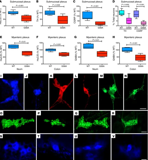
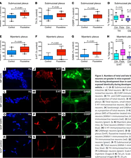
R e s e a R c h a R t i c l ethus appeared to be selectively affected. In SERT Ala56 mice, sub-mucosal TH-expressing (Figure 1B; also compare K with L) and CGRP-expressing neurons (Figure 1C; also compare M with N) were each deficient, not only in absolute terms but as a proportion of total neurons (Figure 1D). In contrast, TH-expressing (Figure 2B; also compare P with Q) and CGRP-expressing neurons (Figure 2C; also compare L with M) were overly abundant in the submucosal plexus of SERTKO animals, again as a proportion of total neurons (Figure 2D). Submucosal TH-expressing (Figure 3B; also compare P with S) and CGRP-expressing neurons (Figure 3C; also compare Q with T) were similarly hyperplastic in fluoxetine-treated mice, once more not only absolutely but also as a proportion of total neurons (Figure 3D). Similarly, SERT Ala56 mice were deficient in GABA-expressing neurons in the myenteric plexus of the ileum (Figure 1G; also compare S with T) and colon (Figure 1H; also com-pare U with V), whereas GABA-expressing neurons were overly abundant in both SERTKO (Figure 2, F and G; also compare N and R with O and S) and fluoxetine-treated mice (Figure 3, G and H; also compare K with N). Expression of SERT Ala56 thus leads to a hypoplastic ENS with selective impairment of development/ survival of late-born neurons, the development of which is also 5-HT–promoted (22). In contrast, in SERTKO mice and in ani-mals subjected to developmental fluoxetine exposure, the ENS is hyperplastic and the development/survival of those neurons that are impaired in SERT Ala56 mice is selectively enhanced.
The placenta has recently been found to be a transient source of 5-HT in early embryos that is able to influence CNS develop-ment prior to the endogenous synthesis of 5-HT in central sero-tonergic neurons (38). The presence of 5-HT in early embryos thus makes it possible for the hyperfunctional SERT Ala56 mutation to decrease enteric neurogenesis even before 5-HT is synthesized in the fetal gut. In fact, the density of serotonergic neurons in the SERT Ala56 intestine was only 36.3% ± 5.6% (P < 0.01; n = 6) of that found in the intestines of WT littermates (see Supplemental Figure 4, A and B; supplemental material available online with this article; doi:10.1172/JCI84877DS1). In contrast, the density of serotonergic neurons in the intestines of fluoxetine-treated mice was significantly greater than that of untreated control animals (Figure 3, F, J, and M). The numbers of serotonergic neurons in SERTKO mice could not accurately be determined, because the content of the marker for these neurons, 5-HT, is reduced (due to lack of reuptake) in neurons of SERTKO animals. Serotonergic neurites, however, were readily visible, and appeared to ramify more extensively in SERTKO than in either G56A or WT gut (Sup-plemental Figure 4, C and D).
Because of the ENS hyperplasia in SERTKO and fluoxetine- treated mice, transverse sections of the bowel were examined to determine whether neurons might be located ectopically in the mucosa or within smooth muscle coats, as occurs in mice lack-ing Hox11l1, which also display ENS hyperplasia (39). No ectopic neurons were found either in SERTKO or in fluoxetine-treated mice (not shown), suggesting that 5-HT affects neuronal devel-opment/survival but not the migration of neural precursors or patterns of gangliogenesis.
The SERT Ala56 mutation leads to slow GI transit and impair-ment of the peristaltic reflexes. Experiimpair-ments were carried out to determine whether the long-lasting ENS hypoplasia associated icits due to altered 5-HT signaling that can be prevented through
mechanism-guided interventions. We assessed the structure and function of the gut in SERT Ala56 animals, comparing results with those obtained in mice either lacking SERT or treated from gesta-tion to weaning with the selective 5-HT reuptake inhibitor fluox-etine. We detected significant, reciprocal abnormalities in these models related to enteric neuronal development, regulation of GI motility, and mucosal growth. Transcription of both Tph isoforms was also abnormal. The SERT Ala56–associated deficits were prevented if the animals were treated during development with a 5-HT4 agonist, prucalopride (34). Our findings reveal that adult bowel function is exquisitely sensitive to SERT activity during ontogeny, and they confirm that 5-HT signaling is critical for the formation of the ENS. Therapies that perturb SERT activity dur-ing development, therefore, may have long-term consequences for bowel function. Observations also suggest that defective 5-HT signaling could provide a common mechanism contributing to peripheral and central defects in ASD.
Results
Numbers of enteric neurons are inversely related to SERT activity dur-ing development. 5-HT promotes enteric neuronal development (22, 32); therefore, we tested the hypothesis that SERT hyperactivity in SERT Ala56 mice would impede enteric neurogenesis (Figure 1). Parallel comparative experiments were carried out with Slc6a4–/–
(herein referred to as SERTKO) animals (Figure 2) and mice exposed to the SERT antagonist fluoxetine from E1 to P21 (Figure 3). Total numbers of enteric neurons and subsets were identified immunocytochemically and quantified in whole mounts of laminar preparations dissected from the gut wall of mice at 6–8 weeks of age. Neuronal numbers were quantified as a function of ganglionic area, which is a parameter that is relatively resistant to stretching of the tissue (35). SERT Ala56 and SERTKO mice were compared with WT littermates. Fluoxetine-treated mice were compared with untreated control animals. Because serotonergic neurons are born early and 5-HT enhances development of late-born rons expressing tyrosine hydroxylase (TH) (dopaminergic neu-rons), γ-aminobutyric acid (GABA), and calcitonin gene–related peptide (CGRP) (22, 36, 37), these phenotypes were chosen for study. GABA-expressing neurons were quantified in the myenteric plexus where they are located, whereas TH- and CGRP-express-ing neurons were quantified in the submucosal plexus where most are found. Total enteric neurons were significantly less abundant in SERT Ala56 than in WT mice (Figure 1). This difference was observed in both the submucosal (Figure 1A; also compare I with J) and the myenteric plexus of the ileum (Figure 1E; also compare O with P) and colon (Figure 1F; also compare Q with R). In contrast, in SERTKO mice, total neurons were significantly more abundant in both the submucosal (Figure 2A; also compare H with I) and the myenteric plexus (Figure 2E; also compare J with K) of the small intestines than in WT animals. The bowel of fluoxetine-treated mice was similar to that of SERTKO animals; again, more neurons were found in both the submucosal (Figure 3A; also compare O with R) and the myenteric plexus (Figure 3E; also compare I with L) of the fluoxetine-treated than the control mice.
The Journal of Clinical Investigation
R e s e a R c h a R t i c l eThe Journal of Clinical Investigation
R e s e a R c h a R t i c l ewere also investigated in vitro to determine whether changes in intestinal transit in SERT Ala56 mice are due to a defect that is intrinsic to the ENS. The extrinsic innervation of the gut is severed in isolated preparations, and the intrinsic circuits of the ENS mediate CMMCs (40).
[image:5.585.53.522.54.510.2]with the SERT Ala56 mutation is reflected in GI motility. Mice were examined at 6–8 weeks of age. Measurements were made in vivo of total GI transit time, gastric emptying, small intes-tinal transit (SIT), and propulsive colorectal motility. Colonic migrating motor complexes (CMMCs; peristaltic reflexes)
The Journal of Clinical Investigation
R e s e a R c h a R t i c l eSERT Ala56 mice. SIT was significantly slower in SERT Ala56 than in WT mice after the injection of exogenous 5-HT (Figure 4D), con-sistent with the hypothesis that the SERT Ala56 is hyperactive and reduces enteric effects even of exogenous 5-HT in vivo.
Intraluminal pressure was raised in isolated preparations of colon to initiate CMMCs, preparations were video-imaged, and spatiotemporal maps of contractile activity patterns were con-structed (Figure 4, E and F). Analysis of these maps revealed that CMMC frequency (Figure 4G; P < 0.0001), velocity (Figure 4H; Total GI transit time in SERT Ala56 mice was significantly
[image:6.585.54.507.51.597.2]lon-ger than that of WT animals (Figure 4A; P < 0.01); however, the rate of gastric emptying (Figure 4B) and SIT (not illustrated) in SERT Ala56 and WT animals did not differ significantly. In contrast to the stomach and SIT, propulsive colorectal motility was signifi-cantly slower in SERT Ala56 than in WT mice (Figure 2C; P < 0.01). Exogenous 5-HT administration increases motility in the small intestine of humans and lower animals (41, 42). We therefore com-pared SIT after exogenous 5-HT injection (1.0 mg/kg) in WT and
The Journal of Clinical Investigation
R e s e a R c h a R t i c l eP < 0.01), and length of propagation (Figure 4I; P < 0.001) were all significantly lower in SERT Ala56 than in WT mice. These observations suggest that the ENS hypoplasia of SERT Ala56 mice impairs generation and conduction of peristaltic reflexes.
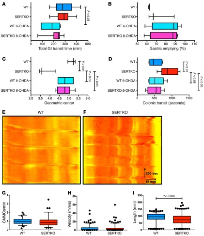
GI motility was analyzed in SERTKO mice and in animals given fluoxetine during development to compare the effects of the SERT Ala56–associated ENS hypoplasia with those of ENS hyperplasia. In vivo measurements of GI motility in SERTKO mice, however, are complicated by the desensitization of 5-HT receptors that has been found to occur spontaneously in these animals (18). Diarrhea and constipation thus alternate in individual SERTKO mice. In contrast to SERT Ala56 animals, total GI transit time (Figure 5A) in SERTKO mice was not significantly different from that of WT ani-mals, although gastric emptying was also not significantly different (Figure 5B). Surprisingly, as in SERT Ala56 mice, SIT (Figure 5C) and propulsive colorectal motility (Figure 5D) were significantly slower in SERTKO than in WT mice. Parameters of in vivo GI tran-sit in mice exposed to fluoxetine during development, moreover, slowed even more than they did in SERTKO animals. Total intes-tinal transit time in fluoxetine-treated animals was significantly
increased (Figure 6A), SIT was significantly slower (Figure 6B), and the time required to eject beads from the rectum was signifi-cantly greater (Figure 6C) than in vehicle-treated control mice.
[image:7.585.48.526.54.399.2]We tested the idea that the unexpectedly slow transit in SERTKO and fluoxetine-treated mice was due to enhanced sym-pathetic input from the CNS. This possibility is plausible because SERTKO mice are highly vulnerable to stress (43) and sympathetic stimulation, which slows GI transit, might result from the stress of measuring GI motility. We thus reinvestigated GI transit after chemical sympathectomy with 6-hydroxydopamine (6-OHDA) and also analyzed ENS-dependent CMMCs in isolated colon, which lacks a functional sympathetic innervation. Although sym-pathectomy accelerated transit, GI transit time and gastric empty-ing were not significantly different in WT and SERTKO mice (Fig-ure 5, A and B). Sympathectomy, however, normalized SIT (Fig(Fig-ure 5C) and propulsive colorectal motility (Figure 5D) in SERTKO mice. Similarly, sympathectomy eliminated the slowing of all mea-sures of in vivo motility in fluoxetine-treated mice without affect-ing any of these parameters in matched control animals (Figure 6, A–C). These observations suggest that sympathetic slowing of GI
The Journal of Clinical Investigation
R e s e a R c h a R t i c l etransit occurs and that both the deletion of SERT and its inhibition during development increase sympathetic input to the intestine. Analyses of the ENS-dependent CMMC in isolated preparations of colon from WT (Figure 5E), SERTKO (Figure 5F), control (Fig-ure 6D), and fluoxetine-treated mice (Fig(Fig-ure 6E) supported these ideas. CMMC frequency (Figure 5G) and velocity (Figure 5H) in
SERTKO were not slower than in WT mice, although the propa-gation length (Figure 5I) of CMMCs was significantly shorter. In isolated preparations of colon from mice that were exposed to fluoxetine during development, moreover, CMMC frequency (Figure 6F), velocity (Figure 6G), and propagation length (Figure 6H) were all significantly greater than in those from control
[image:8.585.88.509.54.540.2]The Journal of Clinical Investigation
R e s e a R c h a R t i c l ewith the SERT Ala56 mutation. 5-HT4 agonists increase enteric neurogenesis, and postnatal enteric neurogenesis is deficient in 5-HT4 KO (Htr4–/–) mice (32). If SERT Ala56 hyperactivity were
to impede enteric neurogenesis by diminishing 5-HT availability at 5-HT4 receptors, an exogenously supplied 5-HT4 agonist that is not a substrate for SERT ought to countermand the defect. We tested this hypothesis by administering the 5-HT4 agonist prucalo-pride (15 mg/kg), which was given orally to dams from gestation through weaning. Offspring were then allowed to mature without further treatment. Treatment with prucalopride was found to pre-vent the hypoplasia that occurs in total neurons (ANNA-1–immu-noreactive) of SERT Ala56 mice in both the submucosal (Figure 7A) and the myenteric plexus (Figure 7D). Prucalopride treatment also prevented the SERT Ala56–associated deficiencies of the sub-mucosal late-born subsub-mucosal dopaminergic (TH-immunoreac-tive; Figure 7B) and CGRP-immunoreactive neurons (Figure 7C) as well as the SERT Ala56–associated deficiency of the myenteric late-born GABA-immunoreactive neurons (Figure 7E). Prucalo-mals. These observations suggest that ENS hyperplasia in mice
exposed during development to fluoxetine is associated with facilitated generation, conduction, and propagation of peristaltic reflexes, which is the opposite of the effect of the ENS hypoplasia occurring in SERT Ala56 mice. Conceivably, the effects of SERT inhibition during development and SERT knockout are different because SERT is functional at the time that CMMCs are analyzed in fluoxetine-treated but not in SERTKO animals. SERT may be required during the generation of CMMCs to inactivate 5-HT, which participates in peristaltic reflex mediation (44). Spontane-ous receptor desensitization in SERTKO mice (18) may also inter-fere with CMMCs. In contrast, chemical sympathectomy prevents slow in vivo transit in both SERTKO and fluoxetine-treated mice, suggesting that enhanced sympathetic input to the bowel occurs in each and thus is probably due to a developmental disturbance of sympathetic control in the CNS.
[image:9.585.46.531.55.387.2]Administration of a 5-HT4 agonist during development rescues mice from the abnormalities of the ENS and GI motility associated
Figure 6. Chemical sympathectomy prevents in vivo slow intestinal transit of fluoxetine-treated mice. Chemical sympathectomy prevented in vivo slow intestinal transit in fluoxetine-treated mice (n = 8–13/group for in vivo motility assays, 2 trials; 4/group for peristalsis). Control and fluoxetine-treated animals were treated, respectively, with vehicle or 6-OHDA. Mice were examined within 2 weeks after the last injection. Data are from 4 mice of each type. (A) Total GIT. Fluoxetine treatment caused slow GIT. 6-OHDA corrected the defect in fluoxetine-treated mice but did not alter total GIT in control mice. (B) Small intestinal transit (SIT). Fluoxetine-treatment caused slow SIT. 6-OHDA corrected the deficit in fluoxetine-treated mice but did not affect SIT in control animals. (C) Colonic motility. Fluoxetine treatment increased the bead ejection time. 6-OHDA corrected the deficit in fluoxetine-treated mice but did not significantly alter ejection time in control animals. §P < 0.05; *P < 0.001; †P was NS. (D and E) WT (D) and fluoxetine-treated (E) mice. Typical
The Journal of Clinical Investigation
R e s e a R c h a R t i c l eThe SERT Ala56 mutation decreases crypt epithelial cell prolifer-ation, stunts growth of villi, and decreases mucosal permeability. The ENS, and specifically 5-HT, have been linked to mucosal mainte-nance (33); myenteric serotonergic neurons innervate submucosal cholinergic neurons that regulate proliferation of transit-amplify-ing cells. As a result, the cell proliferation index, crypt depth, and villus height are all greater in SERTKO than in WT mice. Neuronal, rather than mucosal, 5-HT mediates these effects; SERTKO and double knockout SERTKO/TPH1KO (Slc6a4–/– Tph1–/–) mice are
equivalent, but enhanced mucosal growth does not occur in dou-ble knockout SERTKO/TPH2KO (Slc6a4–/– Tph2–/–) animals. We
therefore compared villus height, crypt depth, and proliferation in SERT Ala56, SERTKO, and fluoxetine-treated mice (Figure 8 and Supplemental Figure 3).
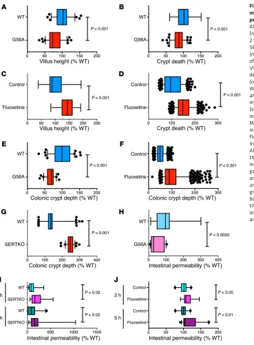
Small intestinal villus height (Figure 8A) and crypt depth (Fig-ure 8B) were both significantly less in SERT Ala56 mice than in WT littermates (Supplemental Figure 3, A and B). In contrast, in fluoxetine-treated mice, small intestinal villus height (Figure 8C) pride rescue of neuronal numbers was accompanied by
[image:10.585.43.395.54.505.2]elimina-tion of the SERT Ala56–associated slowing of total GI transit time (Figure 7F) and of colonic motility (Figure 7G). The prucalopride-mediated rescue of in vivo motility was also manifest in a rescue of CMMCs measured in vitro; prucalopride treatment prevented the SERT Ala56–associated decreases in CMMC frequency (Fig-ure 7H), velocity (Fig(Fig-ure 7I), and length of propagation (Fig(Fig-ure 7J). These in vitro observations suggest that prucalopride rescue is an intrinsic property of the ENS and is not mediated through altered CNS control of the gut. Interestingly, administration of prucalo-pride to WT mice during development led to a decrease in num-bers of GABAergic neurons (Figure 7E) and a decrease in CMMC frequency (Figure 7H) that were not seen when SERT Ala56 pups were exposed to prucalopride. These observations are consistent with the possibility that excessive stimulation of 5-HT4 receptors, as might occur when prucalopride adds to endogenously released 5-HT, can lead to receptor desensitization or, alternatively, to overstimulation and neurotoxicity.
The Journal of Clinical Investigation
R e s e a R c h a R t i c l eimmunoreactive cells per small intestinal crypt (26.1 ± 0.7, n = 30 crypts) of SERT Ala56 mice was significantly less than that of WT mice (45.0 ± 4.2, n = 31 crypts; P < 0.001; Supplemental Figure 3, C and D). Similarly, in the colon, the mean number of Ki67- immunoreactive cells per crypt (21.4 ± 0.5, n = 112 crypts) of SERT Ala56 mice was significantly less than that of WT mice (30.5 ± 0.5, n = 28 crypts; P < 0.001). Crypt epithelial cells have previously been shown to proliferate more rapidly in SERTKO than in WT mice (33). Interestingly, proliferation in the intestinal crypts of fluoxetine-treated mice was similar to that previously observed in SERTKO animals and opposite to that of SERT Ala56 mice (Sup-plemental Figure 3, G–J). The mean number of Ki67-immunore-active cells per small intestinal crypt (22.3 ± 0.3, n = 78 crypts) of fluoxetine-treated mice was significantly greater than that of and crypt depth (Figure 8D) were all significantly greater than
those of WT littermates (Supplemental Figure 3, E and F). As previously reported (33), in SERTKO mice, small intestinal villus height (142% ± 6% WT; n = 90 sections from 6 mice) and crypt depth (128% ± 3% WT; n = 90 sections from 6 mice) were signifi-cantly larger than in WT littermates. Similar results were obtained in measurements of colonic crypt depth, which was significantly shorter than WT in G56A mice (Figure 8E) but significantly larger than that of controls in fluoxetine-treated mice (Figure 8F) and larger than WT in SERTKO animals (Figure 8G).
[image:11.585.43.412.52.553.2]Immunostaining of proliferating cells in crypts with Abs to Ki67 was used to verify whether altered proliferation of tran-sit-amplifying cells was responsible for the differences observed in villus height and crypt depth. The mean number of Ki67-
The Journal of Clinical Investigation
R e s e a R c h a R t i c l ewhen animals were analyzed. Despite changes in abundance of transcripts encoding TPH1 and the density of EC cells, no signifi-cant change was seen in the concentration of 5-HT in the gut of either SERT Ala56 or fluoxetine-treated mice (data not shown). Secretion or intracellular metabolism of 5-HT, which were not measured, may thus balance changes in biosynthesis and uptake.
Discussion
SERT Ala56 is the most common of the rare hyperactive SERT vari-ants that are overtransmitted to children with ASD (10, 11). SERT Ala56 mice exhibit hyperserotonemia and behaviors that align broadly with the core diagnostic features of ASD (12, 13, 45). These traits support the use of SERT Ala56 mice as a model for analyzing potential contributions of 5-HT signaling abnormalities to ASD. Both hyperserotonemia (4, 5) and GI dysfunction (2) are commonly seen in ASD. Because platelets derive their 5-HT from the gut (3) and 5-HT is an important multifunctional GI signaling molecule (17), we postulated that SERT hyperactivity would not only cause hyperserotonemia but also perturb GI structure and function in SERT Ala56 mice. Abnormal 5-HT signaling, moreover, impacts both CNS (38, 46–48) and ENS (22) development. We thus tested the hypothesis that ENS formation and ENS-dependent functions are perturbed in SERT Ala56 mice because of the reduction of endogenous 5-HT availability (13). To do so, we comprehensively analyzed SERT Ala56 mice and, for comparison, SERTKO and fluoxetine-treated animals, to provide, respectively, mice in which SERT is nonfunctional or is inhibited only during ontogeny. These studies of altered SERT activity also provided unique insights into roles of 5-HT signaling in enteric physiology and pathophysiology.
The ENS was strikingly hypoplastic in SERT Ala56 mice. Numbers of neurons were reduced in both plexuses of small and large intestines, and neurons generated after serotonergic neu-rons during ontogeny (expressing TH, CGRP, or GABA) were more deficient than ENS neurons in general. In contrast, the ENS of SERTKO and fluoxetine-treated animals was hyperplas-tic, and the late-born neurons that were deficient in SERT Ala56 mice were especially abundant in these animals. The similarity between SERTKO and fluoxetine-treated mice suggests that the developmental period is a critical time for SERT function in neu-rogenesis. These observations are consistent with the ideas that defective 5-HT signaling due to the increased 5-HT clearance of SERT Ala56 mice interferes with enteric neurogenesis, whereas potentiated serotonergic signaling due to the absence of SERT or its inhibition during development enhances neurogenesis. The sensitivity of late-born neurons to SERT activity is consistent with the ideas that serotonergic neurons, which are early-born, regulate enteric neurogenesis and thus help sculpt the ENS. These obser-vations confirm that 5-HT is an ENS growth factor (22, 49) and that serotonergic signaling is essential for normal neurogenesis (22, 32). The data are also consistent with the hypothesis that a defect common to the ENS and CNS could be responsible in ASD for comorbid GI disturbances.
Motility of the SERT Ala56 bowel was impaired both in vivo (slowed GI transit time and colonic transit) and in vitro (decreased velocity, frequency, and length of conduction of CMMCs). Because CMMCs, which are ENS-dependent, were defective in isolated preparations of SERT Ala56 bowel, the motor abnor-control animals (14.7 ± 00.3, n = 31 crypts; P < 0.001). Similarly,
in the colon, the mean number of Ki67-immunoreactive cells per crypt (12.5 ± 0.3, n = 80 crypts) of fluoxetine-treated mice was significantly greater than that of control mice (9.1 ± 0.2, n = 74 crypts; P < 0.001). These observations support the hypothesis that mucosal growth is stunted in SERT Ala56 mice because SERT regulation of 5-HT signaling is important for epithelial prolifer-ation and mucosal maintenance. The effects of SERT inhibition during development, moreover, are remarkably persistent, since the fluoxetine-treated animals were given at least 6 weeks after fluoxetine withdrawal for SERT activity to recover.
Macromolecular permeability was investigated to determine how the alterations in mucosal maintenance observed in SERT Ala56, SERTKO, and fluoxetine-treated mice affected the integ-rity of mucosal barrier function. Permeability to FITC-dextran in SERT Ala56 mice was found to be significantly less than that of WT animals (Figure 8H). In contrast, permeability of the mucosa of SERTKO (Figure 8I) and fluoxetine-treated mice (Figure 8J) was more than that of WT animals. Electron microscopic analysis after i.v. injection of HRP failed to reveal a loss of tight junctional integrity in any of the mice tested (Supplemental Figure 1).
The Journal of Clinical Investigation
R e s e a R c h a R t i c l ethe hypoplastic mucosae of SERT Ala56 mice and greatest in the hyperplastic mucosae of SERTKO and fluoxetine-treated mice.
The SERT Ala56 mutation increased the GI expression of each of the 5-HT biosynthetic enzymes TPH1 and TPH2. These obser-vations are consistent with the idea that the decreased availabil-ity of 5-HT in the SERT Ala56 bowel triggers a feedback increase in 5-HT biosynthetic capacity in both EC cells (TPH1) and neu-rons (TPH2). We also found that EC cells were more abundant in SERT Ala56 mice than in WT animals, which may account for the increased abundance of transcripts encoding TPH1. Feed-back through 5-HT availability, however, may not account for the increase seen in total EE cells in SERT Ala56 animals, because the EE population includes cells that do not produce 5-HT. The dimin-ished mucosal growth that occurs in SERT Ala56 animals may thus lead to an increase in the proportion of cells that differenti-ate along a neurogenin 3–dependent pathway and acquire an EE phenotype (52). In SERTKO mice, transcripts encoding TPH1 and TPH2 were each less abundant than in WT animals and the den-sities of both EC and total EE cells were decreased, a phenotype opposite to that seen in SERT Ala56 animals. These observations are consistent both with a feedback decrease in 5-HT biosynthesis due to enhanced 5-HT bioavailability in the absence of SERT and with a diminished acquisition of the EE phenotype when mucosal growth speeds up. The decrease in transcripts encoding TPH1 was mimicked in fluoxetine-treated mice, as was the decreased den-sity of EC and total EE cells. Fluoxetine-treated mice, however, did not exhibit the decrease in abundance of transcripts encoding TPH2 that was seen in SERTKO animals; therefore, reduced TPH2 expression probably reflects the ongoing enhanced 5-HT bioavail-ability associated with SERT deletion. The mucosal effects of fluoxetine treatment, on the other hand, outlasted the period of acute SERT inhibition and are likely to be due to the rearranged ENS in these animals.
The GI impact of developmental administration of a SERT inhibitor persisted into adult life, long after the inhibitor was withdrawn. Depression occurs in up to 23% of pregnant women, and SERT inhibition is a common feature of most antidepressants used to treat it (15). Selective serotonin reuptake inhibitor (SSRI) use during pregnancy has been linked to a 2-fold increase in con-genital defects (53). Children exposed to SSRIs or tricyclic antide-pressants in utero were found to be 10-fold more likely to require laxatives for constipation, which is consistent with the idea that antenatal SERT inhibition disturbs ENS development and leads to persistent defects in GI motility (54). The ENS contributes to many GI disorders in adults, including irritable bowel syndrome (55) and inflammatory bowel disease (23, 56). Given the importance of 5-HT signaling to ENS development and the long-lasting effects of fluoxetine treatment on sympathetic output, further investigation is probably warranted of the administration to pregnant or lactat-ing women of antidepressants that affect SERT or 5-HT function.
ASD is severe in its prevalence, impact, and costs to society. Symptoms, more than etiology, define the syndrome; neverthe-less, peripheral abnormalities, such as GI dysfunction (2), also occur. The complexity of the genetic contributions to ASD (57) encourages efforts to identify biomarkers, such as hyperserotone-mia (4, 5), for convergent pathways to account for the pathophys-iology. Because platelets acquire their 5-HT from the GI tract (3), mality is an intrinsic property of the ENS. This consideration is
important because a global defect in SERT affects the CNS and well as the ENS. In both SERTKO and fluoxetine-treated mice, for example, the absence or inhibition of SERT during develop-ment was found to enhance sympathetic activity, which slowed GI motility in vivo. Both the ENS hypoplasia of SERT Ala56 mice and the hyperplasia seen in SERTKO and fluoxetine-treated ani-mals thus have functional consequences.
Two subtypes of 5-HT receptor have been linked to 5-HT–pro-moted enteric neurogenesis, 5-HT2B (49) and 5-HT4 (32). Agonists of each promote enteric neurogenesis; moreover, postnatal enteric neurogenesis is defective when 5-HT4 receptors are deleted (32). To test the hypothesis that 5-HT4 receptors are critical in medi-ating the enteric neurodevelopmental actions of 5-HT, we deter-mined whether a 5-HT4 agonist, prucalopride (34), prevents the development of the SERT Ala56 ENS phenotype. Because pruca-lopride is not a substrate for SERT, its signaling should not be ter-minated prematurely, like that of 5-HT, by an overly active SERT. Prucalopride did, in fact, overcome the SERT Ala56 abnormal-ity, preventing the ENS hypoplasia, deficiencies of late-develop-ing neurons, the slowlate-develop-ing of GI transit in vivo, and the defects in CMMCs that occur in vitro. Systemic agonist administration to dams precludes an absolute conclusion that the actions of prucalo-pride derive from direct stimulation of enteric neurogenesis in off-spring, though this seems likely and is consistent with the known receptor contributions to developing enteric neurons. Regardless, the striking ability of prucalopride to prevent SERT Ala56–associ-ated ENS neuronal hypoplasia and its consequences supports the idea that SERT and 5-HT4 receptors play important roles in 5-HT– promoted neurogenesis and also supports the overall hypothe-sis that the defects seen in SERT Ala56 mice are, in fact, due to the premature clearance of endogenous 5-HT by the hyperactive SERT variant these animals express.
The Journal of Clinical Investigation
R e s e a R c h a R t i c l eGI motility. Colonic motility was measured as the time required to
expel a 3 mm glass bead inserted 2 cm into the rectum (69). Total GI transit time was estimated as the time required for a nonabsorbable dye (carmine red) to appear in stool after its gavage into the stomach (70). Gastric emptying and small bowel transit were evaluated follow-ing the gavage of rhodamine B dextran in methylcellulose. The gut was removed 15 minutes after gavage, and the percentage of dye remain-ing in the stomach as well as the geometric center of the rhodamine B dextran in the intestine was determined (71). Video imaging was used to analyze CMMCs in isolated colon (72, 73). Internal pressure was raised to initiate CMMCs, and spatiotemporal maps of colon diame-ter as a function of time were constructed. CMMC frequency, velocity, and length of conduction were measured (74, 75). See Supplemental Methods for further details.
Quantitation of transcripts. Methods used to extract RNA, reverse-
transcribe it to DNA, and quantify transcript abundance with real-time PCR have been described previously (22, 23) and are detailed in Supple-mental Methods. Transcript expression was normalized to that of glyc-eraldehyde-3-phosphate dehydrogenase (Gapdh).
In vivo permeability. The absorption of FITC-dextran (4.4 kDa;
22 mg/ml in PBS; Sigma-Aldrich) was evaluated by measurement of its concentration in blood (fluorescence at 520 nm) after gavage (76). Submandibular puncture was used to obtain blood (100 μl) 2 and 5 hours after the administration of FITC-dextran.
Statistics. Student’s unpaired t test and 1-way ANOVA were used,
respectively, to compare single and multiple means. A P value of less than 0.05 was considered significant.
Study approval. Animal studies were approved by the IACUC of
Columbia University Medical Center.
Author contributions
KGM designed and conducted the studies, analyzed data, and wrote the manuscript. ZL, KS, VS, and NI conducted experiments and acquired data. GMA conducted experiments and acquired and analyzed data. IS acquired data. JV-V created and provided the Ala56 mice, conducted experiments, and analyzed data. RDB cre-ated and provided the Ala56 mice, conducted experiments, ana-lyzed data, and critically reviewed the manuscript. MDG designed the studies, analyzed data, and wrote the manuscript.
Acknowledgments
This work was supported by NIH grants NS15547, DK093786, and MH096972, the Einhorn Charitable Trust, and the Autism Research Institute.
Address correspondence to: Kara Gross Margolis, Morgan Stanley Children’s Hospital, Columbia University College of Physicians and Surgeons, Department of Pediatrics, Division of Pediatric Gas-troenterology, 620 West 168th Street, New York, New York 10032, USA. Phone: 212.305.5903; E-mail: [email protected]. the ASD-associated increase in platelet 5-HT links GI dysfunction
to ASD. The gene encoding SERT (SLC6A4) has been associated with both platelet 5-HT content and susceptibility to ASD (58–61). The abnormalities of GI function that we have characterized in SERT Ala56 mice are also similar to those that occur at high fre-quency in ASD (2, 62). Our observations thus support the idea that a defect in 5-HT–sensitive neurogenic pathways could be a com-mon feature, describing a novel ASD subtype defined by “reverse phenotyping” (63), in which behavioral and enteric abnormalities due to abnormal serotonergic signaling underlie the condition. A prospective human study will be needed to confirm this sugges-tion. Interestingly, the 5-HT4 receptor, which prucalopride rescue suggests is insufficiently stimulated in SERT Ala56 mice, also promotes hippocampal neurogenesis and has been postulated to underlie the neurogenesis induced by SSRIs (64). Prucalopride and other 5-HT4 agonists were developed for treating chronic con-stipation and concon-stipation-predominant irritable bowel syndrome (34). Whether 5-HT4 agonists might also be helpful therapeutic agents for ASD requires further study.
Methods
Animals. C57BL/6 mice were used for studies of Slc6a4 deletion
(SERTKO) and inhibition during development with fluoxetine. Fluox-etine (7.5 mg/kg/d) was administered to pregnant dams from E1 (the time a mucous plug was detected) through weaning at P21. Animals were tested at 6–8 weeks of age. SERT Ala56 mice were generated on a 129S6 background (12). Experiments were carried out with confirmed
homozygous WT and SERT Ala56 littermates. The selective 5-HT4
agonist prucalopride (15 mg/kg; Shire Pharmaceuticals) was given to dams daily by gavage. Prucalopride was begun at E1 and was continued until P21. Control dams received autoclaved water, also by gavage. The E1–P21 period covers most of enteric neurogenesis (37). The effects of prucalopride were determined 3–5 weeks after cessation of therapy, ensuring that prucalopride itself was no longer present in the animals.
Immunocytochemistry. The immunoreactivities of the neuronal
marker ANNA-1 (65–68) as well as tyrosine hydroxylase (TH), γ-amino-butyric acid (GABA), 5-HT, and calcitonin gene–related peptide (CGRP) were located to quantify the abundance of total, dopaminergic, GABAergic, 5-HT–expressing, and CGRP-expressing enteric neurons. Myenteric and submucosal plexuses were examined in whole mounts of laminar preparations of the gut wall. Methods used have been described previously (22, 23) and are detailed in Supplemental Methods.
Images were obtained with a cooled CCD camera and analyzed with computer assistance (Volocity 6.0 software, Improvision/Perkin-Elmer Life and Analytical Sciences). To count the numbers of labeled cells, a computer-controlled motorized stage was used to scan and
collect images with a ×20 objective covering the entirety of a 10-mm2
area. Collected images were computer-processed (Volocity 6.0 soft-ware) to estimate numbers of immunoreactive cells of each type (cells per square millimeter of ganglionic area).
1. Posar A, Resca F, Visconti P. Autism according to diagnostic and statistical manual of mental disor-ders 5(th) edition: the need for further improve-ments. J Pediatr Neurosci. 2015;10(2):146–148. 2. McElhanon BO, McCracken C, Karpen S, Sharp
WG. Gastrointestinal symptoms in autism
spectrum disorder: a meta-analysis. Pediatrics. 2014;133(5):872–883.
3. Matondo RB, et al. Deletion of the seroto-nin transporter in rats disturbs serotoseroto-nin homeostasis without impairing liver regener-ation. Am J Physiol Gastrointest Liver Physiol.
2009;296(4):G963–G968.
The Journal of Clinical Investigation
R e s e a R c h a R t i c l e2004;43(4):491–499.
5. Ritvo ER, Yuwiler A, Geller E, Ornitz EM, Saeger K, Plotkin S. Increased blood serotonin and plate-lets in early infantile autism. Arch Gen Psychiatry. 1970;23(6):566–572.
6. Morrissey JJ, Walker MN, Lovenberg W. The absence of tryptophan hydroxylase activ-ity in blood platelets. Proc Soc Exp Biol Med. 1977;154(4):496–499.
7. Lesch KP, Wolozin BL, Murphy DL, Riederer P. Primary structure of the human platelet serotonin (5-HT) uptake site: Identity with the brain 5-HT transporter. J Neurochem. 1993;60(6):2319–2322. 8. Hughes FB, Brodie BB. The mechanism of
seroto-nin and catecholamine uptake by platelets.
J Pharmacol Exp Ther. 1959;127:96–102.
9. Prasad HC, et al. Human serotonin trans-porter variants display altered sensitivity to protein kinase G and p38 mitogen-activated protein kinase. Proc Natl Acad Sci U S A. 2005;102(32):11545–11550.
10. Sutcliffe JS, et al. Allelic heterogeneity at the serotonin transporter locus (SLC6A4) confers susceptibility to autism and rigid-compulsive behaviors. Am J Hum Genet. 2005;77(2):265–279. 11. Prasad HC, Steiner JA, Sutcliffe JS, Blakely RD.
Enhanced activity of human serotonin transporter variants associated with autism. Philos Trans R Soc
Lond B Biol Sci. 2009;364(1514):163–173.
12. Veenstra-Vanderweele J, et al. Modeling rare gene variation to gain insight into the oldest biomarker in autism: construction of the serotonin trans-porter Gly56Ala knock-in mouse. J Neurodev
Disord. 2009;1(2):158–171.
13. Veenstra-VanderWeele J, et al. Autism gene variant causes hyperserotonemia, serotonin receptor hypersensitivity, social impairment and repetitive behavior. Proc Natl Acad Sci U S A. 2012;109(14):5469–5474.
14. Gaspar P, Cases O, Maroteaux L. The devel-opmental role of serotonin: news from mouse molecular genetics. Nat Rev Neurosci. 2003;4(12):1002–1012.
15. Kepser LJ, Homberg JR. The neurodevelopmental effects of serotonin: a behavioural perspective.
Behav Brain Res. 2015;277:3–13.
16. Liu C, Maejima T, Wyler SC, Casadesus G, Herlitze S, Deneris ES. Pet-1 is required across different stages of life to regulate serotonergic function. Nat Neurosci. 2010;13(10):1190–1198. 17. Gershon MD. 5-Hydroxytryptamine (serotonin)
in the gastrointestinal tract. Curr Opin Endocrinol
Diabetes Obes. 2013;20(1):14–21.
18. Chen JJ, et al. Maintenance of serotonin in the intestinal mucosa and ganglia of mice that lack the high affinity serotonin transporter (SERT): abnor-mal intestinal motility and the expression of cation transporters. J Neurosci. 2001;21(16):6348–6361. 19. Wade PR, Chen J, Jaffe B, Kassem IS, Blakely
RD, Gershon MD. Localization and function of a 5-HT transporter in crypt epithelia of the gastro-intestinal tract. J Neurosci. 1996;16(7):2352–2364. 20. Blakely RD, et al. Cloning and expression of a
functional serotonin transporter from rat brain.
Nature. 1991;354(6348):66–70.
21. Chen JX, Pan H, Rothman TP, Wade PR, Gershon MD. Guinea pig 5-HT transporter: cloning, expression, distribution and function
in intestinal sensory reception. Am J Physiol. 1998;275(3 pt 1):G433–G448.
22. Li Z, et al. Essential roles of enteric neuronal serotonin in gastrointestinal motility and the development/survival of enteric dopaminergic neurons. J Neurosci. 2011;31(24):8998–9009. 23. Margolis KG, et al. Pharmacological
reduc-tion of mucosal but not neuronal serotonin opposes inflammation in mouse intestine. Gut. 2014;63(6):928–937.
24. Heredia DJ, Gershon MD, Koh SD, Corrigan RD, Okamoto T, Smith TK. Important role of mucosal serotonin in colonic propulsion and peristaltic reflexes: in vitro analyses in mice lacking tryptophan hydroxylase 1. J Physiol. 2013;591(23):5939–5957.
25. Hillsley K, Grundy D. Sensitivity to 5-hydroxy-tryptamine in different afferent subpopulations within mesenteric nerves supplying the rat jeju-num. J Physiol. 1998;509(pt 3):717–727. 26. Blackshaw LA, Grundy D. Effects of
5-hydroxy-tryptamine on discharge of vagal mucosal affer-ent fibres from the upper gastrointestinal tract of the ferret. J Auton Nerv Syst. 1993;45(1):41–50. 27. Lang IM. Noxious stimulation of emesis. Dig Dis
Sci. 1999;44(8 suppl):58S–63S.
28. Pan H, Gershon MD. Activation of intrinsic afferent pathways in submucosal ganglia of the guinea pig small intestine. J Neurosci. 2000;20(9):3295–3309. 29. Bertrand PP, Kunze WAA, Furness JB, Bornstein
JC. The terminals of myenteric intrinsic primary afferent neurons of the guinea pig ileum are excited by 5-HT acting at 5-HT3 receptors.
Neu-roscience. 2000;101(2):459–469.
30. Bülbring E, Crema A. The release of 5-hydroxy-tryptamine in relation to pressure exerted on the intestinal mucosa. J Physiol. 1959;146(1):18–28. 31. Cooke HJ, Christofi FL. Enteric regulation of
mucosal secretion. In: Johnson LR, ed. Physiology
of the Gastrointestinal Tract. 4th ed. New York,
New York, USA: Academic Press; 2006:737–762. 32. Liu MT, Kuan YH, Wang J, Hen R, Gershon MD.
5-HT4 receptor-mediated neuroprotection and neurogenesis in the enteric nervous system of adult mice. J Neurosci. 2009;29(31):9683–9699. 33. Gross ER, Gershon MD, Margolis KG, Gertsberg ZV, Cowles RA. Neuronal serotonin regulates growth of the intestinal mucosa in mice.
Gastroenterology. 2012;143(2):408–417.
34. Briejer MR, et al. The in vitro pharmacological profile of prucalopride, a novel enterokinetic compound. Eur J Pharmacol. 2001;423(1):71–83. 35. Karaosmanoglu T, Aygun B, Wade PR, Gershon
MD. Regional differences in the number of neurons in the myenteric plexus of the guinea pig small intestine and colon: an evaluation of markers used to count neurons. Anat Rec. 1996;244(4):470–480.
36. Chalazonitis A, et al. Bone morphogenetic pro-tein regulation of enteric neuronal phenotypic diversity: relationship to timing of cell cycle exit.
J Comp Neurol. 2008;509(5):474–492.
37. Pham TD, Gershon MD, Rothman TP. Time of origin of neurons in the murine enteric nervous system. J Comp Neurol. 1991;314(4):789–798. 38. Bonnin A, et al. A transient placental source
of serotonin for the fetal forebrain. Nature. 2011;472(7343):347–350.
39. Yamataka A, et al. Intestinal neuronal dysplasia-like pathology in Ncx/Hox11L. J Pediatr Surg. 2001;36(8):1293–1296.
40. Spencer NJ, Bywater RA, Taylor GS. Evidence that myoelectric complexes in the isolated mouse colon may not be of myogenic origin. Neurosci
Lett. 1998;250(3):153–156.
41. Misiewicz JJ, Waller SL, Eisner M. Motor responses of human gastrointestinal tract to 5-hydroxytryptamine in vivo and in vitro. Gut. 1966;7(3):208–216.
42. Oosterbosch L, von der Ohe M, Valdovinos MA, Kost LJ, Phillips SF, Camilleri M. Effects of sero-tonin on rat ileocolonic transit and fluid transfer in vivo: possible mechanisms of action. Gut. 1993;34(6):794–798.
43. Adamec R, Burton P, Blundell J, Murphy DL, Holmes A. Vulnerability to mild predator stress in serotonin transporter knockout mice. Behav
Brain Res. 2006;170(1):126–140.
44. Smith TK, Gershon MD. CrossTalk proposal: 5-HT is necessary for peristalsis. J Physiol. 2015;593(15):3225–3227.
45. Kerr TM, et al. Genetic background modulates phenotypes of serotonin transporter Ala56 knock-in mice. Mol Autism. 2013;4(1):35. 46. Kawasaki H. Spatio-temporal regulation of the
formation of the somatosensory system. Dev
Growth Differ. 2015;57(3):193–199.
47. Chen X, Ye R, Gargus JJ, Blakely RD, Dobrenis K, Sze JY. Disruption of transient serotonin accu-mulation by non-serotonin-producing neurons impairs cortical map development [published online ahead of print January 14, 2015]. Cell Rep. doi:10.1016/j.celrep.2014.12.033.
48. Deneris ES. Molecular genetics of mouse sero-tonin neurons across the lifespan. Neuroscience. 2011;197:17–27.
49. Fiorica-Howells E, Maroteaux L, Gershon MD. Serotonin and the 5-HT(2B) receptor in the development of enteric neurons. J Neurosci. 2000;20(1):294–305.
50. Furness JB, Costa M. Neurons with 5-hydroxytryp-tamine-like immunoreactivity in the enteric ner-vous system: Their projections in the guinea pig small intestine. Neuroscience. 1982;7(2):341–350. 51. Qu ZD, Thacker M, Castelucci P, Bagyanszki M,
Epstein ML, Furness JB. Immunohistochemical analysis of neuron types in the mouse small intestine. Cell Tissue Res. 2008;334(2):147–161. 52. Clevers H. The intestinal crypt, a prototype stem
cell compartment. Cell. 2013;154(2):274–284. 53. Wogelius P, et al. Maternal use of selective
seroto-nin reuptake inhibitors and risk of congenital mal-formations. Epidemiology. 2006;17(6):701–704. 54. Nijenhuis CM, ter Horst PG, van Rein N, Wilffert
B, de Jong-van den Berg LT. Disturbed devel-opment of the enteric nervous system after in utero exposure of selective serotonin re-uptake inhibitors and tricyclic antidepressants. Br J Clin
Pharmacol. 2012;73(1):126–134.
55. Gershon MD. Nerves, reflexes, and the enteric nervous system: pathogenesis of the irrita-ble bowel syndrome. J Clin Gastroenterol. 2005;39(5 suppl 3):S184–S193.
The Journal of Clinical Investigation
R e s e a R c h a R t i c l e57. Persico AM, Napolioni V. Autism genetics. Behav
Brain Res. 2013;251:95–112.
58. Devlin B, et al. Autism and the serotonin trans-porter: the long and short of it. Mol Psychiatry. 2005;10(12):1110–1116.
59. Huang CH, Santangelo SL. Autism and serotonin transporter gene polymorphisms: a systematic review and meta-analysis. Am J Med Genet B
Neu-ropsychiatr Genet. 2008;147B(6):903–913.
60. Kistner-Griffin E, Brune CW, Davis LK, Sutcliffe JS, Cox NJ, Cook EH Jr. Parent-of-origin effects of the serotonin transporter gene associated with autism. Am J Med Genet B Neuropsychiatr Genet. 2011;156(2):139–144.
61. Veenstra-VanderWeele J, et al. Transmission disequilibrium studies of the serotonin 5-HT2A receptor gene (HTR2A) in autism. Am J Med
Genet. 2002;114(3):277–283.
62. Hsiao EY. Gastrointestinal issues in autism spectrum disorder. Harv Rev Psychiatry. 2014;22(2):104–111.
63. Schulze TG, McMahon FJ. Defining the phe-notype in human genetic studies: forward genetics and reverse phenotyping. Hum Hered. 2004;58(3–4):131–138.
64. Imoto Y, et al. Role of the 5-HT4 receptor in chronic fluoxetine treatment-induced neuro-genic activity and granule cell dematuration in
the dentate gyrus. Mol Brain. 2015;8:29. 65. King PH, Redden D, Palmgren JS, Nabors LB,
Lennon VA. Hu antigen specificities of ANNA-I autoantibodies in paraneoplastic neurological disease. J Autoimmun. 1999;13(4):435–443. 66. Lennon VA. The case for a descriptive generic
nomenclature: clarification of immunostaining criteria for PCA-1, ANNA-1, and ANNA-2 autoan-tibodies. Neurology. 1994;44(12):2412–2415. 67. Chu G, Wilson PC, Carter CD, Lennon VA,
Rob-erts-Thomson IC. Intestinal pseudo-obstruction, type 1 anti-neuronal nuclear antibodies and small-cell carcinoma of the lung. J Gastroenterol
Hepatol. 1993;8(6):604–606.
68. Chalk CH, Lennon VA, Stevens JC, Windebank AJ. Seronegativity for type 1 antineuronal nuclear antibodies (‘anti-Hu’) in subacute sensory neu-ronopathy patients without cancer. Neurology. 1993;43(11):2209–2211.
69. Li ZS, Schmauss C, Cuenca A, Ratcliffe E, Ger-shon MD. Physiological modulation of intestinal motility by enteric dopaminergic neurons and the D2 receptor: analysis of dopamine receptor expression, location, development, and function in wild-type and knock-out mice. J Neurosci. 2006;26(10):2798–2807.
70. Kimball ES, Palmer JM, D’Andrea MR, Hornby PJ, Wade PR. Acute colitis induction by oil of mustard
results in later development of an IBS-like acceler-ated upper GI transit in mice. Am J Physiol
Gastro-intest Liver Physiol. 2005;288(6):G1266–G1273.
71. Miller MS, Galligan JJ, Burks TF. Accurate mea-surement of intestinal transit in the rat. J
Pharma-col Methods. 1981;6(3):211–217.
72. Roberts RR, Bornstein JC, Bergner AJ, Young HM. Disturbances of colonic motility in mouse models of Hirschsprung’s disease. Am J Physiol
Gastroin-test Liver Physiol. 2008;294(4):G996–G1008.
73. Roberts RR, Murphy JF, Young HM, Bornstein JC. Development of colonic motility in the neonatal mouse-studies using spatiotemporal maps. Am J Physiol Gastrointest Liver Physiol. 2007;292(3):G930–G938.
74. Gwynne RM, Thomas EA, Goh SM, Sjovall H, Bornstein JC. Segmentation induced by intralu-minal fatty acid in isolated guinea-pig duodenum and jejunum. J Physiol. 2004;556(pt 2):557–569. 75. Welch MG, Margolis KG, Li Z, Gershon MD.
Oxytocin regulates gastrointestinal motility, inflammation, macromolecular permeability, and mucosal maintenance in mice. Am J Physiol
Gas-trointest Liver Physiol. 2014;307(8):G848–G862.