Copyright © 2004, American Society for Microbiology. All Rights Reserved.
Simultaneous Detection of Marine Fish Pathogens by Using Multiplex
PCR and a DNA Microarray
Santiago F. Gonza´lez,
1,2Melissa J. Krug,
3,4Michael E. Nielsen,
2Ysabel Santos,
1and Douglas R. Call
3,4*
Department of Microbiology and Parasitology, University of Santiago de Compostela, 15706 Santiago de Compostela, Spain
1;
Department of Veterinary Microbiology and Pathology
3and WSU and UI Center for Reproductive Biology,
4Washington
State University, Pullman, Washington; and Section of Fish Diseases, Department of Veterinary Microbiology,
Royal Veterinary and Agricultural University, DK-1870 Frederiksberg C, Denmark
2Received 27 August 2003/Returned for modification 12 October 2003/Accepted 18 December 2003
We coupled multiplex PCR and a DNA microarray to construct an assay suitable for the simultaneous
detection of five important marine fish pathogens (
Vibrio vulnificus
,
Listonella anguillarum
,
Photobacterium
damselae
subsp.
damselae
,
Aeromonas salmonicida
subsp.
salmonicida
, and
Vibrio parahaemolyticus
). The array
was composed of nine short oligonucleotide probes (25-mer) complementary to seven chromosomal loci (
cyt
,
rpoN
,
gyrB
,
toxR
,
ureC
,
dly
, and
vapA
) and two plasmid-borne loci (
fatA
and A.sal). Nine primer sets were
designed to amplify short fragments of these loci (100 to 177 bp) in a multiplex PCR. PCR products were
subsequently labeled by nick translation and hybridized to the microarray. All strains of the five target species
(
n
ⴝ
1 to 21) hybridized to at least one species-specific probe. Assay sensitivities ranged from 100% for seven
probes to 83 and 67% for the two remaining probes. Multiplex PCR did not produce any nonspecific
ampli-fication products when tested against 23 related species of bacteria (
n
ⴝ
40 strains; 100% specificity). Using
purified genomic DNA, we were able to detect PCR products with <20 fg of genomic DNA per reaction
(equivalent to four or five cells), and the array was at least fourfold more sensitive than agarose gel
electro-phoresis for detecting PCR products. In addition, our method allowed the tentative identification of virulent
strains of
L
.
anguillarum
serotype O1 based on the presence of the
fatA
gene (67% sensitivity and 100%
specificity). This assay is a sensitive and specific tool for the simultaneous detection of multiple pathogenic
bacteria that cause disease in fish and humans.
Vibriosis and furunculosis are two fish diseases responsible
for considerable economic hardship to mariculture operations
worldwide (3). Vibriosis, mainly caused by
Listonella
anguilla-rum
,
Vibrio vulnificus
, and
Photobacterium damselae
subsp.
damselae
, is a systemic bacterial infection affecting more than
48 fish species in widely distributed regions (3, 35). Other
halophilic
Vibrio
spp., such as
Vibrio parahaemolyticus
, and
V. vulnificus
have been identified as causing vibriosis in humans
(22, 29) and have been isolated from many species of fish,
shellfish, and crustaceans.
Aeromonas salmonicida
is the causal
agent of furunculosis, a disease of major significance in the
culturing of salmonid fish and other valuable marine fish
spe-cies (3).
Conventional microbiological methods needed to identify
these organisms are often limited by the length of time
re-quired to complete the assays. In recent years, enzyme-linked
immunosorbent assays and molecular methods based on DNA
probes or PCR have overcome problems associated with
cul-ture-based techniques, enabling the detection of
microorgan-isms directly in clinical samples without the need for previous
culturing. Molecular diagnosis protocols have been the most
effective methods for the diagnosis of bacterial agents in
mari-cultures because they permit more specific and sensitive
de-tection than do serological assays. Many PCR methods have
been developed for the identification of bacterial pathogens in
aquacultures (30). Although many of these protocols are based
on the amplification of 16S and 23S rRNA genes (2, 19, 24, 25,
31), which are found in all eubacteria, there is a high degree of
genetic similarity for these genes across taxa; therefore, the
specificity of the detection method can be compromised (21,
37). Alternatively, bacterium-specific genes (e.g., virulence
loci) can be used as targets for PCR amplification to permit
more specific detection (16) as well as subspecies and strain
differentiation (9, 28, 32). Conventional PCR is used to amplify
a single gene target, whereas multiplex PCR involves
amplify-ing multiple gene products in a samplify-ingle reaction; the latter
method has been used successfully to detect fish pathogens (4,
14, 32). Agarose gel electrophoresis is typically used to assess
results from multiplex PCRs, but DNA microarrays offer a
more discriminating means to examine reaction products for
specific sequences.
DNA microarrays are important molecular tools that have
been applied to studies of gene expression (38), phylogenetic
classification (12), ecological studies (15), and the detection
and genotyping of bacterial (9, 17) and viral (11) pathogens.
DNA microarrays consist of ordered sets of DNA fixed to solid
surfaces; generally on glass but sometimes on nylon substrates.
Each spot in a microarray is composed of many identical
probes that are complementary to a gene of interest.
Microar-rays can be used to detect cDNA (38), genomic DNA (5), and
plasmid DNA (7) in the context of gene expression analysis
and comparative genomics. They can also be used as end-point
detectors to examine complex mixtures of PCR products (8).
* Corresponding author. Mailing address: Department of
Veteri-nary Microbiology and Pathology, Washington State University, 402
Bustad Hall, P.O. Box 647040, Pullman, WA 99164-7040. Phone: (509)
335-6313. Fax: (509) 335-8529. E-mail: [email protected].
1414
on May 15, 2020 by guest
http://jcm.asm.org/
For the latter application, PCR products are hybridized to
complementary probes and are usually detected by
fluores-cence imaging systems. The objectives of this work included
the design and evaluation of a multiplex PCR coupled with a
low-density microarray for the detection of selected marine
pathogens.
MATERIALS AND METHODS
A total of 75 strains of bacteria from seven genera, mainly isolated from marine fish in the United States, Europe, and Japan, were included in this study (Table 1). The bacterial strains were obtained from the American Type Culture Collection (ATCC; Manassas, Va.); the National Collection of Industrial Marine Bacteria (NCIMB; Aberdeen, Scotland); the Japan Collection of Microorgan-isms (JCM; Tokyo, Japan); the Czechoslovak Collection of Microorganism (CCM); and the collection of the Department of Microbiology and Parasitology, University of Santiago de Compostela, Santiago de Compostela, Spain. The bacteria were grown on tryptic soy agar (Oxoid) supplemented with 1% (vol/vol) NaCl for 24 to 48 h at 25°C.Tenacibaculumin maritimumandFlavobacterium psychophilumstrains were cultured at the appropriate temperatures inFlexibacter maritimusmedium (34) and on modified Anacker-Ordal agar (40). Genomic DNA was extracted with two commercial systems, InstaGene matrix (Bio-Rad, Hercules, Calif.) and Dynabeads DNA DIRECT (Dynal, Oslo, Norway), and quantified by spectrophotometry.
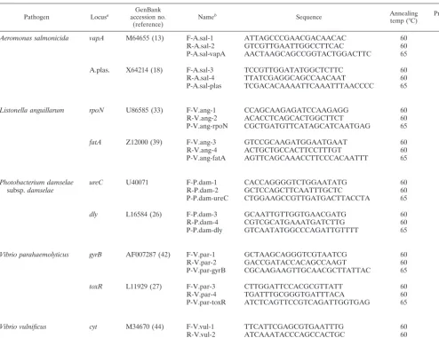
Probes and primers.Nine PCR primer sets and nine internal probe sequences were designed by using the Primer3 program (36). PCR products ranged from 100 to 177 bp in length. Seven specific loci from chromosomal DNA (cyt,rpoN, gyrB,toxR,ureC,dly, andvapA) and two loci from plasmid DNA (fatAand A.sal) were selected for the probe and primer targets (Table 2). All oligonucleotides were purchased from Invitrogen (Carlsbad, Calif.) and were desalted without further modification.
Microarray construction.Slides were prepared by following the methods of Call et al. (6). Briefly, 12-well Teflon-masked slides (Erie Scientific, Portsmouth, N.H.) were sonicated for 2 min in a prewarmed solution of 2.5% Conrad 70 detergent (Fisher Scientific, Fair Lawn, N.J.) and rinsed three times in distilled
H2O. After being dried with compressed air, the slides were immersed for 1 h in an acid bath (3 N HCl), rinsed three times in deionized H2O, and dried again. The slides then were derivatized by immersion in 2% epoxysilane (3-glyci-doxypropyltrimethoxysilane; Sigma-Aldrich, Milwaukee, Wis.) in methanol for 15 min, rinsed twice in methanol, and dried. Oligonucleotide probes were diluted in print buffer (0.1 M Na2HPO4, 0.2 M NaCl, 0.01% sodium dodecyl sulfate) to a final concentration of 60M and spotted onto the slides in quadruplicate by using a MicroGrid II spotter (BioRobotics, Inc., Woburn, Mass.). Printed slides were baked for 60 min at 130°C in a vacuum oven and stored at room temper-ature.
Multiplex PCR.Multiplex PCR mixtures (50-l volume) each contained 50 to 100 ng of purified genomic DNA, 200M each deoxynucleoside triphosphate, 400 nM each primer, 2.5 mM MgCl2, 1⫻reaction buffer, and 2 U ofTaq polymerase (Fisher Scientific, Pittsburgh, Pa.). Thermal cycling was performed with a Mastercycler (Eppendorf, Hamburg, Germany) and included an initial incubation at 95°C for 3 min followed by 30 amplification cycles. Cycling included denaturation for 30 s at 95°C followed by annealing for 1 min at 52, 54, 56, 58, 60, or 62°C. Extension was done for 45 s at 72°C, and cycling was concluded with a final elongation for 5 min at 72°C. All multiplex products were checked by electrophoresis on 1% agarose gels and stained with ethidium bromide (0.5g ml⫺1). Negative test strains that did not show a PCR band upon checking of gels were considered negative for all nine loci and were not labeled or hybridized to the array. PCR mixtures were ethanol precipitated, resuspended in 40 l of sterile water, and labeled by nick translation with a BioNick labeling system (Invitrogen). The labeled products were ethanol precipitated, and the pellets were resuspended in 75l of hybridization buffer (4⫻SSC [60 mM NaCl, 0.6 mM Na citrate] [pH 7.0], 5⫻Denhardt’s solution [0.1% polyvinylpyrrolidone, 0.1% bovine serum albumin, 0.1% Ficoll]).
[image:2.603.44.536.77.360.2]Hybridization and detection.We used a combination of a Tyramide signal amplification (TSA) biotin system (Perkin-Elmer, Boston, Mass.) and fluores-cence to detect hybridized targets (7). Slide wells were incubated with 35l of TNB buffer (0.1 M Tris-HCl, 0.15 M NaCl, 0.5% blocking reagent [TSA biotin system]) for 30 min at room temperature. A 1:10 dilution of the labeled PCR product was prepared in hybridization buffer, heat denatured (2 min at 95°C), and rapidly chilled to 4°C. After aspiration of the TNB buffer from the wells, 35 l of each target was added to each of two wells on the printed slides. The slides
TABLE 1. Test isolates used in this study
aSpecies Strain(s)
Aeromonas caviae
...1.25
Aeromonas hydrophila
...80-A1, B-32, B-35
Aeromonas salmonicida
subsp.
salmonicida
...MT-004, RSP 7.1, SEG-9.1
Aeromonas sobria
...P.33
Flavobacterium psychrophilum
...NCIMB 1947
Listonella anguillarum
...775, 11008, 43-F, 96-F, ATCC 14181, ATCC 43305 to ATCC 43314, ET-208,
NCIMB 571, R-82, RG-111, RV-22, TM-14
Listonella pelagia
...ATCC 25916, NCIMB 1900, NF-182, RPM-87.1, RPM-138.1, ST-11
Photobacterium damselae
subsp.
damselae
...ATCC 33539, CDC2227-81, JCM 8968, RG-91, RM-51, RM-71
Photobacterium damselae
subsp.
piscicida
...ATCC 17911, DI-21, EPOY-8803-II
Photobacterium leiognathi
...ATCC 25521
Photobacterium phosphoreum
...ATCC 11040
Streptococcus parauberis
...RA 99.1
Tenacibaculum maritimum
...LPV 1.7, NCIMB 2154, NCIMB 2158
Vibrio aestuarianus
...ATCC 35048
Vibrio alginolyticus
...CCM 2578
Vibrio campbellii
...ATCC 25920
Vibrio cholerae
...ATCC 14935, V-69
Vibrio fischeri
...ATCC 7744
Vibrio harveyi
...ATCC 14126
Vibrio metschnikovi
...ATCC 7708
Vibrio natriegens
...ATCC 14048
Vibrio nereis
...ATCC 25917
Vibrio ordalii
...NCIMB 2167
Vibrio parahaemolyticus
...ATCC 25969
Vibrio proteolyticus
...ATCC 15338
Vibrio splendidus
...ATCC 25914, ATCC 33125, RM-77, PC 399.1
Vibrio tubiashii
...ATCC 19106, EX-1
Vibrio vulnificus
...ATCC 27562, ATCC 33149, NCIMB 2136, NCIMB 2137
aThe test isolates used in this study originated from multiple countries (Denmark, Japan, Portugal, Scotland, Spain, Thailand, the United Kingdom, and the United
States).
on May 15, 2020 by guest
http://jcm.asm.org/
were placed in a humidified chamber and incubated overnight by being sub-merged in a water bath at 50 or 55°C. After incubation, the hybridization solution was removed by aspiration, and the slides were washed in TNT buffer (0.1 M Tris-HCl [pH 7.5], 0.15 M NaCl, 0.05% Tween 20) three times for 1 min each time with agitation. The wells were incubated for 30 min with streptavidin conjugated to horseradish peroxidase (1:100 in TNB buffer; TSA biotin system). After another washing step, the wells were incubated for 30 min with 10% equine serum albumin (Sigma-Aldrich) in 2⫻SSC for 30 min. The slides were washed again and incubated with biotinylated Tyramide (1:50 in amplification buffer; TSA biotin system) for 10 min. After another washing step, the wells were incubated with streptavidin (2g ml⫺1) conjugated to Alexa Fluor 546 (Molec-ular Probes, Eugene, Oreg.) in 1⫻SSC–5⫻Denhardt’s solution for 60 min. After a final wash in TNT buffer, the slides were spun dry and then imaged with an arrayWoRxescanner (Applied Precision, Issaquah, Wash.).
Microarray image analysis.Image analysis software (softWoRx Tracker; Ap-plied Precision) was used to quantify hybridization signals. The contour function was used to accommodate variations in spot shape and size. To objectively determine whether a spot was positive, we used a variant of a k-means algorithm. Replicate spots were averaged for each hybridization experiment, and the aver-ages were sorted from low to high. The lowest and highest values were used as “seeds” for low and high clusters, respectively. The next lowest value then was compared with the two seeds to determine to which cluster it belonged (i.e., most proximal), and the values for this cluster subsequently were pooled to calculate a new average. This process was continued until all spots were assigned to the low
cluster or the high cluster, followed by calculation of final cluster averages and standard deviations. When final cluster averages differed by⬎3 standard devia-tions, we considered members of the high cluster to represent positive hybrid-ization. In practice, we also imposed a minimum intensity requirement such that the low cluster average could not exceed 25,000 (out of a maximum of 65,535) and the high cluster average could not drop below 10,000. If either condition was not met, then the sample was reprocessed.
Assay specificity and sensitivity.Purified DNA from 75 strains (28 species or subspecies) was used as a DNA template for multiplex PCR followed by hybrid-ization to the microarray. In total, 21L. anguillarum(serotypes O1 to O10), 4V. vulnificus, 1V. parahaemolyticus, 6P. damselaesubsp.damselae, and 3A. salmo-nicidastrains were included as positive test strains, and 40 strains of taxonomi-cally or ecologitaxonomi-cally related bacteria were included as negative test strains. Statistical software from NCSS, Kaysville, Utah (2004 edition), was used to calculate sensitivity and specificity parameters as well as associated 95% confi-dence intervals (CIs; determined by the Wilson method [1]).
[image:3.603.45.540.80.461.2]16S ribosomal DNA (rDNA) PCR.When multiplex PCR failed to amplify any products, we used universal PCR to verify that a template was present and that the reaction was not inhibited by extraction impurities. Using primers UnivRvs_517 (ATTACCGCGGCTGCTGG) and UnivFwd_008 (AGAGTTT GATCMTGGCTCAG), we amplified a ca. 530-bp fragment in 50-l reaction volumes containing reaction buffer (Fisher Scientific), 2 mM MgCl2, a 200M concentration of each deoxynucleoside triphosphate, a 400 nM concentration of each primer, 1 U ofTaqpolymerase, and 100 ng of DNA template. The cycling
TABLE 2. Genes targeted for multiplex PCR and microarray hybridization
Pathogen Locusa accession no.GenBank
(reference) Name
b Sequence Annealing
temp (°C) Product(bp)
Aeromonas salmonicida vapA M64655 (13) F-A.sal-1 ATTAGCCCGAACGACAACAC 60 177
R-A.sal-2 GTCGTTGAATTGGCCTTCAC 60
P-A.sal-vapA AACTAAGCAGCCGGTACTGGACTTC 65
A.plas. X64214 (18) F-A.sal-3 TCCGTTGGATATGGCTCTTC 60 101
R-A.sal-4 TTATCGAGGCAGCCAACAAT 60
P-A.sal-plas TCGACACAAAATTCAAATTTAACCCC 65
Listonella anguillarum rpoN U86585 (33) F-V.ang-1 CCAGCAAGAGATCCAAGAGG 60 125
R-V.ang-2 ACACCTCAGCACTGGCTTCT 60
P-V.ang-rpoN CGCTGATGTTCATAGCATCAATGAG 65
fatA Z12000 (39) F-V.ang-3 GTCCGCAAGATGGAATGAAT 60 137
R-V.ang-4 ACTGCTGCCACTTCCTTTGT 60
P-V.ang-fatA AGTTCAGCAAACCTTCCCACAATTT 65
Photobacterium damselae
subsp.damselae ureC U40071 F-P.dam-1R-P.dam-2 CACCAGGGGTCTGGAATATGGCTCCAGCTTCAATTTGCTC 6060 127
P-P.dam-ureC CTGGAAGCCGTTGATGACTTACCTA 65
dly L16584 (26) F-P.dam-3 GCAATTGTTGGTGAACGATG 60 137
R-P.dam-4 CGTCGCATGAAATGATCTTG 60
P-P.dam-dly GTCAATATGGCCCAGATTGTTTT 65
Vibrio parahaemolyticus gyrB AF007287 (42) F-V.par-1 GCTAAGCAGGGTCGTAATCG 60 145
R-V.par-2 GACCGATACCACAGCCAAGT 60
P-V.par-gyrB CGCAAGAAGTTGCAACGCTTATTAC 65
toxR L11929 (27) F-V.par-3 CTTGGATTCCACGCGTTATT 60 147
R-V.par-4 TGATTTGCGGGTGATTTACA 60
P-V.par-toxR ATCTCAGTTCCGTCAGATTGGTGAG 65
Vibrio vulnificus cyt M34670 (44) F-V.vul-1 TTCATTCGAGCGTGAATTTG 60 100
R-V.vul-2 ATCAAATACCCAGCCACTGC 60
P-V.vul-cyt CCAAGAGCTTGGATGCTATTTCACC 66
aGenetic locus targeted by the described PCR primers and probes:cyt, cytolysine;gyrB, gyrase B;ureC, urease C;dly, phospholipase D; A.plas.,A. salmonicida
plasmid.
bF, sequence of the forward primer; R, sequence of the reverse primer; P, oligonucleotide probe.
on May 15, 2020 by guest
http://jcm.asm.org/
conditions included an initial incubation for 2 min at 95°C followed by 28 cycles that included denaturation for 30 s at 95°C, annealing for 1 min at 62°C, and extension for 1 min at 72°C. Samples were incubated for 10 min at 72°C for a final extension. An aliquot (20l) was checked on gels to confirm amplification. In several instances, we sequenced the resulting product to verify identity (Ampli-con Express, Pullman, Wash.).
Detector sensitivity.To assess overall detection sensitivity under ideal condi-tions, template DNA (P.damselaesubsp.damselae) was diluted 10-fold from 2⫻ 10⫺8g to 2 ⫻10⫺16g and subjected to multiplex PCR. Subsequent PCR products from these dilutions were hybridized to the array. To assess detector sensitivity relative to that of conventional agarose gel electrophoresis,ureCand dly PCR products (fromP.damselae subsp.damselae) were nick translated, diluted twofold, and hybridized to the array. A parallel dilution series was prepared without nick translation for detection by agarose gel electrophoresis and ethidium bromide staining.
RESULTS
We tested PCR annealing at 54, 56, 58, 60, and 62°C with a
gradient thermal cycler. The highest annealing temperature
that was compatible with all primer sets in the multiplex
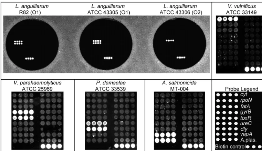
reac-tion was 60°C. Microarray hybridizareac-tion was tested at 50 and
55°C. At 55°C, all multiplex PCR products from the target
bacteria
L
.
anguillarum
,
V
.
parahaemolyticus
,
V
.
vulnificus
,
P
.
damselae
subsp.
damselae
, and
A
.
salmonicida
subsp.
salmo-nicida
produced specific and clear hybridization signals on the
array (Fig. 1).
We tested 75 strains of bacteria representing 28 species
(Table 1). All test strains of the five target species were
cor-rectly detected by at least one species-specific marker. Because
two
L
.
anguillarum
strains were negative for the
fatA
gene and
one
P
.
damselae
subsp.
damselae
strain was negative for the
dly
gene, the calculated sensitivities for these probes were reduced
(Table 3). The large CIs for all of the sensitivity calculations
reflected the limited number of positive test strains that we
could obtain for this study. Multiplex PCR for the 23 nontarget
species produced no amplification products; thus, the
specific-ity of the assay for the panel of strains tested in this study was
100%.
[image:4.603.82.502.71.314.2]To verify that the failure to produce products was not an
artifact of PCR inhibition, all multiplex PCR-negative strains
were also tested by universal 16S rDNA PCR, and an
appro-priately sized product was produced in all cases. The minimum
FIG. 1. Positive control hybridizations. (Upper left panel) Specificity for
L
.
anguillarum
with the multiplex PCR and microarray hybridization.
Genotypes for R-82 (O1), ATCC 43305 (O1), and ATCC 43306 (O2) match the respective genotypes for
rpoN
and
fatA
. (Upper right panel and
lower left panel) Hybridization with probes complementary to
V
.
vulnificus
(
cyt
),
V
.
parahaemolyticus
(
gyrB
and
toxR
),
P
.
damselae
(
ureC
and
dly
),
and
A
.
salmonicida
(
vapA
and A.plas.) in four different PCRs. (Lower right panel) Positions of oligonucleotide probes and the biotin control on
the microarray.
TABLE 3. Results of multiplex PCR and microarray hybridization
Genetic marker
No. of samplesa
Sensitivityb
(95% CI) Specificity
c
(95% CI) Concordant Discordant
Positive Negative Positive Negative
vapA 3 72 0 0 1.0 (0.44–1.0) 1.0 (0.95–1.0)
A.sal 3 72 0 0 1.0 (0.44–1.0) 1.0 (0.95–1.0)
rpoN 21 54 0 0 1.0 (0.85–1.0) 1.0 (0.93–1.0)
fatA 4 69 0 2 0.67 (0.3–0.9) 1.0 (0.95–1.0)
ureC 6 59 0 0 1.0 (0.61–1.0) 1.0 (0.95–1.0)
dly 5 69 0 1 0.83 (0.44–0.97) 1.0 (0.95–1.0)
gyrB 1 74 0 0 1.0 (0.21–1.0) 1.0 (0.95–1.0)
toxR 1 74 0 0 1.0 (0.21–1.0) 1.0 (0.95–1.0)
cyt 4 71 0 0 1.0 (0.51–1.0) 1.0 (0.95–1.0)
aConcordant refers to the number of samples that hybridized correctly to their
respective probes (positive) or the number of samples that were correctly scored as negative by multiplex PCR (negative). Discordant refers to the number of samples that showed positive, nonspecific hybridization (positive) or false-nega-tive results (negafalse-nega-tive).
bNumber of concordant positive samples divided by number of concordant
positive samples plus number of discordant negative samples.
cNumber of concordant negative samples divided by number of concordant
negative samples plus number of discordant positive samples.
on May 15, 2020 by guest
http://jcm.asm.org/
[image:4.603.301.541.524.651.2]DNA template required for the positive detection of multiplex
products (
P
.
damselae
subsp.
damselae
in this case) was 20 fg of
genomic DNA, which is equivalent to four or five cells.
Trip-licate serial dilutions of the
ureC
and
dly
PCR products
dem-onstrated that the
ureC
product was detectable below 1:32,
whereas the
dly
product was detectable only to 1:16 (based on
the detection cluster algorithm). These two combined products
were not visible below a 1:4 dilution when agarose gel
electro-phoresis was used for detection.
DISCUSSION
This is the first microarray technique described for the
de-tection of marine fish pathogens. The availability of rapid,
sensitive, and specific diagnostic methods for the detection of
bacterial pathogens causing diseases is very important in
aquaculture. Nevertheless, existing methods are restricted by
the number of pathogens that can be detected simultaneously
and by overall assay sensitivity or specificity. Like many PCR
assays, the assay described here was suitable for detecting
⬃
5
cell equivalents under optimal conditions. Unlike conventional
multiplex PCR assays, microarray detectors do not require
clear length differences between PCR products; thus, the PCR
can be designed around short, equally sized fragments that are
amplified with similar efficiencies. In addition, because
detec-tion is based on hybridizadetec-tion to specific sequences rather than
product length, time-consuming sequencing or blot-and-probe
techniques are not necessary to confirm product identity (9, 10,
43). Products of various lengths also present a challenge for
developing optimal PCR conditions (primer annealing
temper-atures and similar MgCl2
concentrations). While the dilution
experiments presented here suggest that unequal PCR
ampli-fication efficiencies or unequal hybridization efficiencies exist
for the
ureC
and
dly
targets, the current assay is sufficient for
simultaneous screening for all nine pathogenic markers.
Our prototype assay was highly specific, with no
false-posi-tive detections for a battery of test strains (23 nontarget species
or subspecies). The sensitivity was 100% for seven of the nine
markers. The
fatA
marker hybridized only to four
L
.
anguilla-rum
strains, although these were all serovar O1. Two
addi-tional serovar O1 strains were negative for
fatA
. The
fatA
gene
is harbored on a virulence plasmid (pJM1) that encodes an
iron-sequestering system, and an estimated 90% of serotype
O1 strains harbor this plasmid. Thus, we would not expect all
serovar O1 strains to hybridize to both
L
.
anguillarum
probes.
No other serovars hybridized to the
fatA
marker.
One test strain of
P
.
damselae
subsp.
damselae
(JCM 8968)
did not hybridize to the
dly
probe, although the
ureC
probe was
positive for this strain. This particular strain was originally
classified as
Photobacterium histaminum
(20); thus, the failure
to hybridize is consistent with some degree of genetic
diver-gence. Although all three
A
.
salmonicida
strains were positive
for both plasmid-borne markers (
vapA
and A.sal), not all
strains are expected to harbor these genes (41); thus, the
sen-sitivity reported here (100%) does not accurately reflect what
would likely be encountered in a diagnostic or surveillance
application.
The specificity and sensitivity estimates reported here apply
to the microarray detector only. Both of these variables can be
affected by numerous events “upstream” of the actual
microar-ray hybridization. For example, during the course of this study,
we encountered five instances when a strain of bacteria did not
hybridize as expected to one or more probes. In all of these
instances, partial sequencing of the 16S rRNA gene
demon-strated that the test strains were not correctly identified, and
the microarray hybridization results were consistent with the
species identified by 16S rRNA gene sequencing (these strains
were not included in the present analysis). Either the initial
strain identification was incorrect or subsequent sample
pro-cessing led to an error. In another instance, two test strains
were found to be negative when first hybridized to the array
but were found to be positive when checked a second time (i.e.,
a 2.7% error rate during the hybridization step). These errors
are examples of process-level errors that can be minimized by
using stringent controls and standard operating procedures in
a diagnostic laboratory setting.
The high degree of specificity reported here suggests that
this assay format is not prone to generating false-positives; as
with any assay, if any unusual positive results are detected, then
additional confirmation is advisable. A larger problem is that
of false-negatives. False-negatives can arise due to naturally
occurring sequence polymorphisms in PCR primer or probe
hybridization sequenced. This is not a significant issue if all
polymorphisms are known and can be included on the
microar-ray or if relatively conserved genes are selected. If an armicroar-ray is
dependent on many sequence polymorphisms within the same
probe region (e.g., selected regions of the 16S rRNA gene),
then naturally occurring mutations in these regions could lead
to false-negatives when these variable sequences are tested
with the microarray.
During the execution of any PCR assay, false-negatives can
also result when coprecipitates from the template extraction
interfere with the PCR (23). In the format described here, we
used post hoc PCR amplification of the 16S rRNA gene to
verify that PCR failure was not due to template impurities. It
is clear that if prokaryotic bacterial DNA were used in the
reaction, we could include a 10th primer set targeting the 16S
rRNA gene as a positive control for the PCR. Nevertheless,
the choice of an internal control depends on the matrix that is
sampled. If tissue samples are assayed, then samples without
prokaryotic DNA will still appear negative for a prokaryotic
16S rDNA marker; a eukaryotic positive control could be
in-corporated for this application. A partial solution would be to
spike the reaction with control DNA, but this strategy can
reduce sensitivity if the spiked template is preferentially
am-plified during the PCR (unpublished data). For the survey of
environmental samples, it is appropriate to add control DNA
to separate dilutions of the original extract so that PCR
inhi-bition can be quantified (23). Consequently, the assay
de-scribed here should accommodate multiple matrices (purified
DNA, tissue samples, or environmental samples) with modest
assay or procedural modifications.
This is the first microarray technique described for the
de-tection of bacteria pathogenic for marine fish. The sensitivity
and specificity of the described method and the simultaneous
detection of five bacterial species make it suitable for
prelim-inary diagnoses or confirmation of vibriosis and furunculosis as
well as for the detection of potential human pathogens in sea
farming products.
on May 15, 2020 by guest
http://jcm.asm.org/
ACKNOWLEDGMENTS
This work was supported by the Agricultural Animal Health
Pro-gram (College of Veterinary Medicine, Washington State University)
and the USFWS (through the WSU and UI Center for Reproductive
Biology, Washington State University). S.F.G. thanks the University of
Santiago de Compostela for a research fellowship in support of this
work.
REFERENCES
1. Agresti, A., and B. Coull.1998. Approximate is better than⬘exact’ for interval estimation of binomial proportions. Am. Stat.52:119–126.
2. Arias, C. R., E. Garay, and R. Aznar.1995. Nested PCR method for rapid and sensitive detection ofVibrio vulnificusin fish, sediments, and water. Appl. Environ. Microbiol.61:3476–3478.
3. Austin, B., and D. Austin.1999. Bacterial fish pathogens: disease of farmed and wild fish, 3rd ed. Springer-Praxis, London, England.
4. Brasher, C. W., A. DePaola, D. D. Jones, and A. K. Bej.1998. Detection of microbial pathogens in shellfish with multiplex PCR. Curr. Microbiol.37:
101–107.
5. Call, D., M. Borucki, and T. Besser.2003. Mixed-genome microarrays reveal multiple serotype and lineage-specific differences among strains ofListeria monocytogenes. J. Clin. Microbiol.41:632–639.
6. Call, D., D. Chandler, and F. Brockman.2001. Fabrication of DNA microar-rays using unmodified oligonucleotide probes. BioTechniques30:368–379. 7. Call, D. R., M. Bakko, M. Krug, and M. Roberts.2003. Identifying
antimi-crobial resistance genes using DNA microarrays. Antimicrob. Agents Che-mother.47:3290–3295.
8. Call, D. R., M. Borucki, and F. Loge.2003. Detection of bacterial pathogens in environmental samples using DNA microarrays. J. Microbiol. Methods
53:235–243.
9. Call, D. R., F. J. Brockman, and D. P. Chandler.2001. Detecting and genotypingEscherichia coliO157:H7 using multiplexed PCR and nucleic acid microarrays. Int. J. Food Microbiol.67:71–80.
10. Chizhikov, V., A. Rasooly, K. Chumakov, and D. Levy.2001. Microarray analysis of microbial virulence factors. Appl. Environ. Microbiol.67:3258– 3263.
11. Chizhikov, V., M. Wagner, A. Ivshina, Y. Hoshino, A. Z. Kapikian, and K. Chumakov.2002. Detection and genotyping of human group A rotaviruses by oligonucleotide microarray hybridization. J. Clin. Microbiol.40:2398– 2407.
12. Cho, J. C., and J. M. Tiedje.2001. Bacterial species determination from DNA-DNA hybridization by using genome fragments and DNA microarrays. Appl. Environ. Microbiol.67:3677–3682.
13. Chu, S., S. Cavaignac, J. Feutrier, B. M. Phipps, M. Kostrzynska, W. W. Kay, and T. J. Trust.1991. Structure of the tetragonal surface virulence array protein and gene ofAeromonas salmonicida. J. Biol. Chem.266:15258– 15265.
14. Del Cerro, A., I. Marquez, and J. A. Guijarro.2002. Simultaneous detection ofAeromonas salmonicida,Flavobacterium psychrophilum, andYersinia ruc-keri, three major fish pathogens, by multiplex PCR. Appl. Environ. Micro-biol.68:5177–5180.
15. Gibson, G.2002. Microarrays in ecology and evolution: a preview. Mol. Ecol.
11:17–24.
16. Gonza´lez, S., C. R. Osorio, and Y. Santos.2003. Development of a PCR-based method for the detection ofListonella anguillarumin fish tissues and blood samples. Dis. Aquat. Org.55:109–115.
17. Guschin, D. Y., B. K. Mobarry, D. Proudnikov, D. A. Stahl, B. E. Rittmann, and A. D. Mirzabekov.1997. Oligonucleotide microchips as genosensors for determinative and environmental studies in microbiology. Appl. Environ. Microbiol.63:2397–2402.
18. Hiney, M., M. T. Dawson, D. M. Heery, P. R. Smith, F. Gannon, and R. Powell.1992. DNA probe forAeromonas salmonicida. Appl. Environ. Mi-crobiol.58:1039–1042.
19. Hoie, S., M. Heum, and O. Thorensen.1997. Evaluation of a polymerase chain reaction-based assay for the detection of Aeromonas salmonicida subsp.salmonicidain Atlantic salmon,Salmo salar. Dis. Aquat. Org.30:27– 35.
20. Kimura, B., S. Hokimoto, H. Takahashi, and T. Fujii.2000.Photobacterium histaminumOkuzumi et al. 1994 is a later subjective synonym of Photobac-terium damselaesubsp.damselae(Love et al. 1981) Smith et al. 1991. Int. J. Syst. Evol. Microbiol.50:1339–1342.
21. Kita-Tsukamoto, K., H. Oyaizu, K. Nanba, and U. Simidu.1993. Phyloge-netic relationships of marine bacteria, mainly members of the family Vibri-onaceae, determined on the basis of 16S rRNA sequences. Int. J. Syst. Bacteriol.43:8–19.
22. Klontz, K. C., S. Lieb, M. Schreiber, H. T. Janowski, L. M. Baldy, and R. A. Gunn.1988. Syndromes ofVibrio vulnificusinfections. Clinical and epide-miologic features in Florida cases, 1981–1987. Ann. Intern. Med.109:318– 323.
23. Loge, F. J., D. E. Thompson, and D. R. Call.2002. PCR detection of specific pathogens in water: a risk-based analysis. Environ. Sci. Technol.36:2754– 2759.
24. Magnusson, H. B., O. H. Fridjonsson, O. S. Andresson, E. Benediktsdottir, S. Gudmundsdottir, and V. Andresdottir.1994.Renibacterium salmonina-rum, the causative agent of bacterial kidney disease in salmonid fish, detect-ed by nestdetect-ed reverse transcription-PCR of 16S rRNA sequences. Appl. Environ. Microbiol.60:4580–4583.
25. Marshall, S., S. Heath, V. Henriquez, and C. Orrego.1998. Minimally inva-sive detection ofPiscirickettsia salmonisin cultivated salmonids via the PCR. Appl. Environ. Microbiol.64:3066–3069.
26. McNamara, P. J., W. A. Cuevas, and J. G. Songer.1995. Toxic phospho-lipases D ofCorynebacterium pseudotuberculosis,C.ulceransand Arcanobac-terium haemolyticum: cloning and sequence homology. Gene156:113–118. 27. Miller, V. L., R. K. Taylor, and J. J. Mekalanos.1987. Cholera toxin
tran-scriptional activator ToxR is a transmembrane DNA binding protein. Cell
48:271–279.
28. Mooney, J., E. Powell, C. Clabby, and R. Powell.1995. Detection of Aero-monas salmonicidain wild Atlantic salmon using a specific DNA probe test. Dis. Aquat. Org.21:131–135.
29. Nakashima, S., and K. Takimoto.1987. The epidemiological data of food poisoning in 1986. Food Sanit. Res.37:50–76.
30. Osorio, C., and A. Toranzo.2002. DNA-based diagnostics in sea farming, p. 253–310.InM. Fingerman and R. Nagabhushanam (ed.), Seafood safety and human health. Science Publishers, Inc., Enfield, N.H.
31. Osorio, C. R., M. D. Collins, A. E. Toranzo, J. L. Barja, and J. L. Romalde.
1999. 16S rRNA gene sequence analysis ofPhotobacterium damselaeand nested PCR method for rapid detection of the causative agent of fish pas-teurellosis. Appl. Environ. Microbiol.65:2942–2946.
32. Osorio, C. R., A. E. Toranzo, J. L. Romalde, and J. L. Barja.2000. Multi-plexed PCR assay forureCand 16S rRNA genes clearly discriminates be-tween both subspecies ofPhotobacterium damselae. Dis. Aquat. Org.40:177– 183.
33. O’Toole, R., D. L. Milton, P. Horstedt, and H. Wolf-Watz.1997.rpoNof the fish pathogenVibrio(Listonella)anguillarumis essential for flagellum pro-duction and virulence by the water-borne but not intraperitoneal route of inoculation. Microbiology143:3849–3859.
34. Pazos, F., Y. Santos, Macías, S. Nu´n˜ez, and A. E. Toranzo.1996. Evaluation of media for the successful culture ofFlexibacter maritimus. J. Fish Dis.
19:193–197.
35. Pedersen, K., B. Austin, D. A. Austin, and J. L. Larsen.1999. Vibrios associated with mortality in cultured plaicePleuronectes platessafry. Acta Vet. Scand.40:263–270.
36. Rozen, S., and H. Skaletsky.2000. Primer3 on the WWW for general users and for biologist programmers, p. 365–386.InS. Krawetz and S. Misener (ed.), Bioinformatics methods and protocols: methods in molecular biology. Humana Press, Totowa, N.J.
37. Ruimy, R., V. Breittmayer, P. Elbaze, B. Lafay, O. Boussemart, M. Gauthier, and R. Christen.1994. Phylogenetic analysis and assessment of the genera Vibrio,Photobacterium,Aeromonas, andPlesiomonasdeduced from small-subunit rRNA sequences. Int. J. Syst. Bacteriol.44:416–426.
38. Schena, M.2000. Microarray biochip technology. Eaton Publishing, Natick, Mass.
39. Tolmasky, M. E., L. A. Actis, and J. H. Crosa.1993. A single amino acid change in AngR, a protein encoded by pJM1-like virulence plasmids, results in hyperproduction of anguibactin. Infect. Immun.61:3228–3233. 40. Toranzo, A. E., and J. L. Barja.1993. Fry mortality syndrome (FMS) in
Spain. Isolation of the causativeFlexibacter psychrophilus. Bull. Eur. Assoc. Fish Pathol.13:30–32.
41. Toranzo, A. E., Y. Santos, S. Nun˜ez, and J. Barja.1991. Biochemical and serological characteristics, drug resistance and plasmid profiles of Spanish isolates ofAeromonas salmonicida. Gyobyo Kenkyu26:55–60.
42. Venkateswaran, K., N. Dohmoto, and S. Harayama.1998. Cloning and nucleotide sequence of thegyrBgene of Vibrio parahaemolyticusand its application in detection of this pathogen in shrimp. Appl. Environ. Micro-biol.64:681–687.
43. Volokhov, D., A. Rasooly, K. Chumakov, and V. Chizhikov.2002. Identifi-cation of Listeriaspecies by microarray-based assay. J. Clin. Microbiol.
4:4720–4728.
44. Yamamoto, K., A. C. Wright, J. B. Kaper, and J. G. Morris.1990. The cytolysin gene ofVibrio vulnificus: sequence and relationship to theVibrio choleraeEl Tor hemolysin gene. Infect. Immun.58:2706–2709.