Gram-Positive Organisms and Resistance Markers in Positive
Blood Cultures
Linoj P. Samuel, Robert J. Tibbetts, Adam Agotesku, Margaret Fey, Rhonda Hensley, Frederick A. Meier Henry Ford Health System, Department of Pathology, Detroit, Michigan, USA
Rapid identification of pathogens directly from positive blood cultures can play a major role in reducing patient mortality rates. We evaluated the performance of the Verigene Gram-Positive Blood Culture (BC-GP) assay (Nanosphere Inc., Northbrook, IL) for detection of commonly isolated Gram-positive organisms as well as associated resistance markers from positive blood cul-tures. Positive blood cultures (VersaTREK; Trek Diagnostic Systems, Independence, OH) from 203 patients with Gram-positive organism infections were analyzed using the BC-GP assay within 12 h for the detection of 12 different organisms, including staphylococci, streptococci, and enterococci, as well as for the presence of 3 resistance markers (mecA,vanA, andvanB). Results were compared to those of routine laboratory methods for identification and susceptibility testing. For identification of organ-isms and detection of resistance markers in 178 monomicrobial positive blood cultures, the BC-GP assay showed 94% and 97% concordance, respectively, with routine methods. After 25 polymicrobial cultures were included, the results showed 92% and 96% agreement for identification and resistance markers, respectively, for a total of 203 positive cultures. In 6/25 polymicrobial cultures, at least 1 isolate was not detected. Concordance levels for detection of major pathogens suchStaphylococcus aureus (nⴝ45) and enterococci (nⴝ19) were 98% and 95%, respectively. Agreement levels for detection of resistance markers such as mecAandvanA/Bwere 92% and 100%, respectively. The BC-GP assay is capable of providing rapid identification of Gram-posi-tive cocci as well as detection of resistance markers directly from posiGram-posi-tive blood cultures at least 24 to 48 h earlier than conven-tional methods.
R
apid detection and identification of bloodstream pathogensare crucial to timely therapeutic intervention and patient management. Early administration of appropriate antibiotics
im-proves survival of bacteremic patients (1–3), but improvement
depends on rapid identification and susceptibility testing of pathogens from positive blood cultures. This process may take 1 to 3 days, leading to potential delays in administering appropriate therapy. This delay is significant, because each additional day re-quired for definitive identification of pathogens in blood cultures
has been shown to increase mortality rates (4). Strategies that
re-duce the time required for reporting positive blood cultures such as prompt Gram staining and notification processes have been
demonstrated to reduce length of hospital stay and mortality (2,5,
6). The use of molecular technologies for early identification of
pathogens and resistance determinants directly from positive blood cultures may be able to reduce the time required for labo-ratory processes and significantly impact patient management.
Gram-positive organisms are implicated as the primary
patho-gens in the majority of bacteremic episodes (6). Rapid
identifica-tion of these isolates directly from positive blood cultures has been shown to improve patient outcomes as well as reduce
inappropri-ate antimicrobial therapy and decrease hospital charges (7–11).
Rapid detection of nonpathogens contaminating blood cultures is also valuable. Beekmann et al. demonstrated that whereas coagu-lase-negative staphylococci (CoNS) represented 22% of all
Gram-positive isolates from blood cultures,⬃80% of the isolates were
subsequently determined to represent contamination (12). Early
identification of these isolates can significantly reduce
unneces-sary antimicrobial usage and decrease length of stay (8). In
addi-tion, rapid detection of resistance markers such asmecA, which
confers methicillin resistance in staphylococci, directly from
pos-itive blood cultures can have a similar pospos-itive impact on
mortal-ity, hospital costs, and length of stay (10,13).
Early efforts toward rapid detection of pathogens from positive blood cultures involved peptide nucleic acid-based fluorescence
in situhybridization (PNA-FISH) (7–9). Other approaches in-cluded molecular amplification methods coupled with
probe-based detection (10,13,14). Matrix-associated laser desorption
ionization–time of flight (MALDI-TOF) analysis for identifica-tion of bacterial isolates from positive blood cultures is another
recent development (15–18). Each methodology has limitations,
for example, the inability to detect resistance markers (PNA-FISH and MALDI-TOF) or problems with detecting polymicrobial
in-fections (MALDI-TOF) (16,17). The use of microarrays in this
setting has been previously described and offers the advantages of being able to detect multiple targets and associated resistance
markers simultaneously (19–21). Here we evaluate a
microarray-based assay, the Verigene Gram-Positive Blood Culture (BC-GP) nucleic acid test (Nanosphere Inc., Northbrook, IL), which is per-formed on the Verigene system for detection of Gram-positive microorganisms and associated resistance markers in positive
aer-obic blood culture bottles. The assay is capable of detecting
Staph-ylococcusspp.,Streptococcusspp.,Listeriaspp.,Staphylococcus
au-Received10 November 2012Returned for modification14 December 2012 Accepted26 January 2013
Published ahead of print30 January 2013
Address correspondence to Linoj P. Samuel, [email protected].
Copyright © 2013, American Society for Microbiology. All Rights Reserved.
doi:10.1128/JCM.02982-12
on May 16, 2020 by guest
http://jcm.asm.org/
Downloaded from
on May 16, 2020 by guest
http://jcm.asm.org/
Downloaded from
on May 16, 2020 by guest
http://jcm.asm.org/
reus, Staphylococcus epidermidis, Staphylococcus lugdunensis,
Streptococcus pneumoniae, Streptococcus pyogenes, Streptococcus agalactiae, Streptococcus anginosus group, Enterococcus faecalis, andEnterococcus faecium. In addition, the assay is capable of
de-tecting the presence ofmecAifS. aureusorS. epidermidisor both
are present andvanA andvanB(vanA/B) ifE. faecium andE.
faecalisor both are detected.
(The study data were presented in poster format at the 52nd Interscience Conference on Antimicrobial Agents and Chemo-therapy [ICAAC], 2012.)
MATERIALS AND METHODS
Patient samples.Clinical samples for this study were blood cultures sub-mitted as part of routine patient care to the Henry Ford Health System (HFHS) Core Microbiology laboratory, which serves the 900-bed Henry Ford Hospital in Detroit, MI, as well as 3 acute care hospitals and 32 medical centers in southeast Michigan. Blood cultures were collected in REDOX EZ Draw 40-ml aerobic and anaerobic culture bottles (Trek Di-agnostic Systems, Cleveland, OH) and incubated on a VersaTREK instru-ment (Trek Diagnostic Systems). The instruinstru-ment detects organism growth by measuring changes in headspace pressure of the blood culture bottles. Aerobic bottles from nonconsecutive cultures that flagged posi-tive were Gram stained and, if posiposi-tive for Gram-posiposi-tive cocci or bacilli, were included in the study. Only one positive blood culture per patient was included in the study. Laboratory staff performing the BC-GP assay were not aware of the organism identification at the time of testing. A total of 203 positive blood cultures containing Gram-positive organisms were included in this study. This study was approved by the Institutional Re-view Board at HFHS.
Blood culture processing.The aerobic bottles from blood cultures selected for inclusion in the study were sampled aseptically in a biosafety cabinet and inoculated into blood agar and MacConkey and chocolate agar for overnight incubation at 37°C and 5% CO2. An additional aliquot of 1.5 ml was removed for study purposes, and 350l of the aliquot was utilized for testing on the BC-GP assay within 12 h of the blood culture flagging positive. The remainder was stored at⫺70°C for additional test-ing as needed. If a valid result was not obtained in the initial run, an additional aliquot was taken from the blood culture bottle (stored at room temperature) and retested within 24 h of the positive blood culture signal. If the test could not be performed within 24 h, an aliquot was frozen at ⫺70°C for later testing.
Subcultured isolates were identified by the use of a combination of rou-tine identification tests and automated platforms such as the Vitek 2 (bio-Mérieux, Durham, NC). Susceptibility testing was performed using the Vitek 2, disk diffusion methods (CLSI M100-S22), and E-tests (bioMérieux, Dur-ham, NC). In the case of discordant results, a combination of routine labora-tory methods such as Vitek 2 and API (bioMérieux, Durham, NC) and/or bidirectional sequencing was performed at a reference laboratory (Dynacare Laboratories, Milwaukee, WI) as needed for resolution.
Microarray testing. The Verigene platform includes the Verigene ProcessorSPand Verigene Reader. The Verigene ProcessorSPcarries out extraction of nucleic acid from specimens using magnetic glass beads. Patient samples are loaded into an extraction tray, which is then loaded into the processor along with the utility tray, pipette tip holder assembly, and test cartridge. These items are all single-use disposable components that contain all the reagents required for testing. The Verigene Reader controls the processor and is responsible for specimen tracking, test selec-tion, imaging, and analysis of test cartridges and display of the results.
For testing on the Verigene platform using the BC-GP assay, 350l of blood culture media from the positive aerobic bottle is loaded into the extraction tray, which is then placed into the processorSPalong with all other consumables. The instrument extracts nucleic acid from the sample which is then mixed with the appropriate buffer and transferred to the test cartridge. The target analyte, if present, hybridizes to synthetic gene-spe-cific oligonucleotide capture strands on the test cartridge substrate slide.
Another synthetic mediator target-specific nucleotide is introduced to form a hybridization sandwich with the gene of interest. At this point, a gold nanoparticle-labeled probe is introduced with oligonucleotides com-plementary to the intermediate oligonucleotide bound to the gene of in-terest. Finally, the gold nanoparticles are coated with silver to enhance the optical signal. The test cartridge is then removed from the ProcessorSP, and the substrate slide is inserted into the Verigene Reader for analysis. The Verigene Reader projects white light across the substrate slide, detects the relative brightness of each spot due to gold nanoparticles bound to target-specific probes and provides a “Detected” or “Not Detected” result for each of the panel members.
Statistical analysis.Results of routine laboratory testing were not available to laboratory testing personnel at the time of BC-GP testing. Final discrepant result analysis was also not made available until the con-clusion of sample testing. Concordance was determined in comparison to results of local laboratory methods, with discrepancies resolved by testing at a reference laboratory using either routine laboratory methods or bidi-rectional sequencing as described previously.
RESULTS
A total of 203 positive blood cultures containing 227 Gram-posi-tive organisms were tested using the BC-GP assay on the Verigene system. Over 12% (25/203) of positive samples yielded two or more organisms during routine laboratory culture. The distribu-tion of microorganisms that were isolated from the positive blood
cultures is shown inTable 1. At least 91% (207/227) of the isolates
were members of the panel of targets on the BC-GP assay.
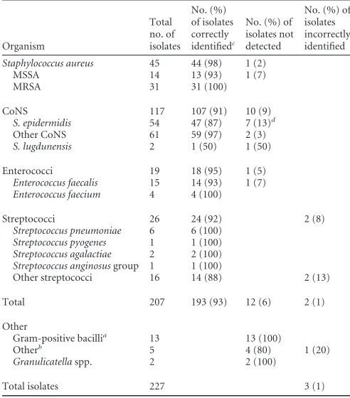
Staph-TABLE 1Distribution of isolates encountered during the study and numbers of isolates correctly identified, misidentified, or not detected
Organism
Total no. of isolates
No. (%) of isolates correctly identifiedc
No. (%) of isolates not detected
No. (%) of isolates incorrectly identified
Staphylococcus aureus 45 44 (98) 1 (2)
MSSA 14 13 (93) 1 (7)
MRSA 31 31 (100)
CoNS 117 107 (91) 10 (9)
S. epidermidis 54 47 (87) 7 (13)d
Other CoNS 61 59 (97) 2 (3)
S. lugdunensis 2 1 (50) 1 (50)
Enterococci 19 18 (95) 1 (5)
Enterococcus faecalis 15 14 (93) 1 (7)
Enterococcus faecium 4 4 (100)
Streptococci 26 24 (92) 2 (8)
Streptococcus pneumoniae 6 6 (100)
Streptococcus pyogenes 1 1 (100)
Streptococcus agalactiae 2 2 (100)
Streptococcus anginosusgroup 1 1 (100)
Other streptococci 16 14 (88) 2 (13)
Total 207 193 (93) 12 (6) 2 (1)
Other
Gram-positive bacillia 13 13 (100)
Otherb 5 4 (80) 1 (20)
Granulicatellaspp. 2 2 (100)
Total isolates 227 3 (1)
aIncludesBacillusspp.,Corynebacteriumspp., etc. b
Includes one isolate each ofE. casseliflavus,Dermacoccusspp., andLeuconostocspp. and two isolates ofRothiaspp.
c
Correct identification to the species level.
dA total of 4 of 7 isolates ofS. epidermidiswere correctly identified to the genus level
but not to the species level.
on May 16, 2020 by guest
http://jcm.asm.org/
[image:2.585.297.543.87.367.2]ylococci, enterococci, and streptococci represented 72% (162/ 227), 8% (19/227), and 11% (26/227) of total isolates, respectively. NoListeriaspp. were detected during the course of this study.
S. aureusrepresented 28% (45/162) of the staphylococcal iso-lates detected. The BC-GP assay accurately detected 98% (44/45)
of these isolates (Table 1). The remaining staphylococcal isolates
wereS. epidermidisisolates (54/117), S. lugdunensisisolates (2/ 117), or other CoNS isolates (61/117). The BC-GP assay correctly
identified 87% (47/54) of theS. epidermidisisolates. Of the
re-maining 7 isolates, 4/7 were correctly identified as being staphylo-cocci but were not identified to species level and 3/7 isolates were
not detected (Table 1). Only 2 isolates ofS. lugdunensiswere
en-countered during the course of the study, and 1/2 was present in a polymicrobial culture and was not detected. The remaining CoNS isolates were identified to the genus level, with only 3% (2/61) of
isolates not detected (Table 1).
A total of 19 enterococcal isolates were detected using the
BC-GP assay, including 4/19 isolates ofE. faeciumand 15/19
iso-lates ofE. faecalis(Table 1). At least 95% (18/19) of the
enterococ-cal isolates were captured by the BC-GP assay, with the sole
excep-tion being anE. faecalisisolate that was present in a mixed culture
withEscherichia coli. A total of 26 streptococcal isolates were also encountered during the course of the study, and 92% (24/26) were detected and accurately identified to either the genus or species
level (Table 1). The remaining isolates (2/26) were identified by
the reference laboratory asStreptococcus oralisandStreptococcus
mitisbut were misidentified by the BC-GP assay asS. anginosus
group andS. pneumoniae, respectively. Other streptococcal
iso-lates that were correctly identified includedS. pneumoniae(6/6),
S. pyogenes(1/1),S. agalactiae(2/2), andS. anginosusgroup (1/1). The positive blood cultures tested using the BC-GP assay included 20 Gram-positive isolates that were not part of the bacterial panel of targets in the assay. These isolates included Gram-positive
ba-cilli (n⫽13),Granulicatellaspp. (n⫽2),Rothiaspp. (n⫽2), and
one each ofDermacoccus nishinomiyaensis,Enterococcus
casselifla-vus, and Leuconostoc mesenteroides subsp. cremoris (Table 1).
None of these isolates were detected by the BC-GP assay, as
antic-ipated, with the exception of theLeuconostocisolate, which was
misidentified asS. epidermidis.
The BC-GP assay was also evaluated for its ability to detect the
presence of the resistance markersmecAandvanA/B. The
pres-ence ofmecAwas confirmed by routine laboratory methods in
69% (31/45) and 78% (42/54) of isolates ofS. aureusandS.
epi-dermidis, respectively (Table 2). The BC-GP assay detected the
presence ofmecAin 97% (30/31) and 88% (37/42) of blood
cul-tures positive forS. aureusandS. epidermidis, respectively (Table
2). The BC-GP assay also detected the presence ofvanA/Bin 100%
(9/9) of blood cultures positive for enterococcal isolates that were later determined to be vancomycin resistant by routine laboratory
methods (Table 2).
The BC-GP assay showed overall concordance of 93% (193/ 207) for detection and identification of Gram-positive bacterial
isolates in positive blood cultures (Table 1). Agreement was better
with monomicrobial blood cultures (94%, 168/178) than with
polymicrobial blood cultures (76%, 19/25) (Table 3). The BC-GP
assay detected 93% (75/81) of resistance markers (mecA and
vanA/B) in positive blood cultures withS. aureus,S. epidermidis,
orE. faeciumorE. faecalis(Table 2). Concordance levels for de-tection of resistance markers in monomicrobial and polymicro-bial cultures were 97% and 84%, respectively.
DISCUSSION
The use of microarray- or DNA probe-based assays for the detec-tion of bacterial pathogens and resistance markers in positive
blood cultures has been previously described (19–23). We
evalu-ated the ability of the BC-GP assay to detect 12 different Gram-positive bacterial targets and 3 resistance markers in 203 Gram-positive aerobic REDOX 40-ml blood culture bottles. The BC-GP assay is run on the Verigene platform, which is comprised of one or more processor units linked to a reader unit that also serves as the user interface. The testing is cartridge based, making it more amenable to random-access testing rather than batched processing. Each unit can run one sample in 2.5 h, with hands-on time of 5 to 10 min. Timely use of the BC-GP assay on positive blood cultures for identification of blood culture isolates and detection of resistance markers offers a potential time savings of 1 to 3 days over routine laboratory methods. The assay showed overall agreement of 93% and 93% for the detection and identification of Gram-positive bacterial isolates and associated resistance markers, respectively, compared to reference methods.
The ability to identify potential contaminants such as CoNS directly from blood cultures offers the potential advantages of
de-escalation of therapy (8). The BC-GP assay was able to identify
91% (107/117) of CoNS isolates to either the species or genus
level. Of the 10 discrepant results in this group, 4/10 wereS.
epi-dermidisisolates that were correctly identified as staphylococci but
TABLE 2Performance of the BC-GP assay for detection of resistance markers in positive blood cultures
Isolate category
Total no. of isolates
No. (%) of isolates with gene detected
No. (%) of isolates with gene not detected
mecApositive
S. aureus 31 30 (97) 1 (3)
S. epidermidis 42 37 (88) 5 (12)
TotalmecApositive 73 67 (92) 6 (8)
TotalvanA/Bpositive 9 9 (100) 0 (0)
Total 82 76 (93) 76 (7)
TABLE 3Agreement between the BC-GP assay and reference methods for identification of positive blood cultures
Blood culture category and parameter
Total no. of cultures
No. (%) of cultures with concordant results
No. (%) of cultures with discrepant results
Monomicrobial 178
Identification 178 168 (94) 10 (6)
Resistance markers 178 173 (97) 5 (3)
Polymicrobial 25
Identification 25 19 (76) 6 (24)
Resistance markers 25 21 (84) 4 (16)
Total agreement
Identification 203 187 (92) 16 (8)
Resistance markers 203 194 (96) 9 (4)
on May 16, 2020 by guest
http://jcm.asm.org/
were not identified to the species level. The assay performed well
with regard to detection ofS. aureus, with 98% (44/45) of isolates
correctly identified (Table 1). The performance of the BC-GP
as-say for detection ofmecAwas better inS. aureusthan inS.
epider-midis(97% versus 88%) (Table 2). This may be accounted for by
the fact thatS. epidermidiswas more likely to be present in
poly-microbial positive cultures thanS. aureus. Three of seven cultures
withS. epidermidisand discrepantmecAresults were
polymicro-bial. In addition, 3 positive cultures containingS. epidermidiswere
falsely positive formecAusing the BC-GP assay. A potential
limi-tation of the assay is that in mixed cultures containing bothS.
aureusandS. epidermidis, themecAgene target, if positive, cannot be assigned specifically to isolates of either organism.
The assay performed well for the detection ofE. faeciumandE.
faecalisandvanA/B. Only 1 isolate ofE. faecalisin a polymicrobial
positive culture was not detected, and 9/9vanA/B-positive
cul-tures were detected (Table 2). Another limitation of the assay is
that it does not detect enterococci other thanE. faeciumorE.
faecalis, as was evidenced in a positive blood culture that
con-tainedE. casseliflavus. The assay also accurately identified 24/26
streptococcal isolates, with the most notable exception being anS.
mitisisolate that was misidentified asS. pneumoniae. The difficulty in distinguishing between these two species has been noted with other rapid blood culture identification methods such as
MALDI-TOF analysis (16,17) and is another potential limitation, although
in this study, 6/6S. pneumoniaeisolates were accurately identified
(Table 1).
Other rapid methods for detection of pathogens in positive blood cultures such as MALDI-TOF analysis perform better with Gram-negative than Gram-positive isolates; they show significant variability in performance, with percentages of isolates accurately detected and identified ranging from 37% to 86.3% depending on
the protocol (16–18). Similar issues have been associated with
DNA probe-based assays that target multiple pathogens, with dis-cordant results ranging from 11% to 21% for Gram-positive cocci
(22,23). The microarray-based Prove-it sepsis assay (Mobidiag
Ltd., Helsinki, Finland) was able to detect 93% (123/133) of CoNS isolates from positive blood cultures, but an additional 24 CoNS isolates were isolated by the reference method that were not part of
the sepsis panel targets (21). The BC-GP assay was able to correctly
identify 93% (193/207) of isolates for which targets were available
on the panel (Table 1). Another 7 isolates were correctly identified
to the genus level (data not shown), and only 1% (3/227) of the
total isolates were misidentified (Table 1).
Another limitation of the BC-GP assay relates to its perfor-mance with polymicrobial blood cultures. The assay correctly identified all isolates in 76% (19/25) of the polymicrobial blood cultures and at least one isolate in 83% (5/6) of the remaining
polymicrobial cultures (Table 3). In addition, 4/9 of the
discrep-ancies relating to detection of resistance markers were in
polymi-crobial blood cultures (Table 3). The issues that hinder accurate
identification of all isolates in positive polymicrobial blood cul-tures are not, however, limited to this assay. A study by Kok et al., using MALDI-TOF analysis for direct identification of polymicro-bial blood cultures, yielded no isolates detected in 32% of cultures
and only 1 isolate detected in the remaining cultures (16). Similar
studies by Schubert et al. and La Scola and Raoult using probe-based methods yielded a single organism identification in 25/27 and 18/22 positive polymicrobial blood cultures, respectively,
with no organisms detected in the remaining cultures (17,18).
The microarray-based Prove-it sepsis assay failed to identify any bacteria in 25% of polymicrobial blood cultures and did not detect
all isolates in almost 50% of these cultures (21). The majority of
these discrepancies were related to the presence of Gram-positive
cocci, includingS. aureusand enterococci (21). While
polymicro-bial blood cultures may represent contamination, the potential for significant mortality, particularly in the presence of known
patho-gens such asS. aureus, cannot be dismissed (24). In order for
physicians to adopt and act upon results generated by these tech-nologies, performance with regard to polymicrobial blood cul-tures needs to show improvement. Still, the BC-GP assay per-formed favorably compared to other rapid identification assays for detection of isolates involved in polymicrobial bacteremia.
A limitation of this study is that the majority of isolates (72%) were staphylococci and the remaining BC-GP panel targets were
underrepresented in the final tally (Table 1). In addition, only
aerobic bottles were tested, although the manufacturer’s package insert indicates that anaerobic bottles demonstrate adequate ana-lytic performance. In comparison to DNA amplification- and probe-based assays, the BC-GP assay appears to have the advan-tage of ease of use due to automation, with minimal hands-on time. This convenience, along with the random-access nature of the platform, reduces the need for batch processing and thus fa-cilitates the rapid identification of positive blood cultures with Gram-positive organisms within 2.5 h from the time of positive blood culture. In addition, the assay performed better than its peers in detection of Gram-positive cocci and resistance markers, including those in polymicrobial positive cultures, although the need for improvement remains. Unlike some of the comparator methods, including MALDI-TOF analysis, the BC-GP assay is de-pendent on initially obtaining the Gram stain and the results are limited to Gram-positive organisms and resistance markers pres-ent on the panel. The MALDI-TOF approach, however, does not currently have a rapid and reliable means for detection of resis-tance markers from positive blood cultures. In conclusion, the BC-GP assay represents a useful tool for the rapid and accurate detection of Gram-positive pathogens and resistance markers in positive blood cultures.
ACKNOWLEDGMENTS
This study was supported by Nanosphere Inc., Northbrook, IL. In addi-tion, Nanosphere Inc. provided partial funding for L.P.S. to travel to and present the study data in poster format at ICAAC 2012.
REFERENCES
1.Leibovici L, Shraga I, Drucker M, Konigsberger H, Samra Z, Pitlik SD. 1998. The benefit of appropriate empirical antibiotic treatment in patients with bloodstream infection. J. Intern. Med.244:379 –386.
2.Cunney RJ, McNamara EB, Alansari N, Loo B, Smyth EG.1997. The impact of blood culture reporting and clinical liaison on the empiric treat-ment of bacteraemia. J. Clin. Pathol.50:1010 –1012.
3.Gaieski DF, Mikkelsen ME, Band RA, Pines JM, Massone R, Furia FF, Shofer FS, Goyal M.2010. Impact of time to antibiotics on survival in patients with severe sepsis or septic shock in whom early goal-directed therapy was initiated in the emergency department. Crit. Care Med.38: 1045–1053.
4.Bouza E, Sousa D, Munoz P, Rodriguez-Creixems M, Fron C, Lechuz JG.2004. Bloodstream infections: a trial of the impact of different meth-ods of reporting positive blood culture results. Clin. Infect. Dis.39:1161– 1169.
5.Barenfanger J, Graham DR, Kolluri L, Sangwan G, Lawhorn J, Drake CA, Verhulst SJ, Peterson R, Moja LB, Ertmoed MM, Moja AB, Shevlin DW, Vautrain R, Callahan CD. 2008. Decreased mortality associated
on May 16, 2020 by guest
http://jcm.asm.org/
with prompt Gram staining of blood cultures. Am. J. Clin. Pathol.130: 870 – 876.
6.Beekmann SE, Diekema DJ, Chapin KC, Doern GV.2003. Effects of rapid detection of bloodstream infections on length of hospitalization and hospital charges. J. Clin. Microbiol.41:3119 –3125.
7.Ly T, Gulia J, Pyrgos V, Waga M, Shoham S.2008. Impact upon clinical outcomes of translation of PNA FISH-generated laboratory data from the clinical microbiology bench to bedside in real time. Ther. Clin. Risk Manag.4:637– 640.
8.Forrest GN, Mehta S, Weekes E, Lincalis DP, Johnson JK, Venezia RA. 2006. Impact of rapid in situ hybridization testing on coagulase-negative staphylococci positive blood cultures. J. Antimicrob. Chemother.58:154 – 158.
9.Forrest GN, Roghmann MC, Toombs LS, Johnson JK, Weekes E, Lincalis DP, Venezia RA.2008. Peptide nucleic acid fluorescent in situ hybridization for hospital-acquired enterococcal bacteremia: delivering earlier effective antimicrobial therapy. Antimicrob. Agents Chemother. 52:3558 –3563.
10. Bauer KA, West JE, Balada-Llasat JM, Pancholi P, Stevenson KB, Goff DA.2010. An antimicrobial stewardship program’s impact with rapid polymerase chain reaction methicillin-resistant Staphylococcus aureus/S. aureus blood culture test in patients with S. aureus bacteremia. Clin. In-fect. Dis.51:1074 –1080.
11. Nguyen DT, Yeh E, Perry S, Luo RF, Pinsky BA, Lee BP, Sisodiya D, Baron EJ, Banaei N. 2010. Real-time PCR testing for mecA reduces vancomycin usage and length of hospitalization for patients infected with methicillin-sensitive staphylococci. J. Clin. Microbiol.48:785–790. 12. Beekmann SE, Diekema DJ, Doern GV.2005. Determining the clinical
significance of coagulase-negative staphylococci isolated from blood cul-tures. Infect. Control Hosp. Epidemiol.26:559 –566.
13. Brown J, Paladino JA.2010. Impact of rapid methicillin-resistant Staph-ylococcus aureus polymerase chain reaction testing on mortality and cost effectiveness in hospitalized patients with bacteraemia: a decision model. Pharmacoeconomics28:567–575.
14. Pasko C, Hicke B, Dunn J, Jaeckel H, Nieuwlandt D, Weed D, Wood-ruff E, Zheng X, Jenison R.2012. Staph ID/R: a rapid method for deter-mining staphylococcus species identity and detecting the mecA gene di-rectly from positive blood culture. J. Clin. Microbiol.50:810 – 817. 15. Tan KE, Ellis BC, Lee R, Stamper PD, Zhang SX, Carroll KC.2012.
Prospective evaluation of a matrix-assisted laser desorption ionization– time of flight mass spectrometry system in a hospital clinical microbiology
laboratory for identification of bacteria and yeasts: a bench-by-bench study for assessing the impact on time to identification and cost-effectiveness. J. Clin. Microbiol.50:3301–3308.
16. Kok J, Thomas LC, Olma T, Chen SC, Iredell JR.2011. Identification of bacteria in blood culture broths using matrix-assisted laser desorption-ionization Sepsityper and time of flight mass spectrometry. PLoS One 6:e23285. doi:10.1371/journal.pone.0023285.
17. La Scola B, Raoult D.2009. Direct identification of bacteria in positive blood culture bottles by matrix-assisted laser desorption ionisation time-of-flight mass spectrometry. PLoS One4:e8041. doi:10.1371/journal.pone .0008041.
18. Schubert S, Weinert K, Wagner C, Gunzl B, Wieser A, Maier T, Kostrzewa M.2011. Novel, improved sample preparation for rapid, direct identification from positive blood cultures using matrix-assisted laser de-sorption/ionization time-of-flight (MALDI-TOF) mass spectrometry. J. Mol. Diagn.13:701–706.
19. Cleven BE, Palka-Santini M, Gielen J, Meembor S, Kronke M, Krut O. 2006. Identification and characterization of bacterial pathogens causing bloodstream infections by DNA microarray. J. Clin. Microbiol.44:2389 – 2397.
20. Fishbain JT, Sinyavskiy O, Riederer K, Hujer AM, Bonomo RA.2012. Detection of extended-spectrum beta-lactamase and Klebsiella pneu-moniae carbapenemase genes directly from blood cultures by use of a nucleic acid microarray. J. Clin. Microbiol.50:2901–2904.
21. Tissari P, Zumla A, Tarkka E, Mero S, Savolainen L, Vaara M, Aitta-korpi A, Laakso S, Lindfors M, Piiparinen H, Maki M, Carder C, Huggett J, Gant V.2010. Accurate and rapid identification of bacterial species from positive blood cultures with a DNA-based microarray plat-form: an observational study. Lancet375:224 –230.
22. Karumaa S, Karpanoja P, Sarkkinen H. 2012. PCR identification of bacteria in blood culture does not fit the daily workflow of a routine microbiology laboratory. J. Clin. Microbiol.50:1031–1033.
23. Steindor M, Weizenegger M, Harrison N, Hirschl AM, Schweickert B, Gobel UB, Mackenzie CR.2012. Use of a commercial PCR-based line blot method for identification of bacterial pathogens and the mecA and van genes from BacTAlert blood culture bottles. J. Clin. Microbiol.50:157– 159.
24. Park SY, Park KH, Bang KM, Chong YP, Kim SH, Lee SO, Choi SH, Jeong JY, Woo JH, Kim YS.2012. Clinical significance and outcome of polymicrobial Staphylococcus aureus bacteremia. J. Infect.65:119 –127.
on May 16, 2020 by guest
http://jcm.asm.org/
Evaluation of a Microarray-Based Assay for Rapid Identification of
Gram-Positive Organisms and Resistance Markers in Positive Blood
Cultures
Linoj P. Samuel, Robert J. Tibbetts, Adam Agotesku, Margaret Fey, Rhonda Hensley, Frederick A. Meier Henry Ford Health System, Department of Pathology, Detroit, Michigan, USA
Volume 51, no. 4, p. 1188 –1192, 2013. Page 1190, Table 2, last row, last column: “76 (7)” should read “6 (7).”
Copyright © 2013, American Society for Microbiology. All Rights Reserved.