R E S E A R C H A R T I C L E
Open Access
IL-6 signaling blockade increases inflammation
but does not affect muscle function in the
mdx
mouse
Matthew C Kostek
1,4*, Kanneboyina Nagaraju
2, Emidio Pistilli
2,3, Arpana Sali
2, San-Huei Lai
2, Brad Gordon
1and Yi-Wen Chen
2,5*Abstract
Background:IL-6 is a pleiotropic cytokine that modulates inflammatory responses and plays critical roles in muscle maintenance and remodeling. In the mouse model (mdx) of Duchenne Muscular Dystrophy, IL-6 and muscle inflammation are elevated, which is believed to contribute to the chronic inflammation and failure of muscle regeneration in DMD. The purpose of the current study was to examine the effect of blocking IL-6 signaling on the muscle phenotype including muscle weakness and pathology in themdxmouse.
Methods:A monoclonal antibody against the IL-6 receptor (IL-6r mAb) that blocks local and systemic IL-6 signaling was administered tomdxand BL-10 mice for 5 weeks and muscle function, histology, and inflammation were examined.
Results:IL-6r mAb treatment increasedmdxmuscle inflammation including total inflammation score and ICAM-1 positive lumens in muscles. There was no significant improvement in muscle strength nor muscle pathology due to IL-6r mAb treatment inmdxmice.
Conclusions:These results showed that instead of reducing inflammation, IL-6 signaling blockade for 5 weeks caused an increase in muscle inflammation, with no significant change in indices related to muscle regeneration and muscle function. The results suggest a potential anti-inflammatory instead of the original hypothesized pro-inflammatory role of IL-6 signaling in themdxmice.
Keywords:IL-6, Muscular dystrophy, Inflammation, Duchenne
Background
A hallmark feature of Duchenne Muscular Dystrophy (DMD) is chronic inflammation, it occurs within the muscle of patients and in the mouse model of the dis-ease (mdx). Several inflammatory factors (cytokines) in-crease in the muscle including 6, TNF-alpha, and IL-1 beta [IL-1]. Additionally inflammatory cell infiltration is found in DMD and mdx muscle, the majority of these cells being macrophages and lymphocytes [2]. And yet, while the primary protein defect (dystrophin) causes the disease, it is now believed that the chronic inflammation
in skeletal muscle hinders the repair or regenerative process that would occur if this inflammation was reduced [1,3,4]. Indeed, corticosteroids are a current standard therapy for boys with DMD, which may act by reducing inflammation. Yet corticosteroid effects are highly variable, last for a limited period of time, and have severe adverse side effects [5]. The exact mechan-ism by which corticosteroids benefit these patients is un-known and somewhat paradoxical; long term use causes muscle wasting in most clinical populations except DMD. If the beneficial effects (e.g. anti-inflammatory) of corticosteroid use could be realized without the side effects it could provide a treatment for those with DMD. As inflammation is seen as a primary contributor to the disease pathology, identifying the molecular mediators (e.g. cytokines, inflammatory cells, growth factors) of
* Correspondence:[email protected];[email protected] 1
Laboratory of Muscle and Translational Therapeutics, Department of Exercise Science, University of South Carolina, Columbia, SC, USA
2
Department of Integrative Systems Biology, George Washington University, Children’s National Medical Center, Washington, DC, USA
Full list of author information is available at the end of the article
inflammation that are beneficial to muscle repair in DMD pathology is likely to lead to new treatments.
Several studies have examined the effect of blocking inflammatory cytokines or depleting inflammatory cells in mdx and have consistently yielded promising results. [2,4,6-10]. For example, in a model of T and B cell deple-tion the scid mouse was recently crossed with the mdx
(scid/mdx) [11]. Scid/mdx mice showed significantly improved muscle pathology as measured by fibrosis, muscle necrosis, and TGF-Beta production. Yet, a report of immune system depletion (RAG2/mdx mouse) pro-duced no improvement in muscle strength or function [12]. Paradoxically, reducing inflammation in healthy animals where muscle damage has occurred is detrimen-tal to tissue repair [13] and satellite cell activation and differentiation [14]. Thus it seems a reduction, not ab-sence of inflammation is ideal. In this regard, specific mediators and markers of the inflammatory process (e.g. IL-6) warrant examination in a dystrophic model.
Il-6 is a ubiquitously expressed cytokine that can have pro and anti-inflammatory effects depending on concen-tration and the local tissue milieu of immune cells and cytokines. IL-6 exerts its biological activities through interaction with specific receptors expressed on the sur-face of target cells. The receptor complex mediating the biological activities consists of two distinct membrane-bound glycoproteins (ligand binding gp80 and non-ligand binding component gp130). As numerous cells of the body express the gp130 transmembrane receptor, the ac-tivity of IL-6 is wide spread. However, the IL-6 receptor is required for gp130 activation. The IL-6 receptor (IL-6r) can be found on the cell surface or in soluble form. IL-6 binding to its soluble receptor and then interacting with the gp130 receptor is known as “trans” signaling and allows amplification of IL-6 signaling to cells not normally expressing the specific receptor. The soluble form of the receptor can also be shed from cells or be expressed by al-ternative splicing in cells where IL-6r is expressed. Thus numerous cell / tissue types can be affected. By blocking the IL-6r binding site, IL-6 cannot activate gp130, which effectively eliminates most IL-6 signaling throughout the body. A monoclonal antibody (mAb) for IL-6r has been successfully used to prevent and reverse Crohn’s disease, rheumatoid arthritis, and other inflammatory diseases by suppressing inflammatory events [15-17]. Indeed, our col-laborators laboratory recently utilized IL-6 blockade through this same antibody to suppress the muscle pro-tein degradation of cancer cachexia [18]. And while all of the mechanistic actions of IL-6 blockade are not yet clear, it is very likely to reduce systemic and local levels of in-flammation, possibly affecting growth factor production (i. e. Insulin like growth factor 1 (IGF1)) and thus tissue growth and maintenance [19-23]. Indeed, transgenic mice overexpressing IL-6 suffer from severe muscle atrophy
which can be ameliorated by blockade of IL-6 with IL-6r antibody [22]. Likewise, in the mdx mouse IL-6 muscle levels are dramatically elevated, while in age matched wild-type IL-6 is negligible; indeed, it’s barely detectable via western blot in wild-type mice [24]. Interestingly, muscle of wild-type mice, when injured, expresses IL-6 at a similar level tomdxmice, but control animal IL-6 levels quickly return to baseline. Thus, increased levels of IL-6 in muscle are a normal response to injury but resting levels are very low in healthy muscle and very high inmdx
muscle. In humans, acute increases in IL-6 will decrease circulating IGF1 (muscle growth factor), while chronic high levels of circulating IL-6 are associated with a decreased skeletal muscle mass [25,26]. The effect on IGF1 is likely direct because IL-6 directly affects liver and muscle IGF1 [27,28]. Finally, primary muscle cell prolif-eration and differentiation (known to be critical for muscle growth) are modulated by IL-6 [27,29,30]. Thus again while some IL-6 is necessary for proper cell func-tion, chronic high level exposure of IL-6 to muscle cells seems to be detrimental. Due to the known effect of IL-6r mono-clonal antibody (mAb) on inflammation, muscle at-rophy, and the relationship of inflammation to muscle function inmdx mice as mentioned, it is an ideal candi-date therapeutic agent to test in themdxmouse.
In this study, we tested the efficacy of this agent in miti-gating muscle disease inmdxmice. As IL-6 is likely to have systemic and local effects, we examined these through dir-ect (Suppressor of cytokine signaling 3 (SOCS3), Serum amyloid a (SAA)) and indirect (TNF-alpha, IGF1) targets of IL-6 signaling in target tissues, liver and muscle. And as chronically elevated levels of IL-6 have most often been reported to be detrimental to skeletal muscle health, we hypothesized that IL-6r mAb treatment will reduce chronic muscle inflammation and necrosis and improve muscle function. Additionally, we hypothesize that these thera-peutic effects will be related to the effect of IL-6 on IGF1 production.
Methods
Experimental protocol
(IL-6r mAb or Kh-5), every third day, 5 mg/kg in 200μl sterile saline solution for five weeks (8 mice/group and four groups). The experimental drugs, anti-IL-6r mAb (clone MR16–1, rat IgG1) or purified rat IgG as control (KH-5) were a kind gift of (Chugai-Roche Co. Ltd).
Animals were housed at the Veterans Affairs Animal Research Facility and all protocols were approved by The Children’s National Medical Center and V.A. IACUC, Washington, DC. Mice were checked daily for signs of dis-tress and were maintained on a 12:12-h light–dark cycle in a low-stress environment (22°C, 50% humidity and low noise) and given food and water ad libitum. Data collec-tion of performance measures were performed within three hours of the end of the active dark cycle (0700). After final data collection (24 hours after final subcutane-ous injection), mice were asphyxiated with CO2 and mus-cles were immediately dissected, washed in PBS, blotted, weighed and frozen in isopentane cooled with liquid nitro-gen. All samples were transferred to chilled tubes on dry ice and stored at -80c until analysis. Diaphragms were carefully extracted and placed directly in 10% buffered for-malin for 24 hrs, transferred to 70% ethanol and shipped to HistoServe inc. (Germantown, MD) for paraffin embed-ding, cross-sectioning and H&E staining.
Rotarod (RR) Performance was used to assess neuro-muscular coordination at the conclusion of the study. All mice underwent 3 days of acclimatization to the protocol before primary data collection. Mice were placed on the rod in the identical forward direction, and then the RR (Life Science Series 8) was started at 0 rpm and increased to 40 rpm at 0.4 rpm/s, after a 60 second acclimatization period at 4 rpm. The latency to fall from the rod was recorded in seconds with a maximum time of 180 seconds. This procedure was repeated for a total of six trials (2 trials per day with > 2 hours of rest time between trials) over three days, and the average of the trials was used in the data analysis.
Grip Strength Measurements was used to assess muscle strength, with measures taken after 5 weeks of treatment. All GSM measures were conducted by one trained investi-gator. For forelimb strength, holding the mice by the tail, the front feet were allowed to grip a grate, and then they were pulled from the grate, generating a force measured by the force transducer (Columbus Instruments). For hind limb strength, both front and back feet were allowed to grip the grate and mice were pulled across the grate. Five measurements were taken, five days consecutively, with the first day used as acclimatization and not included in final data analysis. The average of the measurements was used in the data analysis.
Histology
To assess histopathologic differences of mice for skeletal muscle and diaphragm after treatment, standard measures
of fiber diameter, central nucleation, muscle regeneration and inflammatory cell counts were made, similar to those suggested for mdx drug trials [32]. To quantify muscle histology, transverse frozen muscle sections were cut in a cryostat after partial embedding in OCT. Cryostat sections (12-μm thick) were cut at the mid-belly of the gastrocne-mius of at least 2 sections with a minimum of 50 μm of separation between each section. Sections were stained with H&E, dehydrated in ethanol washes, rinsed in xylene, mounted with Permount, and dried overnight. Stained slides were observed under 400× magnification (Olympus BX41) with 2 experienced, blinded reviewers randomly selecting a total of 20 fields per mouse between at least 2 cut sections for analysis. Diaphragm analysis consisted of identical measures as skeletal muscle though fewer slides of optimally preserved tissue were available, which resulted in lower cell counts in the diaphragms. The Infin-ity Analyze software, 4.4 was used for all analysis. The total number of fibers was counted using the manual counting feature in the software for each of the 20 fields. All cells in the field of view were used in the analysis. Additionally, total myonuclei, centralized myonuclei, and regenerating fibers were counted in 15 fields. To distin-guish inflammation, regenerating fibers, or fibers with central nucleation standardized criteria were used. Regen-erating fibers were identified by small caliber, centralized large nuclei, and basophilic cytoplasm. Fiber diameters were calculated based on the minimal ‘Feret’s diameter’ [33]. Fibers not otherwise identified as regenerating or in-flammatory that contained a centralized nuclei were counted as a centrally nucleated fiber. Finally, an inflam-matory score was assigned to each field of view and aver-aged per slide (mouse), based on the number of observed mononuclear cells: (<5 inflammatory cells = 0, 5–10 in-flammatory cells = 1, 11–15 = 2, and >15 =3). Data were expressed as absolute cell counts, μm (diameter), or in-flammation score 0–3. As a global measure of the muscle damage and repair process (muscle remodeling), we com-bined the inflammation and regeneration data by first ranking the scores for these two variables using a rank-order scale for each mouse. This was based on scores for regeneration and inflammation for each individual animal. The rank-order scale allowed us to combine these data and utilize a non-parametric statistical analysis to compare between treatment groups and determine a percentage difference in rankings between treatment groups.
ICAM-1 staining
(biotin labeled, Biolegend) 1:100 in blocking solution at 4 degrees overnight and washed three times (10 min-utes) in PBST. Slides were then incubated with avidin (HRP, BioLegend) 1:500 in blocking solution for 60 min-utes at room temperature. Tissue staining was com-pleted with DAB substrate (Vector labs) according to the manufacturer’s protocol. DAB reactions were opti-mized and stopped at 3 minutes with cold tap water. Sections were dried briefly and covered with aqueous mounting medium and glass cover slip.
Sections were examined at 400× magnification (Olym-pus BX41). A trained blinded reviewer conducted ana-lysis by selecting 20 random fields from the two muscle sections per mouse. Lumens were scored as positive or negative for staining, intra-tester reliability was deter-mined for the one reviewer analyzing six random slides (Kappa agreement median = 0.93). Total number of lumens and total number of “positive” lumens were quantified. Data are reported as mean number of lumens and positive lumens per mouse. Normalization was con-ducted by dividing the number of positive lumens by the number of negative lumens per mouse and then aver-aging per treatment.
Quantitative RT-PCR
The contralateral (from that used for histology) gastro-cnemius and an ~30 mg section of liver from each mouse was used for RNA extraction and subsequent quantitative real-time PCR analysis. Tissue was homoge-nized with a Polytron homogenizer in 1 ml of Trizol (Invitrogen) and RNA was extracted per manufacturer’s instructions. Isolated RNA pellets were resuspended in 20 μl of DEPC treated water and RNA concentration, purity, and integrity were examined using 1 μl of RNA in the Nanodrop 1000 with a subsequent 800 ng of RNA run in a 1.1% RNase-free agarose gel stained with eth-idium bromide. Pictures of gels were taken on an UV imager (BioSpectrum, UVP).
Total RNA (3 ug) was reverse transcribed to cDNA using oligo dT primer (0.05 ug/μl) and reagents (Super-script II) from Invitrogen, (CA) and according to manu-facturers insturctions, cDNA was amplified in triplicate in SYBRWGreen PCR Master Mix (Applied Biosystems, CA) except for TNF-alpha and IL-10 which were examined using TaqMan Gene Expression Assays per manufacturers instructions. The thermal cycling conditions of our ABI 7300 Real-time PCR system include 94°C for 5 minutes, followed by 40 cycles of amplification at 94°C for 30 sec-onds, followed by 60°C for 1 minute. Each SYBR Green re-action contained 20 ng of template cDNA with 75 nM primer concentration. PCR primers were designed using a Primer Express program v 1.01 (Applied Biosystems, CA). Primer sequences used for murine IGF1 were (forward)
5’ CGCTCTGCTTGCTCACCTTCAC 3’, (reverse) 5’
CACTCATCCACAATGCCTGTCTG 3’; SAA
(for-ward) 5’ CTGCAGAAGTGATCAGC 3’, (reverse) 5’ AT
TGTGTACCCTCTCCCC 3’; 36b4 (forward) 5’ GC
AGACAACGTGGGCTCCAAGCAGAT 3’, (reverse) 5’
GGTCCTCCTTGGTGAACACGAAGCCC 3’; SOCS3
(forward) 5’ GATTTCGCTTCGGGACTAGCTC 3’, (re-verse) 5’TTGAGGCGCAGGCTGGTG 3’; GAPDH (for-ward) 5’ GGAGCCAAACGGGTCATCAT 3, (reverse) 5’
TCACGCCACATCTTTCCAGA 3’.
Standard controls were employed for all qRT-PCR amplifications. Glyceraldehyde-3-phosphate dehydrogen-ase (GAPDH) was used as internal control in the muscle samples while acidic ribosomal phosphoprotein (36b4) was used for liver samples. The primers were purchased from Applied Biosystem, CA. All primers were tested for non-specific amplicons and primer dimers by visualizing PCR products on 2% agarose gels prior to performing qRT-PCR, as well as by dissociation curve analysis fol-lowing the RT-PCR assays. Additionally, no template controls were checked for each reaction by melting curve, agarose gel, and ΔRn. Primer, template, PCR efficiency was checked for each primer pair. The 2^ΔΔCT method was used to determine fold differences and a t-test was used (P < 0.05) to test statistical significance.
Data handling and statistical analysis
Results from body weight are reported as means (SD). All other data are reported as means ± SE. Force produc-tion is reported in Newtons per gram of body mass (N/g) and RR is reported as latency to fall in seconds. Appropriate statistical assumptions (e.g. normality and variances) were examined before hypothesis testing. Data analysis comparing IL-6r mAb treated mice was done independently for mdx and BL-10 mice with two-tailed independent t-tests for analyzing central nucleation, inflammation score, fiber diameter, regenerating fibers, ICAM-1 lumen scores, and qRT-PCR. To examine muscle remodeling, quantification scores for inflamma-tion and regenerainflamma-tion were converted to a rank-order scale, the data was pooled, and a Wilcoxon rank-sum
test compared differences between treated and
Results
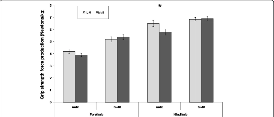
All mice completed the IL-6r mAb trial. No adverse events occurred due to the multiple injections. At baseline, body weights between groups were similar and increased as expected over the 5-week trial. No differences were noted between IL-6r mAb and Kh-5 groups in total body or gastrocnemius dissection weight. Antibody treatment resulted in an 11 % greater strength increase (hindlimb) in IL-6r mAb treated mdx mice compared to Kh-5 treated
mdxmice, though this did not reach statistical significance (6.5 ± 0.2 N/g versus 5.7 ± 0.3 N/g, P = 0.055). No differ-ence was found in forelimb strength of mdx IL-6r mAb
treated mice when compared to mdx Kh-5 treated
(P = 0.28, Figure 1). In control mice (BL-10), no differences were noted between IL-6r mAb and Kh-5 treated mice (P = 0.53, 0.86 forelimb and hindlimb respectively).
Rotarod
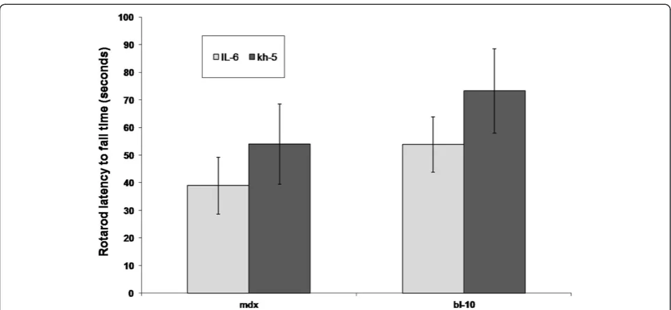
All mice completed RR measures with only one mdx
mouse per group (IL-6 or Kh-5) attaining the 180 s max-imum measure. In contrast, nine BL-10 mice achieved the 180 s maximum, five from the IL-6r mAb group and 4 in the Kh-5 group. No significant differences in RR performance (Figure 2) were noted after treatment be-tween IL-6r mAb and Kh-5 treated mdxmice (P = 0.43) or BL-10 (P = 0.32).
Histology
Blinded reviewers analyzed the randomly selected microscopy fields of H&E stained sections resulting in approximately 840 and 780 muscle fibers analyzed
respectively for each mdx-IL6r mAb and mdx-Kh5 mouse. For comparison, approximately 600 fibers from BL-10 mice were examined (Figure 1). There were no differences between BL-10 treatment groups. Measures of central nucleation (CN) were not significantly differ-ent between treated and un-treated mdx mice, though
mdx-IL6r mAb treated mice had 8% (P = 0.27) fewer CN fibers in skeletal muscle gastrocnemius (Gas) (Table 1). Inflammation scores were nearly 50% higher in IL-6r mAb treatment group in skeletal muscle (Gas) as com-pared tomdx Kh-5 treated mice (P = 0.04). A histogram of muscle diameters (Figure 3) demonstrates little differ-ence between IL-6r mAb treated and Kh-5 treated mdx
mice, though IL-6r mAb treated mouse muscle dia-meters were slightly smaller (Table 1). IL-6r mAb treat-ment demonstrated no change in the number of regenerating fibers (P = 0.07) (Table 1). To understand the muscle damage and regeneration process from a glo-bal perspective, as IL-6 plays a complex role in the process of muscle degeneration/regeneration, we com-bine the inflammation and regeneration scores by first converting them to a rank-order scale, combining the two ranks, followed by a Wilcoxon rank-sum test. The results showed that the pooled variable of muscle re-modeling increased 37% in IL-6r mAb mdx mice com-pared to Kh5 treatedmdx(P = 0.02).
ICAM1 staining
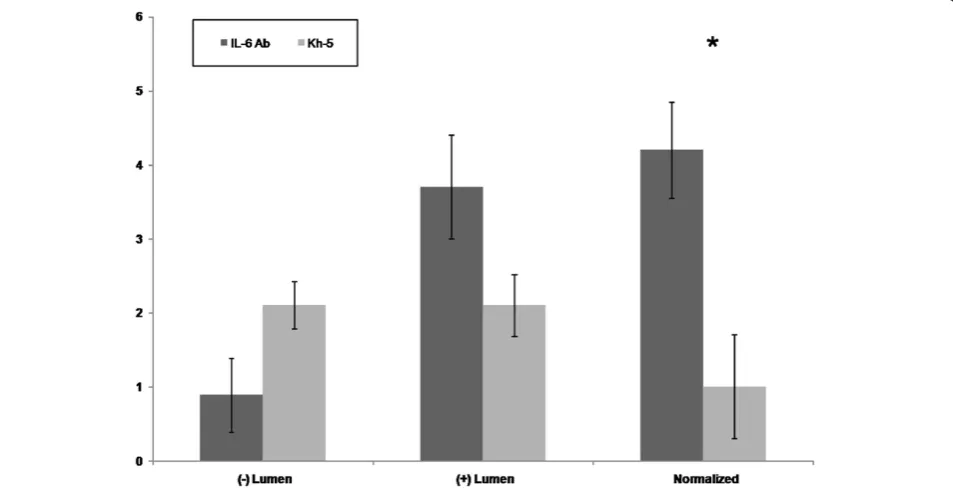
[image:5.595.57.539.488.694.2]A total of 20 fields were randomly selected per mouse. A total of 38 lumens within the skeletal msucle were quan-tified for Kh-5 treated, and 41 lumens for IL-6r mAb treated mdxmice. Figure 4 demonstrates the differences
between mdx-IL-6r mAb treated and mdx-Kh-5 trea-ted. Figure 5 is representative of a positive versus negatively stained lumen for ICAM1. When normal-ized to the number of non-stained lumens, IL-6r mAb treated mdx had 4 positive lumens to Kh-5’s 1 (P = 0.04).
Real-time PCR
Under optimized conditions for all primer pairs, SAA mRNA (marker of systemic IL-6 action) levels in the liver were 3.8 ± 0.4fold lower (P < 0.01) in mdx-IL-6r
mAb treated mice versus mdx-Kh-5 treated mice
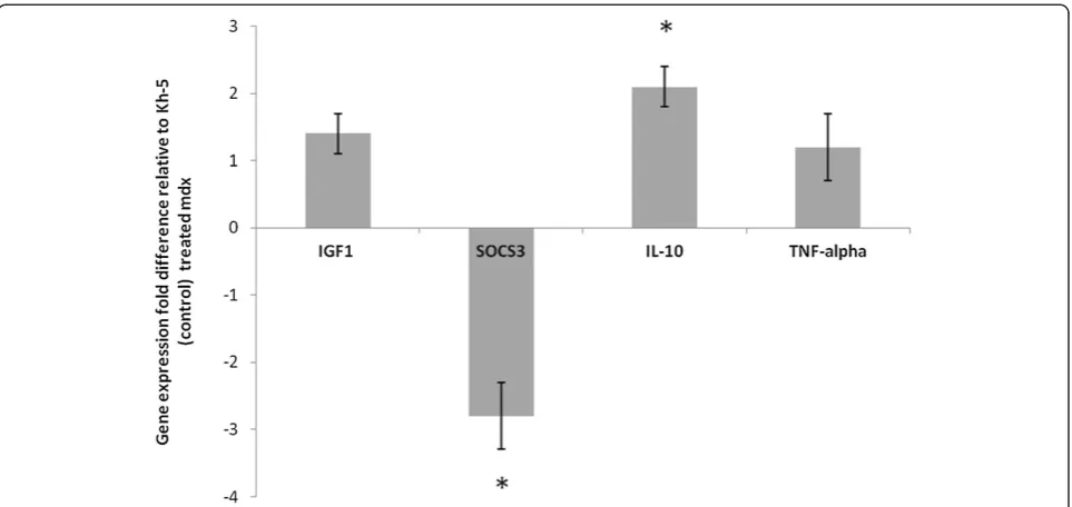
(Figure 6). Gastrocnemius muscle SOCS3 (marker of local IL-6 action, Figure 7) was 2.8 ± 0.5 fold lower (P = 0.05) and IL-10 was 2.1 ± 0.3 fold higher (P = 0.03) in mdx-IL-6r mAb treated mice versus mdx-Kh-5 trea-ted mice (Figure 6). TNF-alpha of treatrea-ted mdx was not different from untreated mdx, (1.2 ± 0.5 fold, P = 0.45). IGF1 mRNA levels were not significantly different be-tween mdx-IL-6r mAb and mdx-Kh-5 treated mice in the liver, (1.1 ± 0.1 fold higher in mdx/IL-6r mAb, P = 0.78) or muscle (1.4 ± 0.3 fold higher in mdx/IL-6r mAb, P = 0.71, Figures 6 & 7).
Discussion
Here we report the first study to block IL-6 signaling in
mdxmice. We hypothesized that overproduction of IL-6 by mdx muscle contributes to the pathologic inflamma-tion of the disease and that by reducing IL-6 signaling we would decrease inflammation and improve muscle pathology. On the contrary, we report that five weeks of IL-6 blockade caused an increase in muscle inflamma-tion, with no change in muscle regeneration. Also con-trary to our expectations, we found no change in IGF1 gene expression by muscle or liver. And although there was a trend for improvements in one measure of grip strength after 5 weeks of IL-6r mAb treatment, no measures of muscle function reached statistical sig-nificance. The increase in inflammation is complex and will require a more specific inquiry into mechan-isms and consequences than our current study pro-vides. The modulation of inflammation we observed could be beneficial (e.g. up-regulation of IL-10) or detrimental.
[image:6.595.57.539.87.310.2]Chronic high levels of circulating IL-6 is associated with muscle wasting in rodents and humans. Exogenous injections of IL-6 induces skeletal muscle protein Figure 2Comparison of rotarod latency to fall time after IL-6r mAb treatment.Comparison of BL-10 and mdx mice after 4 weeks of IL-6 mAb or control Ab (Kh-5) treatment. Data are means and bars represent SEM.
Table 1 Histological analysis of L-6r mAb treated and control (Kh-5) treated mdx and bl/10 mice
Regenerating fibers (n) Diameter (um) Central nucleated fibers (%) Gas Central nucleated fibers (%) Dia Inflamation score
Kh5-mdx 5.6 ± 1.2 34.1 ± 1.0 72.8 ± 2.6 32.8 ± 1.9 14.9 ± 3.4
IL6rab-mdx 9.6 ± 1.7 31.7 ± 1.6 64.5 ± 6.9 29.3 ± 1.4 26.2 ± 3.1
kh5-bI/10 0.4 ± 0.1 38.2 ± 1.1 2.2 ± 0.5 1.8 ± 0.3 1.4 ± 1.2
IL6Ab-bl/10 0.2 ± 0.1 40.1 ± 0.8 1.5 + 0.5 0.9 ± 0.1 3.1 ± 1.1
[image:6.595.56.540.658.726.2]breakdown in rats [34] and mice [35]. In addition, skel-etal muscle growth in young rats is attenuated by chronic high levels of IL-6 [36]. In terms of disease states, overexpression of IL-6 can cause cancer cachexia [37]. In humans, exogenously administered IL-6 up to high physiologic circulating levels, causes a decrease in circulating IGF1 [26]. The decrease in IGF1 is likely due to decreased liver production and this is likely to
[image:7.595.58.539.88.307.2]attenuate muscle growth. Additionally, in cross-sectional studies of the elderly, a decrease in muscle (sarcopenia) correlates with high circulating IL-6 [25]. Yet, while chronic high levels of IL-6 appear detrimental to muscle, a complete absence of IL-6 hinders muscle repair. Knock-out models of IL-6 have shown that it is neces-sary for proper muscle hypertrophy, activation of satel-lite cells, and regulation of extracellular matrix Figure 3Fiber diameter histogram of mdx mice after 4 weeks of IL-6r mAb or control (Kh-5) treatment.Data are means and bars
represent SEM.
[image:7.595.60.537.461.705.2]remodeling [38,39]. Thus the IL-6/muscle interaction is complex but chronic high levels of IL-6, as occurs in the
mdxmouse, is likely detrimental to muscle [24].
Therefore, as chronic inflammation is associated with muscle loss and reducing inflammation in mdx appears to be beneficial, we used a compound that blocks a me-diator of systemic (acute phase response, APR) and muscle inflammation, IL-6. The IL-6r mAb has been used successfully to treat several models of inflammatory diseases [15-17]. In each trial, circulating IL-6 protein levels are not affected but IL-6 signaling is blocked as verified by common liver markers of APR such as serum amyloid A (SAA) expression. Therefore, we verified ef-fectiveness in our model by examining the immediate
downstream effect of IL-6 signaling systemically (SAA in liver) and locally (SOCS3 in muscle). Indeed, IL-6r mAb had an effect both systemically and locally to reduce IL-6 signaling inmdxmice
Contrary to our primary hypothesis, after 5 weeks of
IL-6r mAb treatment in mdx mice, inflammation
[image:8.595.57.539.90.223.2]increased. Because IL-6 can increase or exacerbate the inflammatory process and because mdx mice have ex-tremely high levels of IL-6 expression in skeletal muscle along with inflammation, we hypothesized that by creasing IL-6 signaling, inflammation would also de-crease. Analysis of H&E stained muscle samples found a nearly 50% increase in inflammation. Yet, H&E stained samples only estimate inflammation. To examine a more Figure 5Representative (+) and (−) stained lumens in mdx gastrocnemius muscle. A) Negatively stained lumenB) positively stained lumen; (400× magnification). Scale bar: (A) 50μm.
[image:8.595.58.539.461.683.2]direct marker of tissue inflammation we quantified ICAM-1 as it will be expressed by the vascular endothe-lium where leukocytes will bind ICAM-1 on the endo-thelial surface and transmigrate across the vascular endothelial and into tissues during the inflammatory process [40,41]. ICAM-1 staining confirmed that an in-crease in inflammation occurred in our IL-6r mAb trea-ted mdx mice. As this was contrary to our original hypothesis and due to the ability of IL-6 to modulate pro-inflammatory cytokines (TNF-alpha) while not affecting pro-growth cytokines (TGF-Beta), we sought to examine markers that are consistently shown to be pro-inflammatory or pro-growth in skeletal muscle. TNF-alpha muscle gene expression was not different between treated and untreated mice while IL-10 muscle gene ex-pression increased. Thus it is possible that while overall inflammation increased it could be pro-growth and sup-port increased regeneration (i.e. # of regenerating fibers), [42]. Indeed, IL-10 has been shown to be beneficial in
mdxmice [43] and thus may be increasing regeneration, while no change in TNF-alpha is suggestive that overall pro-inflammatory signals were not affected by our treatment.
Our findings in inflammation are not without prece-dent, in mdx mice, other treatments that should de-crease inflammation have been shown to cause an increase of inflammation [7,8]. In our current studies, this could have been caused by an increase in cytokines that directly bind to gp130 (e.g. IL-11, IL-27). Further-more, IL-6 itself can be pro- and anti-inflammatory [44].
It is possible that IL-6 was increased in response to the high level of inflammation in mdx muscle and that our treatment was blocking the anti-inflammatory effects of IL-6. Also as mentioned, the increased level of inflam-mation may be pro-growth (e.g. 2 C macrophages) as evidenced by an increase in IL-10. The exact cause or implication of the increased inflammation is not clear. Yet, it is seemingly associated with an increase in regen-erating fibers. And while our measures of regenregen-erating fibers (H&E) were not statistically different, it was nearly double when comparing treated to untreated mdx. To examine this process from a global perspective that combines the overall damage/inflammation/repair process, we pooled inflammation and regeneration data as an indicator of muscle remodeling, and found a sig-nificant increase occurred due to IL-6 blockade treat-ment. While the consequences of this increase in remodeling are unknown, it is likely to have a long term effect as it is an indication of the overall tissue damage and repair process including critical aspects of muscle satellite cell activation. Thus while long-term effects are not known, IL-6 blockade did cause an increase in inflammation.
[image:9.595.58.540.88.316.2]A marker of general muscle growth/repair, IGF1, can be directly affected by IL-6 levels [26]. As our mAb treatment should affect cell signaling and thus gene ex-pression, we used IGF1 mRNA as an indicator of down-stream consequence of blocking IL-6 signaling and an indicator of muscle growth. IL-6r mAb did not affect IGF1 gene expression in muscle or liver ofmdx treated Figure 7Gene expression of mdx treated relative to control-Ab treated gastrocnemius muscle.Insulin like growth factor (IGF1),
mice. And though IGF1 did not change, the increased remodeling is suggestive of additional growth factor re-lease. Indeed, IL-10 did increase due to treatment and it is possible that other growth factors may have increased and/or macrophage subtype conversion to a pro-growth phenotype (M2c) could have occurred which leads to satellite cell or fibroblast proliferation [45]. Yet, in the case of fibroblast proliferation inmdx, it is likely to have detrimental consequences of increased collagen produc-tion and fibrosis [46]. The current treatment of 5 weeks is unlikely to produce any definitive results in terms of fibrosis; a longer study is needed. IGF1 gene expression was not affected locally or systemically, contrary to our hypothesis. This suggests that IL-6 was not hindering this growth pathway.
After 5 weeks of IL-6 signaling blockade in mdxmice there was no change in measures of muscle function. And while five weeks is a short period to improve muscle function, our a-priori calculation of statistical power should have allowed us to detect a 10% increase in some measures of muscle function. A post-hoc ana-lysis of our data revealed a greater variance in our grip strength data than was used in our a-priori power calcu-lation, causing a decrease in statistical power. In this re-gard, though the increase was not statistically significant, we note a trend for hind-limb grip strength to increase (11%). The increase in muscle strength, in our data, could be due to an actual increase in muscle force pro-duction or could be due to a repro-duction in body weight and while our mdx did tend to gain weight at a faster rate than BL-10 mice (which is typical of mdx) if body weight is increasing proportionally to the increase in strength, then functionally or in terms of ambulation, it would be of little consequence. Also noteworthy of the muscle strength testing is that testing could in theory cause muscle damage but we are aware of no reports of this affecting these measures inmdxmice. Furthermore, it was recently reported that although susceptible to in-jury, mdx mice respond to muscle damage by a repair process that is at least as efficient as BL-10 mice [47]. Our other measure of muscle function, neuromuscular coordination, could also have been improved by the reduced inflammation alone, though we found no change [48,49]. A more precise measure of muscle force production would have allowed us to detect smaller changes in muscle function and though this was attempted on a small number of soleus muscle samples (ex-vivo) technical difficulties in data collection prohib-ited analysis of. As improved muscle function could be caused by increases in size or number of myofibers, a re-duction in muscle necrosis, or improved muscle repair we note that dissected muscle weights normalized for body weight were not different between groups of trea-ted mice and while muscle fiber counts were greater in
mdx treated mice in randomly selected microscopic fields, the fiber diameter was less and could account for the greater number of fibers counted. Thus as remodel-ing was increased it did not affect standard measures of muscle function.
The strength of the current study is the well character-ized therapeutic agent and model of disease (mdx). The therapeutic agent has been used to treat several inflamma-tory diseases and blocks nearly all IL-6 signaling through-out the body and did so in our current investigation [15-17]. The early life of the mdxmouse (4–12 weeks) is considered the point of greatest inflammation and muscle necrosis and is thus an appropriate target age for studies such as ours [50]. A limitation of our study is having only one time point (5 weeks) of pathologic analysis. Thus, short-term effects beyond inflammation are difficult to interpret in terms of consequences such as fibrosis, lon-gevity, or long term muscle function. Future studies should pursue the inflammation by more direct measures, i.e. cell sorting on fresh tissues, to more precisely quantify inflammation. Furthermore, a long term study could more directly address the effects of modified inflammation, fibrosis, and muscle repair.
Conclusion
The current study showed that while IL-6 signaling is believed to contribute to the chronic inflammation and muscle wasting in DMD, reducing IL-6 signaling using neutralizing antibodies did not reduce the inflammation in mdx mice. Instead, the inflammation was increased by the treatment. Considering the muscle strength and pathology were either slightly improved or no change, the increase in inflammation is likely directly in response to the reduction of IL-6 signaling, which implies an anti-inflammatory role of IL-6 signaling in the mdx mice. In conclusion, while IL-6 is affecting the muscle inflammation and remod-eling process in mdx, short term blockade does not produce statistically significant improvements in dis-ease pathology or muscle function.
Abbreviations
IL-6: Interleukin 6; IL-6r mAb: Monoclonal antibody against the IL-6 receptor; mdx: Dystrophin deficient mouse; TGF-B: Transforming growth factor–beta; IGF1: Insulin-like growth factor 1; RR: Rotarod; Gas: Gastrocnemius; 36b4: Acidic phosphoprotein; SAA: Serum Amyloid A; SOCS3: Suppressor of cytokine signaling 3.
Competing interests
The authors report no competing interest.
Authors’contributions
all aspects of study design, oversaw data collection, and contributed to manuscript preparation. All authors read and approved the final manuscript.
Grant support
NIH/NICHD R01HD048051 (Kostek and Chen), NIH/NIAMS R01 AR052027 (Chen), NIH/NICHD U54 HD053177 (Chen), NIH/NICHD R24HD050846 (Chen).
Author details
1Laboratory of Muscle and Translational Therapeutics, Department of Exercise Science, University of South Carolina, Columbia, SC, USA.2Department of Integrative Systems Biology, George Washington University, Children’s National Medical Center, Washington, DC, USA.3University of Pennsylvania School of Medicine, Philadelphia, Pennsylvania, USA.4Laboratory of Muscle and Translational Therapeutics, 921 Assembly St. / PHRC / 3rd floor, Columbia, SC, USA.5111 Michigan Ave. NW, Department of Integrative Systems Biology, George Washington University School of Medicine, Washington, DC, USA.
Received: 18 October 2011 Accepted: 24 May 2012 Published: 20 June 2012
References
1. Tidball JG:Inflammatory processes in muscle injury and repair.Am J Physiol Regul Integr Comp Physiol2005,288(2):R345–R353.
2. Spencer MJ, Montecino-Rodriguez E, Dorshkind K, Tidball JG:Helper (CD4 (+)) and cytotoxic (CD8(+)) T cells promote the pathology of dystrophin-deficient muscle.Clin Immunol2001,98(2):235–243.
3. Grounds MD:Two-tiered hypotheses for Duchenne muscular dystrophy. Cell Mol Life Sci2008,65(11):1621–1625.
4. Hodgetts S, Radley H, Davies M, Grounds MD:Reduced necrosis of dystrophic muscle by depletion of host neutrophils, or blocking TNFalpha function with Etanercept in mdx mice.Neuromuscul Disord
2006,16(9–10):591–602.
5. Angelini C:The role of corticosteroids in muscular dystrophy: a critical appraisal.Muscle Nerve2007,36(4):424–435.
6. Radley HG, Grounds MD:Cromolyn administration (to block mast cell degranulation) reduces necrosis of dystrophic muscle in mdx mice. Neurobiol Dis2006,23(2):387–397.
7. Andreetta F, Bernasconi P, Baggi F, Ferro P, Oliva L, Arnoldi E, Cornelio F, Mantegazza R, Confalonieri P:Immunomodulation of TGF-beta 1 in mdx mouse inhibits connective tissue proliferation in diaphragm but increases inflammatory response: implications for antifibrotic therapy.J Neuroimmunol2006,175(1–2):77–86.
8. Dumont N, Lepage K, Cote CH, Frenette J:Mast cells can modulate leukocyte accumulation and skeletal muscle function following hindlimb unloading.J Appl Physiol2007,103(1):97–104. 9. Wehling M, Spencer MJ, Tidball JG:A nitric oxide synthase transgene
ameliorates muscular dystrophy in mdx mice.J Cell Biol2001,155(1):123–131. 10. Acharyya S, Villalta SA, Bakkar N, Bupha-Intr T, Janssen PM, Carathers M, Li ZW, Beg AA, Ghosh S, Sahenk Z,et al:Interplay of IKK/NF-kappaB signaling in macrophages and myofibers promotes muscle degeneration in Duchenne muscular dystrophy.J Clin Invest2007,
117(4):889–901.
11. Farini A, Meregalli M, Belicchi M, Battistelli M, Parolini D, D'Antona G, Gavina M, Ottoboni L, Constantin G, Bottinelli R,et al:T and B lymphocyte depletion has a marked effect on the fibrosis of dystrophic skeletal muscles in the scid/mdx mouse.J Pathol2007,213(2):229–238. 12. Golumbek PT, Keeling RM, Connolly AM:Strength and corticosteroid
responsiveness of mdx mice is unchanged by RAG2 gene knockout. Neuromuscul Disord2007,17(5):376–384.
13. Summan M, Warren GL, Mercer RR, Chapman R, Hulderman T, Van Rooijen N, Simeonova PP:Macrophages and skeletal muscle regeneration: a clodronate-containing liposome depletion study.Am J Physiol Regul Integr Comp Physiol2006,290(6):R1488–R1495.
14. Chazaud B, Sonnet C, Lafuste P, Bassez G, Rimaniol AC, Poron F, Authier FJ, Dreyfus PA, Gherardi RK:Satellite cells attract monocytes and use macrophages as a support to escape apoptosis and enhance muscle growth.J Cell Biol2003,163(5):1133–1143.
15. Nishimoto N, Kanakura Y, Aozasa K, Johkoh T, Nakamura M, Nakano S, Nakano N, Ikeda Y, Sasaki T, Nishioka K,et al:Humanized anti-interleukin-6
receptor antibody treatment of multicentric Castleman disease.Blood
2005,106(8):2627–2632.
16. Choy EH, Isenberg DA, Garrood T, Farrow S, Ioannou Y, Bird H, Cheung N, Williams B, Hazleman B, Price R,et al:Therapeutic benefit of blocking interleukin-6 activity with an anti-interleukin-6 receptor monoclonal antibody in rheumatoid arthritis: a randomized, double-blind, placebo-controlled, dose-escalation trial.Arthritis Rheum2002,46(12):3143–3150. 17. Yokota S, Miyamae T, Imagawa T, Iwata N, Katakura S, Mori M, Woo P,
Nishimoto N, Yoshizaki K, Kishimoto T:Therapeutic efficacy of humanized recombinant anti-interleukin-6 receptor antibody in children with systemic-onset juvenile idiopathic arthritis.Arthritis Rheum2005,
52(3):818–825.
18. White JP, Baynes JW, Welle SL, Kostek MC, Matesic LE, Sato S, Carson JA:
The regulation of skeletal muscle protein turnover during the progression of cancer cachexia in the Apc(Min/+) mouse. PLoS One6(9):e24650.
19. Tsujinaka T, Ebisui C, Fujita J, Kishibuchi M, Morimoto T, Ogawa A, Katsume A, Ohsugi Y, Kominami E, Monden M:Muscle undergoes atrophy in association with increase of lysosomal cathepsin activity in interleukin-6 transgenic mouse.Biochem Biophys Res Commun1995,207(1):168–174. 20. Cappola AR, Bandeen-Roche K, Wand GS, Volpato S, Fried LP:Association of
IGF-I levels with muscle strength and mobility in older women.J Clin Endocrinol Metab2001,86(9):4139–4146.
21. Barbieri M, Ferrucci L, Ragno E, Corsi A, Bandinelli S, Bonafe M, Olivieri F, Giovagnetti S, Franceschi C, Guralnik JM,et al:Chronic inflammation and the effect of IGF-I on muscle strength and power in older persons.Am J Physiol Endocrinol Metab2003,284(3):E481–E487. 22. Tsujinaka T, Fujita J, Ebisui C, Yano M, Kominami E, Suzuki K, Tanaka K,
Katsume A, Ohsugi Y, Shiozaki H,et al:Interleukin 6 receptor antibody inhibits muscle atrophy and modulates proteolytic systems in interleukin 6 transgenic mice.J Clin Invest1996,97(1):244–249.
23. De Benedetti F, Alonzi T, Moretta A, Lazzaro D, Costa P, Poli V, Martini A, Ciliberto G, Fattori E:Interleukin 6 causes growth impairment in transgenic mice through a decrease in insulin-like growth factor-I. A model for stunted growth in children with chronic inflammation. J Clin Invest1997,99(4):643–650.
24. Kurek JB, Nouri S, Kannourakis G, Murphy M, Austin L:Leukemia inhibitory factor and interleukin-6 are produced by diseased and regenerating skeletal muscle.Muscle Nerve1996,19(10):1291–1301. 25. Stenholm S, Maggio M, Lauretani F, Bandinelli S, Ceda GP, Di Iorio A,
Giallauria F, Guralnik JM, Ferrucci L:Anabolic and catabolic biomarkers as predictors of muscle strength decline: the InCHIANTI study. Rejuvenation Res,13(1):3–11.
26. Nemet D, Eliakim A, Zaldivar F, Cooper DM:Effect of rhIL-6 infusion on GH–> IGF-I axis mediators in humans.Am J Physiol Regul Integr Comp Physiol2006,291(6):R1663–R1668.
27. Al-Shanti N, Saini A, Faulkner SH, Stewart CE:Beneficial synergistic interactions of TNF-alpha and IL-6 in C2 skeletal myoblasts–potential cross-talk with IGF system.Growth Factors2008,26(2):61–73. 28. Ahmed TA, Buzzelli MD, Lang CH, Capen JB, Shumate ML,
Navaratnarajah M, Nagarajan M, Cooney RN:Interleukin-6 inhibits growth hormone-mediated gene expression in hepatocytes. Am J Physiol Gastrointest Liver Physiol2007,292(6):G1793–G1803. 29. Okazaki S, Kawai H, Arii Y, Yamaguchi H, Saito S:Effects of calcitonin
gene-related peptide and interleukin 6 on myoblast differentiation.Cell Prolif
1996,29(4):173–182.
30. Wang X, Wu H, Zhang Z, Liu S, Yang J, Chen X, Fan M:Effects of interleukin-6, leukemia inhibitory factor, and ciliary neurotrophic factor on the proliferation and differentiation of adult human myoblasts. Cell Mol Neurobiol2008,28(1):113–124.
31. Katsume A, Saito H, Yamada Y, Yorozu K, Ueda O, Akamatsu K, Nishimoto N, Kishimoto T, Yoshizaki K, Ohsugi Y:Anti-interleukin 6 (IL-6) receptor antibody suppresses Castleman's disease like symptoms emerged in IL-6 transgenic mice.Cytokine2002,20(6):304–311.
32. Spurney CF, Gordish-Dressman H, Guerron AD, Sali A, Pandey GS, Rawat R, Van Der Meulen JH, Cha HJ, Pistilli EE, Partridge TA,et al:Preclinical drug trials in the mdx mouse: assessment of reliable and sensitive outcome measures.Muscle Nerve2009,39(5):591–602.
34. Goodman MN:Interleukin-6 induces skeletal muscle protein breakdown in rats.Proc Soc Exp Biol Med1994,205(2):182–185.
35. Zhang L, Du J, Hu Z, Han G, Delafontaine P, Garcia G, Mitch WE:IL-6 and serum amyloid A synergy mediates angiotensin II-induced muscle wasting.J Am Soc Nephrol2009,20(3):604–612.
36. Bodell PW, Kodesh E, Haddad F, Zaldivar FP, Cooper DM, Adams GR:
Skeletal muscle growth in young rats is inhibited by chronic exposure to IL-6 but preserved by concurrent voluntary endurance exercise. J Appl Physiol2009,106(2):443–453.
37. Barton BE, Murphy TF:Cancer cachexia is mediated in part by the induction of IL-6-like cytokines from the spleen.Cytokine2001,
16(6):251–257.
38. Serrano AL, Baeza-Raja B, Perdiguero E, Jardi M, Munoz-Canoves P:
Interleukin-6 is an essential regulator of satellite cell-mediated skeletal muscle hypertrophy.Cell Metab2008,7(1):33–44.
39. White JP, Reecy JM, Washington TA, Sato S, Le ME, Davis JM, Wilson LB, Carson JA:Overload-induced skeletal muscle extracellular matrix remodelling and myofibre growth in mice lacking IL-6.Acta Physiol (Oxf )
2009,197(4):321–332.
40. Yang L, Froio RM, Sciuto TE, Dvorak AM, Alon R, Luscinskas FW:ICAM-1 regulates neutrophil adhesion and transcellular migration of TNF-alpha-activated vascular endothelium under flow.Blood2005,106(2):584–592. 41. Rothlein R, Dustin ML, Marlin SD, Springer TA:A human intercellular
adhesion molecule (ICAM-1) distinct from LFA-1.J Immunol1986,
137(4):1270–1274.
42. Xing Z, Gauldie J, Cox G, Baumann H, Jordana M, Lei XF, Achong MK:IL-6 is an antiinflammatory cytokine required for controlling local or systemic acute inflammatory responses.J Clin Invest1998,101(2):311–320. 43. Villalta SA, Rinaldi C, Deng B, Liu G, Fedor B, Tidball JG:Interleukin-10
reduces the pathology of mdx muscular dystrophy by deactivating M1 macrophages and modulating macrophage phenotype.Hum Mol Genet,
20(4):790–805.
44. Scheller J, Chalaris A, Schmidt-Arras D, Rose-John S:The pro- and anti-inflammatory properties of the cytokine interleukin-6.Biochim Biophys Acta,1813(5):878–888.
45. Villalta SA, Nguyen HX, Deng B, Gotoh T, Tidball JG:Shifts in macrophage phenotypes and macrophage competition for arginine metabolism affect the severity of muscle pathology in muscular dystrophy.Hum Mol Genet2009,18(3):482–496.
46. Reitamo S, Remitz A, Tamai K, Uitto J:Interleukin-10 modulates type I collagen and matrix metalloprotease gene expression in cultured human skin fibroblasts.J Clin Invest1994,94(6):2489–2492.
47. Call JA, Eckhoff MD, Baltgalvis KA, Warren GL, Lowe DA:Adaptive strength gains in dystrophic muscle exposed to repeated bouts of eccentric contraction.J Appl Physiol,111(6):1768–1777.
48. Spiller R:How inflammation changes neuromuscular function and its relevance to symptoms in diverticular disease.J Clin Gastroenterol2006,
40(Suppl 3):S117–S120.
49. Maselli RA, Richman DP, Wollmann RL:Inflammation at the neuromuscular junction in myasthenia gravis.Neurology1991,41(9):1497–1504. 50. Grounds MD, Radley HG, Lynch GS, Nagaraju K, De Luca A:Towards
developing standard operating procedures for pre-clinical testing in the mdx mouse model of Duchenne muscular dystrophy.Neurobiol Dis2008,
31(1):1–19.
doi:10.1186/1471-2474-13-106
Cite this article as:Kosteket al.:IL-6 signaling blockade increases inflammation but does not affect muscle function in themdxmouse.
BMC Musculoskeletal Disorders201213:106. Submit your next manuscript to BioMed Central
and take full advantage of:
• Convenient online submission
• Thorough peer review
• No space constraints or color figure charges
• Immediate publication on acceptance
• Inclusion in PubMed, CAS, Scopus and Google Scholar
• Research which is freely available for redistribution