R E S E A R C H A R T I C L E
Open Access
Elevated leptin levels induce inflammation
through IL-6 in skeletal muscle of aged
female rats
Ryo Tazawa
1, Kentaro Uchida
1*, Hisako Fujimaki
1, Masayuki Miyagi
1, Gen Inoue
1, Hiroyuki Sekiguchi
2,
Kosuke Murata
1, Ken Takata
1, Ayumu Kawakubo
1and Masashi Takaso
1Abstract
Background:Chronic inflammation with aging contributes to sarcopenia. Previous studies have suggested that the accumulation of adipose tissue in skeletal muscle, referred to as intermuscular adipose tissue (IMAT), increases with age and is associated with inflammation. However, the mechanism governing ectopic inflammation in skeletal muscle due to aging is not fully understood. Leptin, an adipocytokine derived from adipose tissue, is an important mediator of inflammatory processes. We examined changes in leptin levels with age and whether leptin contributes to ectopic inflammation.
Methods:To evaluate ectopic inflammation in skeletal muscle, we measured alterations to the expression of inflammatory cytokine genes (Il1b, Il6, andTnfa) and muscle break down-related gene (MuRF1andAtrogin1) in the quadricep muscles of young (10 weeks) and aged (48 weeks) female rats using quantitative reverse-transcription polymerase chain reaction (Q-RT-PCR). Histological examination was performed to identify the extent of IMAT.
LeptinmRNA and leptin protein expression were examined using Q-RT-PCR and enzyme-linked immunosorbent assay, respectively. The effect of leptin on the mRNA expression ofIl1b,Il6, andTnfain quadricep muscle-derived cells was also examined by stimulating the cells with 0 (control), 1, or 10μg/mL rat recombinant leptin using Q-RT-PCR.
Results:Aged rats had significantly higherIl6, MuRF1, and Atrogin1but notIl1bandTnfa,expression and greater levels of IMAT in their quadricep muscles than young rats. Aged rats also had significantly higherleptinexpression and leptin protein concentration in their quadricep muscles than young rats. The addition of exogenous leptin to quadricep muscle-derived cells significantly increased the gene expression ofIl1bandIl6but notTnfa.
Conclusions:Our results suggest that elevated leptin levels due to aging cause ectopic inflammation through IL-6 in the skeletal muscle of aged rats.
Keywords:Leptin, Interleukin-6, Intermuscular adipose tissue, Inflammation
Background
Sarcopenia is the reduction in skeletal muscle mass and function with age, and is a major public health concern. The sarcopenia-related morbidity rate is 5–13% in 60- to 70-year-old individuals, and 11–50% in those above 80 years old [1]. Sarcopenia can lead to a rise in the inci-dence of falls and the risk of fractures in the elderly, and
is therefore linked to physical disability, and increased mortality, morbidity, and health care costs [2, 3]. Al-though a number of factors are implicated in the patho-physiology of sarcopenia, its pathopatho-physiology remains elusive.
Proinflammatory cytokines associated with skeletal muscle metabolism contribute to sarcopenia [4–9]. A previous study reported that older individuals with sar-copenia had higher levels of serum interleukin (IL)-6 than those without sarcopenia [7]. IL-1βconcentrations increase in inflammatory conditions, and elevated levels
© The Author(s). 2019Open AccessThis article is distributed under the terms of the Creative Commons Attribution 4.0 International License (http://creativecommons.org/licenses/by/4.0/), which permits unrestricted use, distribution, and reproduction in any medium, provided you give appropriate credit to the original author(s) and the source, provide a link to the Creative Commons license, and indicate if changes were made. The Creative Commons Public Domain Dedication waiver (http://creativecommons.org/publicdomain/zero/1.0/) applies to the data made available in this article, unless otherwise stated. * Correspondence:[email protected]
1Department of Orthopedic Surgery, Kitasato University School of Medicine,
of IL-1βinhibit myoblast differentiation [8,9]. However, the mechanisms governing the abnormal levels of IL-1β and IL-6 expression in skeletal muscle due to aging is not fully understood.
The accumulation of adipose tissue in skeletal muscle, referred to as intermuscular adipose tissue (IMAT), increases with age. Adipose tissue produces several adi-pocytokines and regulates inflammatory conditions [10]. Leptin, one such adipocytokine derived from adipose tissue, is an important mediator of inflammatory pro-cesses [11]. Previous studies have shown that leptin has pro-inflammatory properties and upregulates Il-6 ex-pression in human synovial fibroblasts [12] and rat microglia [13]. Leptin also stimulates Il1bexpression in human chondrocytes [14] and rat microglia [15]. We hypothesized that IMAT-derived leptin may be differen-tially expressed in the muscle with age and may regulate the expression of inflammatory cytokines.
Here, we investigated the expression of leptin and in-flammatory cytokines with age and the relationship be-tween leptin and inflammatory cytokines in rat muscle.
Methods Animals
This study used female Sprague-Dawley (SD) rats ob-tained from Charles River Laboratories Japan, Inc. (Yoko-hama, Japan). Rats were fed a commercial pelleted diet (CRF-1, Oriental Yeast Industry, Tokyo). All experimental protocols were in accordance with the guidelines of the Animal Ethics Committee of Kitasato University (Permis-sion number: 2018–085).
Quantitative reverse-transcription polymerase chain reaction (Q-RT-PCR) analysis
Preliminary experiments using rats aged 10, 24, 48, and 96 weeks indicated that leptin mRNA expression in 48-and 96-week-old rats was significantly higher than that in 10-week-old rats (Additional file 1: Figure S1). How-ever, a previous study reported that SD rats developed a tumor early or late in life, over the age range of 494 to 798 days (approximately 71 to 114 weeks) at the time of first tumor observation [16]. Therefore, to evaluate changes in cytokine expression due to aging, SD rats were divided into two age groups: the young group (10 weeks) and aged group (48 weeks) (n= 10 each). Animals were anaesthetized first with isoflurane following by an intramuscular injection of a mixture comprising medeto-midine, midazolam, and butorphanol tartrate into the upper limb. Blood was removed to prevent contamin-ation with blood components before sacrificing the rats by cervical dislocation. Bilateral quadricep muscles were removed without the fascia and washed with phosphate buffered saline solution (PBS). TRIzol (Invitrogen, Carls-bad, CA, USA) was used to extract total RNA from the
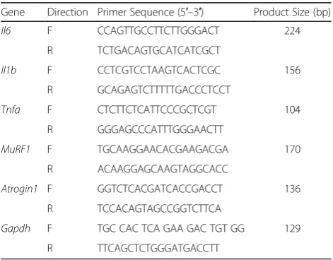
quadricep muscles, based on the manufacturer’s proto-col. The RNA formed the template for first-strand cDNA synthesis, which was performed with SuperScript III RT (Invitrogen) in a reaction (final volume, 25μL) comprising 2μL cDNA, a specific primer set (0.2μM final concentration), and 12.5μL SYBR Premix Ex Taq (Takara, Shiga, Japan). Primers for Il1b, Il6, Tnfa, MuRF1, Atrogin1,andleptinwere designed using Primer Blast and made by Hokkaido System Science Co., Ltd. (Sapporo, Japan). The primer sequences are listed in Table 1. The primer-amplified products were confirmed for specificity using melt curve analysis. Q-RT-PCR was conducted on a CFX-96 Real-Time PCR Detection Sys-tem (Bio-Rad, Hercules, CA, USA). The Q-RT-PCR protocol was as follows: initial denaturation at 95 °C for 1 min, and 40 cycles of 95 °C for 5 s and 60 °C for 30 s. mRNA expression levels of inflammatory cytokine (Il1b, Il6, Tnfa), muscle breakdown-related marker (MuRF1, Atrogin1) and leptin in the quadricep mus-cles were determined by normalizing to that of glyceraldehyde-3-phosphate dehydrogenase (Gapdh) using the delta-delta Ct method. Relative expression was calculated using the mean of all control samples (samples from quadriceps muscles from the young group or vehicle-treated quadriceps muscle-derived cells in vitro).
Histological evaluation
[image:2.595.304.540.548.732.2]To investigate the accumulation of adipose tissue in skeletal muscle due to aging, SD rats were again sepa-rated into two age groups: the young group (10 weeks) and aged group (48 weeks). After anesthetization with isoflurane following by an intramuscular injection of a mixture comprising medetomidine, midazolam, and butorphanol tartrate, rats were sacrificed by cervical dis-location. Quadricep muscles were removed without the fascia and fixed in paraformaldehyde before embedding
Table 1Sequences of the primers used in this study
Gene Direction Primer Sequence (5′–3′) Product Size (bp)
Il6 F CCAGTTGCCTTCTTGGGACT 224
R TCTGACAGTGCATCATCGCT
Il1b F CCTCGTCCTAAGTCACTCGC 156
R GCAGAGTCTTTTTGACCCTCCT
Tnfa F CTCTTCTCATTCCCGCTCGT 104
R GGGAGCCCATTTGGGAACTT
MuRF1 F TGCAAGGAACACGAAGACGA 170
R ACAAGGAGCAAGTAGGCACC
Atrogin1 F GGTCTCACGATCACCGACCT 136
R TCCACAGTAGCCGGTCTTCA
Gapdh F TGC CAC TCA GAA GAC TGT GG 129
in paraffin. The tissue was cut into 3-μm-thick sections and stained with hematoxylin-eosin (HE).
Enzyme-linked immunosorbent assay (ELISA) for leptin To investigate changes in leptin protein expression due to aging, ELISA was performed on tissue obtained from rats separated into the same two age groups as the above experiments: the young group (10 weeks) and aged group (48 weeks) (n= 10 each).
Quadricep muscles harvested from rats as described above were homogenized in radioimmune precipitation (RIPA) buffer (Wako Pure Chemical Co., Inc., Osaka, Japan) containing an added protease inhibitor cocktail (Roche, Madison WI, USA). Total protein concentration in the solution was ascertained by the bicinchoninic acid (BCA) assay (Thermo Fisher Scientific, Rockford IL, USA) and leptin protein concentration by a rat leptin ELISA kit (R&D Systems, Inc., Minneapolis MN, USA).
Effect of leptin onIl1b, Il6andTnfaexpression in quadriceps muscle-derived cells
Our preliminary experiments showed that there was no difference between young and aged rats in response to leptin. Dose of leptin was determined based on previous studies [13,17]. Ten-week-old SD rats were used for this experiment. Quadricep muscles were removed bilaterally as described above and digested with type I collagenase overnight at 37 °C to extract muscle cells. The cells were cultured inα-MEM supplemented with 10% fetal bovine serum in six-well plates for 1 week at 37 °C in a 5% CO2 incubator. After 1 week of incubation, cells were conflu-ent on the wells. Subsequconflu-ently, recombinant rat leptin (0, 1 and 10μg/mL) (Biolegend, San Diego, CA, USA) was added, and the cells were stimulated for 24 h. Total RNA was extracted from treated (1 and 10μg/mL leptin) and control (0μg/mL leptin) cells, andIl1b,Il6 and Tnfa
expression was ascertained using Q-RT-PCR.
Statistical analysis
Differences between the two age groups were compared using the Mann-Whitney U test. Differences among treated and control muscle-derived cells were compared using the one-way ANOVA and Tukey multiple compar-ison’s test. SPSS was used as the statistical software (Ver-sion 19.0; SPSS, Inc., Chicago, IL, USA), and p< 0.05 was used to indicate statistical significance.
Results
Expression of inflammatory cytokines and muscle breakdown-related gene expression in quadriceps muscles
To evaluate ectopic inflammation and muscle break-down in skeletal muscle, we performed Q-RT-PCR to examine changes in the gene expression of inflammatory
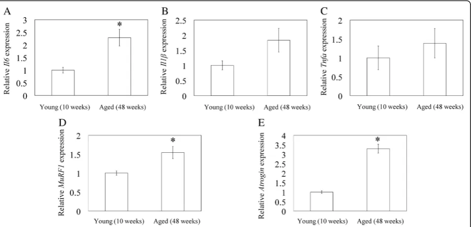
cytokines, Il1b, Il6 and Tnfa, in quadricep muscle. The aged group had significantly higher Il6 mRNA expres-sion than the young group (p= 0.001; Fig. 1a). In con-trast, there was no significant difference in Il1b and Tnfa mRNA expression (p= 0.096 and p= 0.327, re-spectively; Fig. 1b, c). MuRF1 and Atrogin1 mRNA expression were higher in the quadriceps muscles of aged rats than young rats (p= 0.002 and p< 0.001, respectively; Fig.1d, e).
IMAT
To investigate changes in the accumulation of adipose tissue due to aging, we examined the extent of IMAT in histologically-stained sections. Quadricep muscles of aged rats (48 weeks) had greater levels of IMAT than that of young rats (10 weeks) (Fig.2a–d).
Leptinexpression and leptin protein concentration in quadricep muscles
To investigate whether increased IMAT due to aging re-sults in leptin production, we examined leptin mRNA expression and leptin protein expression in the quadri-cep muscles of aged and young rats. Leptin mRNA expression and leptin protein concentration were signifi-cantly higher in the aged group than in the young group (p= 0.049 andp< 0.001, respectively; Fig.3a, b).
Effect of leptin onIl1b, Il6andTnfaexpression in quadricep muscle-derived cells
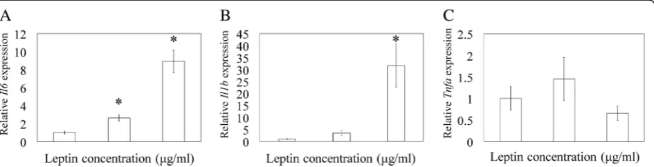
In vitro experiments were performed to elucidate the relationship between IL-6, IL-1β, TNF-α and leptin. Q-RT-PCR analysis showed that the additional of ex-ogenous leptin at 1 and 10μg/ml significantly elevated
Il6 mRNA expression compared to control (p= 0.001 and p< 0.001, respectively; Fig. 4a).Il1b mRNA expres-sion was also significantly elevated in the presence 10μg/ml leptin (p= 0.025; Fig. 4b). There was no differ-ence between the leptin-stimulated and control groups inTnfaexpression (p= 0.279; Fig.4c).
Discussion
This study aimed to examine changes to the expression of leptin and inflammatory cytokines due to aging and to determine the relationship between these factors in the rat quadricep muscle. We showed that IMAT, leptin gene and protein expression, Il6, and MuRF1 and Atro-gin1 mRNA expression were higher in the quadricep muscles of aged rats than young rats. In addition, stimu-lation of muscle-derived cells with exogenous leptin sig-nificantly and dose-dependently increased Il1b and Il6
Fig. 1Quantitative reverse-transcription polymerase chain reaction analysis of inflammatory cytokines and muscle breakdown-related gene expression in quadriceps muscles. Relative gene expression of inflammatory cytokines,Il6(a),Il1b(b),Tnfa(c), and muscle breakdown-related geneAtrogin1(d) andMuRF1(e) in quadriceps muscle extracted from young (10 weeks) and aged (48 weeks) rats. Data represent mean ± SE (n= 10). *p< 0.05, between young and aged groups
[image:4.595.61.539.88.319.2] [image:4.595.55.542.368.695.2]Previous studies have reported that higher IL-6 con-centrations in plasma are associated with lower muscle mass and lower muscle strength in elderly people [6], and that aging is linked to elevated IMAT in the thigh muscle in humans [18, 19]. Additionally, IMAT within the fascia is correlated withIl6expression in subcutane-ous adipose tissue in elderly men [20]. However, these studies analyzed subcutaneous or serum levels of inflam-matory cytokines. Here, we showed that IMAT,Il6, and muscle breakdown-related gene (MuRF1, Atrogin1) ex-pression were increased in the quadricep muscles of aged rats compared to young rats. An experimental study showed that IL-6 administration caused muscles to break down in rats [21]. IL-6 stimulate Atrogin1
mRNA and Atrogin1 protein expression in mice gastro-cnemius muscle [22]. Inhibition of Il-6 suppressesMuRF1
expression and ameliorates tail suspension-induced skel-etal muscle atrophy [23]. Taken together, these findings suggest that changes inIl6expression with age are associ-ated with the formation of micro inflammation environ-ments in ectopic muscle and a reduction in muscle mass.
Serum leptin is positively associated with IMAT in humans [20]. In our study, an increase in IMAT corre-sponded with elevated leptin expression and leptin protein concentrations in the quadriceps of aged rats. In addition, leptin stimulated Il6 and Il1b expression in quadricep-derived cells. Plasma leptin levels are in-creased in individuals with sarcopenia and visceral obes-ity compared to those with sarcopenia or visceral obesobes-ity alone [24]. The development of sarcopenia is correlated with raised serum levels of IL-6, an inflammatory factor [7]. Further, IL-1β impaired myoblast differentiation in the murine myoblast cell line C2C12 [25]. Taken together, our findings and those of previous reports suggest that IMAT-derived leptin induces ectopic inflammation through IL-6 and IL-1β and may contribute sarcopenic pathology.
Several studies showed that TNF-α level increases with aging [26, 27]. In our study, there was no differ-ence between 10- and 48-week-old rats in Tnfa ex-pression level. We excluded 96-week-old rats from evaluation to eliminate the possible effect of tumor. Fig. 3Leptin gene expression by quantitative reverse-transcription polymerase chain reaction analysis and leptin protein concentration by enzyme-linked immunosorbent assay in quadricep muscles.aRelativeleptingene expression in quadricep muscles extracted from young (10 weeks) and aged (48 weeks) rats.bLeptin protein concentration in quadriceps muscles from young (10 weeks) and aged (48 weeks) rats. Data indicate mean ± SE (n = 10). *p< 0.05, between young and aged groups
[image:5.595.58.545.88.242.2] [image:5.595.65.538.573.694.2]However, our preliminary experiment showed that Tnfa
expression in 96-week-old rats was 2.0-hold higher than that in 10-week-old rats. Further investigation using older rats may reveal whether the elevation ofTnfain skeletal muscle contributes to sarcopenic pathology.
Two limitations of this study warrant mention. First, we performed in vitro experiments using cells derived from young rats in standard culture conditions. Leptin resistance was introduced by negative regulators of lep-tin signaling such as inflammatory signals, including IKKβ/NFκB and ER stress [28, 29]. However, to mimic leptin resistance in vivo, a specific condition was needed in vitro [30]. Further investigation under specific condi-tions using aged rat-derived cells is needed to reveal lep-tin resistance in aged rats. Second, we investigated only two time points. A better understanding of the develop-ment of sarcopenia requires analysis of multiple time points.
Conclusions
In conclusion, IMAT and leptin and Il6 expression in-crease with age in rat quadriceps. Our results suggest that IMAT-derived leptin regulates Il6 expression and creates a micro inflammatory environment in ectopic muscle due to aging.
Additional file
Additional file 1:Figure S1. Age-related changes inLeptinmRNA ex-pression. We investigated age-related changes inLeptinmRNA expression in quadriceps muscle of rats aged 10, 24, 48, and 96 weeks using quanti-tative reverse-transcription polymerase chain reaction (Q-RT-PCR) analysis (eachn= 5). Q-RT-PCR analysis indicated that leptin mRNA expression in 48- and 96-week-old rats was higher than that in 10-week-old rats. (TIFF 242 kb)
Abbreviations
ELISA:Enzyme-linked Immunosorbent assay; HE: Hematoxylin-eosin; IL-1β: Interleukin-1β; IL-6: Interleukin-6; IMAT: Intermuscular adipose tissue; Q-RT-PCR: Quantitative reverse-transcription polymerase chain reaction; SD: Sprague dawley; SE: Standard error
Acknowledgements
We thank Ms. Yuko Onuki for her help with Q-RT-PCR analysis.
Funding
This investigation was supported in part by research grants from the Parents’ Association of Kitasato University School of Medicine. The role of the funding included English editing.
Availability of data and materials
The datasets supporting the conclusions of this article are included within the article. The raw data can be requested from the corresponding author.
Authors’contributions
RT, KU and MT designed the study and analyzed the data. RT and KU wrote the manuscript. RT, HF, MM, GI, HS, KM, KT and AK participated in the data collection, analysis, and interpretation. All authors read and approved the final manuscript.
Ethics approval and consent to participate
All experimental protocols were approved by the Kitasato University School of Medicine Animal Care Committee (Permission number: 2018–085).
Consent for publication
Not applicable
Competing interests
The authors declare that they have no competing interests.
Publisher’s Note
Springer Nature remains neutral with regard to jurisdictional claims in published maps and institutional affiliations.
Author details
1
Department of Orthopedic Surgery, Kitasato University School of Medicine, 1-15-1 Minami-ku, Kitasato, Sagamihara City, Kanagawa 252-0374, Japan.
2
Shonan University of Medical Sciences Research Institute, Nishikubo 500, Chigasaki City, Kanagawa 253-0083, Japan.
Received: 10 January 2019 Accepted: 22 April 2019
References
1. von HS, Morley JE, Anker SD. An overview of sarcopenia: facts and numbers on prevalence and clinical impact. J Cachexia Sarcopenia Muscle. 2010;1(2): 129–33.
2. Filippin LI, Teixeira VN, da Silva MP, Miraglia F, da Silva FS. Sarcopenia: a predictor of mortality and the need for early diagnosis and intervention. Aging Clin Exp Res. 2015;27(3):249–54.
3. Murphy RA, Ip EH, Zhang Q, Boudreau RM, Cawthon PM, Newman AB, Tylavsky FA, Visser M, Goodpaster BH, Harris TB. Transition to sarcopenia and determinants of transitions in older adults: a population-based study. J Gerontol A Biol Sci Med Sci. 2014;69(6):751–8.
4. Cesari M, Kritchevsky SB, Baumgartner RN, Atkinson HH, Penninx BW, Lenchik L, Palla SL, Ambrosius WT, Tracy RP, Pahor M. Sarcopenia, obesity, and inflammation--results from the trial of angiotensin converting enzyme inhibition and novel cardiovascular risk factors study. Am J Clin Nutr. 2005; 82(2):428–34.
5. Schrager MA, Metter EJ, Simonsick E, Ble A, Bandinelli S, Lauretani F, Ferrucci L. Sarcopenic obesity and inflammation in the InCHIANTI study. J Appl Physiol (1985). 2007;102(3):919–25.
6. Visser M, Pahor M, Taaffe DR, Goodpaster BH, Simonsick EM, Newman AB, Nevitt M, Harris TB. Relationship of interleukin-6 and tumor necrosis factor-alpha with muscle mass and muscle strength in elderly men and women: the health ABC study. J Gerontol A Biol Sci Med Sci. 2002;57(5):M326–32. 7. Bian AL, Hu HY, Rong YD, Wang J, Wang JX, Zhou XZ. A study on
relationship between elderly sarcopenia and inflammatory factors IL-6 and TNF-alpha. Eur J Med Res. 2017;22(1):25.
8. Cohen TV, Many GM, Fleming BD, Gnocchi VF, Ghimbovschi S, Mosser DM, Hoffman EP, Partridge TA. Upregulated IL-1beta in dysferlin-deficient muscle attenuates regeneration by blunting the response to pro-inflammatory macrophages. Skelet Muscle. 2015;5:24.
9. de AP, Tomazoni SS, Frigo L, de Carvalho PT, Vanin AA, Santos LA, buquerque-Pontes GM, De MT, Tairova O, Marcos RL, Lopes-Martins RA, Leal-Junior EC. What is the best treatment to decrease pro-inflammatory cytokine release in acute skeletal muscle injury induced by trauma in rats: low-level laser therapy, diclofenac, or cryotherapy? Lasers Med Sci. 2014; 29(2):653–8.
10. Tilg H, Moschen AR. Adipocytokines: mediators linking adipose tissue, inflammation and immunity. Nat.Rev.Immunol. 2006;6(10):772–83. 11. La CA, Matarese G. The weight of leptin in immunity. Nat Rev Immunol.
2004;4(5):371–9.
12. Yang WH, Liu SC, Tsai CH, Fong YC, Wang SJ, Chang YS, Tang CH. Leptin induces IL-6 expression through OBRl receptor signaling pathway in human synovial fibroblasts. PLoS One. 2013;8(9):e75551.
14. Simopoulou T, Malizos KN, Iliopoulos D, Stefanou N, Papatheodorou L, Ioannou M, Tsezou A. Differential expression of leptin and leptin's receptor isoform (Ob-Rb) mRNA between advanced and minimally affected osteoarthritic cartilage; effect on cartilage metabolism. Osteoarthritis Cartilage. 2007;15(8):872–83.
15. Pinteaux E, Inoue W, Schmidt L, Molina-Holgado F, Rothwell NJ, Luheshi GN. Leptin induces interleukin-1beta release from rat microglial cells through a caspase 1 independent mechanism. J Neurochem. 2007;102(3):826–33. 16. RK D, GT S, KA B. Tumor incidence in normal Sprague-Dawley female rats.
Cancer Res. 1956;16(3):194–7.
17. Miao D, Zhang L. Leptin modulates the expression of catabolic genes in rat nucleus pulposus cells through the mitogen-activated protein kinase and Janus kinase 2/signal transducer and activator of transcription 3 pathways. Mol Med Rep. 2015;12(2):1761–8.
18. Buford TW, Lott DJ, Marzetti E, Wohlgemuth SE, Vandenborne K, Pahor M, Leeuwenburgh C, Manini TM. Age-related differences in lower extremity tissue compartments and associations with physical function in older adults. Exp Gerontol. 2012;47(1):38–44.
19. Marcus RL, Addison O, Kidde JP, Dibble LE, Lastayo PC. Skeletal muscle fat infiltration: impact of age, inactivity, and exercise. J Nutr Health Aging. 2010; 14(5):362–6.
20. Zoico E, Rossi A, Di FV, Sepe A, Olioso D, Pizzini F, Fantin F, Bosello O, Cominacini L, Harris TB, Zamboni M. Adipose tissue infiltration in skeletal muscle of healthy elderly men: relationships with body composition, insulin resistance, and inflammation at the systemic and tissue level. J Gerontol A Biol Sci Med Sci. 2010;65(3):295–9.
21. Goodman MN. Interleukin-6 induces skeletal muscle protein breakdown in rats. Proc Soc Exp Biol Med. 1994;205(2):182–5.
22. Baltgalvis KA, Berger FG, Pena MM, Davis JM, White JP, Carson JA. Muscle wasting and interleukin-6-induced atrogin-I expression in the cachectic Apc ( min/+ ) mouse. Pflugers Arch. 2009;457(5):989–1001.
23. Yakabe M, Ogawa S, Ota H, Iijima K, Eto M, Ouchi Y, Akishita M. Inhibition of interleukin-6 decreases atrogene expression and ameliorates tail
suspension-induced skeletal muscle atrophy. PLoS One. 2018;13(1): e0191318.
24. Kohara K, Ochi M, Tabara Y, Nagai T, Igase M, Miki T. Leptin in sarcopenic visceral obesity: possible link between adipocytes and myocytes. PLoS One. 2011;6(9):e24633.
25. Broussard SR, McCusker RH, Novakofski JE, Strle K, Shen WH, Johnson RW, Dantzer R, Kelley KW. IL-1beta impairs insulin-like growth factor i-induced differentiation and downstream activation signals of the insulin-like growth factor i receptor in myoblasts. J Immunol. 2004;172(12):7713–20. 26. Greiwe JS, Cheng B, Rubin DC, Yarasheski KE, Semenkovich CF. Resistance
exercise decreases skeletal muscle tumor necrosis factor alpha in frail elderly humans. FASEB J. 2001;15(2):475–82.
27. Phillips T, Leeuwenburgh C. Muscle fiber specific apoptosis and TNF-alpha signaling in sarcopenia are attenuated by life-long calorie restriction. FASEB J. 2005;19(6):668–70.
28. Ozcan L, Ergin AS, Lu A, Chung J, Sarkar S, Nie D, Myers MG Jr, Ozcan U. Endoplasmic reticulum stress plays a central role in development of leptin resistance. Cell Metab. 2009;9(1):35–51.
29. Zhang X, Zhang G, Zhang H, Karin M, Bai H, Cai D. Hypothalamic IKKbeta/ NF-kappaB and ER stress link overnutrition to energy imbalance and obesity. Cell. 2008;135(1):61–73.