R E S E A R C H A R T I C L E
Open Access
Is there a role for Digital X-ray
Radiogrammetry as surrogate marker for
radiological progression and imaging of
structural integrity in rheumatoid arthritis?
Alexander Pfeil
1*, Peter Oelzner
1, Diane M. Renz
2, Andreas Hansch
3, Gunter Wolf
1and Joachim Böttcher
4Abstract
Introduction:The established scoring techniques based on radiographs present limitations in the evaluation of structural integrity due to high effectiveness of innovative therapeutic strategies. The aim of this study was to evaluate the periarticular mineralisation as detected by Digital X-ray Radiogrammetry (DXR) as surrogate marker for structural integrity during the course of rheumatoid arthritis (RA).
Methods:11 centers throughout Germany contributed data of 94 patients with verified RA. The patients were treated with leflunomide or methotrexate during a mean observation period of 22 months. All patients underwent complete computerized calculations of bone mineral density (BMD) and metacarpal index (MCI) by DXR using digitized hand radiographs. The radiological assessment of disease progression was estimated by the Sharp Score.
Results:The Sharp Score revealed no significant change during the study period. DXR-BMD revealed minimal
decrease of−1.4 % (leflunomide group) versus a higher reduction of−4.3 % (methotrexate group). Regarding DXR-MCI, a reduction of−2.2 % (leflunomide group) and−4.9 % (methotrexate group) was observed.
Conclusion:Quantitative data of hand bone mass estimated by the presented DXR-technique may be a complementary
precise tool in the identification of RA-related radiographic changes and in the assessment of structural integrity.
Keywords:Sharp score, Digital X-ray radiogrammetry, Rheumatoid arthritis, Bone mineral density, Structural integrity, Radiological progression
Background
Rheumatoid arthritis (RA) is a systemic inflammatory disease characterized by progressive joint destruction and relevant disability based on the synovitis of large articula-tions and in particular of the peripheral joints of the hand as well as feet [1, 2].
Plain radiography has been employed as the gold stand-ard diagnostic tool for evaluating disease progression and effectiveness of therapy in RA for many years, both in indi-vidual patients and clinical trials. Numerous scoring methods have been proposed and validated to reliably quantify radiographic visible joint damage during the course of RA [3–6].
Periarticular osteoporosis and joint space alterations are the first RA-related morphological signs before bone erosions occur [7–9]. Quantitative hand bone measure-ments by Digital X-ray Radiogrammetry (DXR), which capture periarticular osteoporosis [10], have been pro-posed as outcome measures in RA in cross-sectional studies and also as surrogate marker for radiological disease progression [9, 11–16].
The introduction of effective therapeutic strategies in the treatment of RA focused on the visualization of struc-tural integrity rather than the inhibition of radiographic progression [17]. Acutally, the established scoring tech-niques present upcoming limitations in the evaluation of structural integrity based on the high effectiveness of therapeutic strategies, the recent beginning of study inclu-sion before eroinclu-sions occur and the limited disease-related morphological differences between the study groups. * Correspondence:[email protected]
1
Department of Internal Medicine III, Jena University Hospital–Friedrich Schiller University Jena, Erlanger Allee 101, 07747 Jena, Germany Full list of author information is available at the end of the article
The objective of this prospective longitudinal multi-center study was the quantification of periarticular demineralization by DXR in comparison to the estab-lished Sharp Score in the evaluation of radiological pro-gression and structural integrity.
Methods Patient
In this prospectively planned, comparative, multicenter study with retrospective data analysis, patients suffer-ing from RA were included between February and October 2010 by German rheumatologists based on the LEMERADIX-Register. All patients had to fulfill the following criteria before study inclusion:
Confirmed diagnosis of RA according to the American College of Rheumatology criteria of 1988 [18] with different disease activities.
Monotherapy with either leflunomide (LEF) or methotrexate (MTX) during the entire documentation period.
Treatment with LEF in the case of contraindication of MTX (e. g. gastrointestinal ulcers, active hepatic diseases, bone marrow suppression) concerning European League against rheumatism
recommendations for the treatment of RA [19].
No combination therapy of LEF or MTX with other disease modifying anti-rheumatic drugs (DMARD) or biologicals.
The treatment with oral glucocorticosteroids was allowed for both study cohorts.
No intake of bisphosphonates or hormone replacement therapy during the documentation period.
Prior use of LEF or MTX.
Available X-ray of one hand at start of therapy with LEF or MTX (±3 months).
Available X-ray of the same hand from the time period 1 to 3 years after start of therapy with LEF or MTX.
Age≥18 years.
Patient informed consent prior to inclusion.
Acquisition of hand radiographs
All plain radiographs in anterior-posterior projection were acquired using digital X-ray equipments.
Sharp-score
The severity of RA was evaluated using the Sharp scores with the joint space narrowing as well as the erosion score segment [20] which evaluates joints of the hands by two in-dependent readers (blinded to each other) as follows:
Sharp Erosion Score which evaluates 34 joints of the hands (total sum of points: 170).
Sharp Joint Space Narrowing Score, which evaluates 36 joints of the hands (total sum of points: 144).
The individual sum of scoring points was then divided by the number of evaluated joints. If there was ambigu-ity in the blinded assessment, a third radiologist reviewed the radiographs and provided the final deci-sion. The readers of the radiographs were blinded to the treatment groups.
Measurement of metacarpal bone mass (by digital X-ray radiogrammetry)
Digital X-ray radiogrammetry (Pronosco X-Posure Sys-tem™, Version 2.0; Sectra; Sweden) was applied to deter-mine the bone deter-mineral density (BMD in g/cm2), cortical thickness (CT in cm), metacarpal bone width (W in cm) and metacarpal index (MCI; a dimensionless parameter based on the mean cortical thickness normalized with the mean outer bone diameter of the metacarpals), re-quiring radiographs of the non-dominant hand [21]. The radiographs were subsequently scanned into the system at a resolution of 300 dots per inch, corresponding to 5.9 line pairs/mm. The computer algorithms automatic-ally defined regions of interest around the narrowest bone parts (i. e. diaphysis) of the metacarpals II, III and IV and subsequently determined the outer and inner cortical edges of the cortical metacarpal bone parts. The DXR-technique automatically estimates DXR-BMD in g/ cm2, DXR-MCI (a dimensionless parameter), DXR-CT in cm and DXR-W in cm [22]. Regarding detailed tech-nical information see also Pfeil A et al. 2011 [16].
Ethics
The study is an non-interventional study with an retro-spective analysis of pre-existing data in different centers in Germany. The study protocol was approved by the Ethics Committee of the Friedrich-Schiller-University Jena (number 2714-12/09) for all participating units. All patients received oral and written information prior to inclusion, and consented to participate by signing the informed-consent document. On a special note, the au-thors emphasize that all radiographs used for DXR cal-culations were performed as part of routine clinical care. No additional radiographs were obtained only for study purposes.
Statistical analysis
compared within the groups by Wilcoxon signed-rank tests using a significance level of p < 0.05 respectively. The statistical analysis was performed using SPSS® ver-sion 15.0 (SPSS, Chicago, Illinois, USA), for Windows.
Results Baseline data
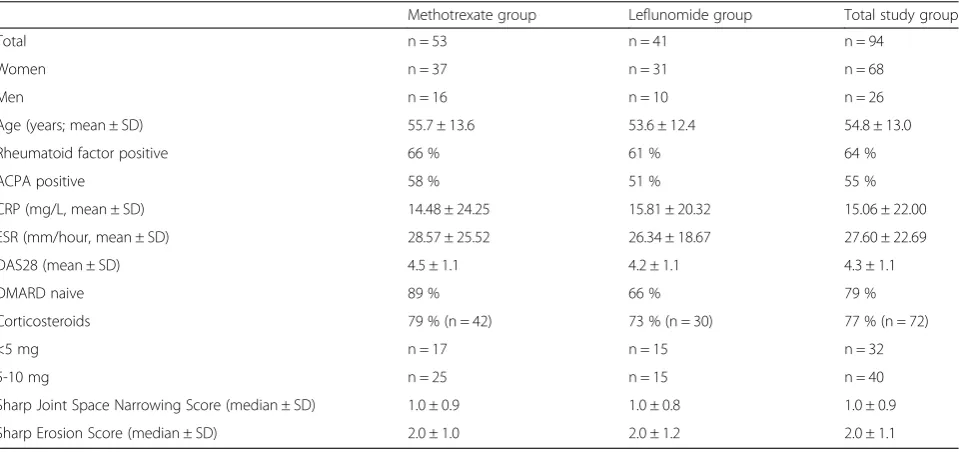
A total of 11 centers throughout Germany contributed data of 94 patients. The detailed clinical patient characteristics are given in Table 1. The mean time from RA symptoms to diagnosis was 56 ± 90 months and the time from the diag-nosis to the inclusion in the study was 29 ± 67 months. The mean observation period was 22 ± 8 months. Of the 94 pa-tients included in the efficacy analysis set, 53 were treated on average with 15 ± 3.5 mg MTX and 41 patients achieved LEF (10 mg: 5 patients, 20 mg: 36 patients).
Influence of inflammatory activity
Erythrocyte Sedimentation Rate (ESR) and C- reactive protein (CRP)
The ESR showed a mean decrease ± SD of 5.59 ± 13.70 mm (from 26.08 mm to 20.49 mm) in the LEF group and a mean decrease of 8.46 ± 18.25 mm (from 28.02 mm to 19.57 mm) in the MTX. The CRP showed a mean de-cline ± SD of 8.04 ± 23.65 mg/l (from 15.81 mg/l to 7.77 mg/l) in the LEF group and a mean decline of 5.64 ± 18.74 mg/l (from 14.48 mg/l to 8.84 mg/l) in the MTX group.
Disease Activity Score (DAS28
The LEF group presented an change of the DAS28 of −1.2 ± 1.4 (from 4.2 to 3.0). Regarding the MTX group a mean decline of −1.1 ± 1.1(from 4.5 to 3.4) was observed.
Radiological progression
For both treatment groups (LEF versus MTX) no signifi-cant changes of the Sharp Erosion Score and Sharp Joint Space Narrowing Score were observed. The median Sharp Erosion Score and Sharp Joint Space Narrowing Score was 1 at baseline. At follow up (month 22) the median Sharp Erosion Score and Sharp Joint Space Nar-rowing Score was also 1.
Methotrexate group (see Table 2 and Figure 1)
DXR-BMD in the MTX group was significantly reduced (-4.3 %) from 0.578 g/cm2± 0.070 g/cm2 (baseline) to 0.553 g/cm2± 0.080 g/cm2 (month 22). Regarding DXR-MCI (−4.9 %) and DXR-CT (−5.4 %), a significant de-crease from 0.447 ± 0.076 (baseline) to 0.425 ± 0.078 (month 22) and from 0.186 cm ± 0.029 cm (baseline) to 0.176 cm ± 0.032 cm (month 22) was revealed. DXR-W presented a mild reduction (−0.6 %) from 0.840 cm ± 0.086 cm (baseline) to 0.835 cm ± 0.093 cm (month 22).
Leflunomide group (see Table 2 and Figure 1)
[image:3.595.59.539.458.684.2]For the 41 patients treated with LEF, DXR-BMD decreased (−1.4 %) from 0.569 g/cm2± 0.088 g/cm2
Table 1Characterization of the study cohort
Methotrexate group Leflunomide group Total study group
Total n = 53 n = 41 n = 94
Women n = 37 n = 31 n = 68
Men n = 16 n = 10 n = 26
Age (years; mean ± SD) 55.7 ± 13.6 53.6 ± 12.4 54.8 ± 13.0
Rheumatoid factor positive 66 % 61 % 64 %
ACPA positive 58 % 51 % 55 %
CRP (mg/L, mean ± SD) 14.48 ± 24.25 15.81 ± 20.32 15.06 ± 22.00
ESR (mm/hour, mean ± SD) 28.57 ± 25.52 26.34 ± 18.67 27.60 ± 22.69
DAS28 (mean ± SD) 4.5 ± 1.1 4.2 ± 1.1 4.3 ± 1.1
DMARD naive 89 % 66 % 79 %
Corticosteroids 79 % (n = 42) 73 % (n = 30) 77 % (n = 72)
<5 mg n = 17 n = 15 n = 32
5-10 mg n = 25 n = 15 n = 40
Sharp Joint Space Narrowing Score (median ± SD) 1.0 ± 0.9 1.0 ± 0.8 1.0 ± 0.9
Sharp Erosion Score (median ± SD) 2.0 ± 1.0 2.0 ± 1.2 2.0 ± 1.1
Notes: ACPA = Antibodies to citrullinated proteins CRP = C-reactive Protein
DAS = Disease Activity Score
(baseline) to 0.561 g/cm2± 0.094 g/cm2(month 22). A re-duction from 0.452 ± 0.095 (baseline) to 0.442 ± 0.098 (month 22) was observed for DXR-MCI (-2.2 %) and also from 0.185 cm ± 0.038 cm (baseline) to 0.181 cm ± 0.040 cm (month 22) for DXR-CT (−2.2 %). Additionally, DXR-W increased (+0.1 %) from 0.822 cm ± 0.073 cm (baseline) to 0.823 cm ± 0.073 cm (month 22).
Discussion
It is well known that irreversible joint damage in RA oc-curs soon after the onset of symptoms, often within the first two years [13]. Early and effective targeted treat-ment, for instance with DMARDs, is required to prevent joint destruction.
Among the different DMARDs currently used in the treatment of RA, MTX is most frequently prescribed.
Several studies also confirmed the efficacy of LEF on in-flammation control and on radiological progression in RA [23–25].
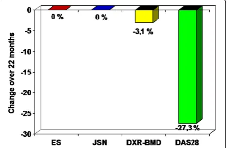
Actually this trial is the first longitudinal study which compares the established Sharp Score and the DXR-technique focusing on the structural integrity. Our study revealed a significantly more pronounced reduction of DXR-BMD (−3.1 %) for the total study cohort during an observation period of 22 months. No difference in the effectiveness of therapy could be illustrated using the Sharp Score. Additionally, the study presented no longi-tudinal change of the Sharp Score for both treatment groups during the observation period. The limitation of the established scoring techniques concerning the detec-tion of structural integrity is also related to the fact that radiological progression based on the use of effective DMARDs as control group resulting in a low radio-logical progression rate which is not visible by a Scoring technique. On the other hand a misinterpretation of ef-fectiveness comparing different therapeutic strategies, in particular consideration of patients with non-erosive RA, exists.
[image:4.595.57.540.100.240.2]These results are confirmed by this study showing dif-ferences between both treatment groups using DXR, but no differences could be documented by detailed scoring. However, DXR-BMD can be recommended as outcome measure and seems to be a valid surrogate marker for structural integrity caused by the similar pathogenetic mechanism as radiographic bone damage, because peri-articular demineralization is still present, even if radio-graphic visible joint damage on X-rays apparently is arrested [26]. Altogether, these recent data showed that the DXR-technique as a precise and reliable tool could distinguish the effectiveness of MTX versus LEF therapy. Otherwise, potential limitation of this study was the Table 2Changes of DXR-parameters between baseline and month 22 for the leflunomide and methotrexate group
Group Baseline Month 22 Difference Relative change p-value
mean (SD) mean (SD) mean (SD)
Leflunomide DXR-BMD in g/cm2 0.569 (0.088) 0.561 (0.094)
−0.008 (0.024) - 1.4 % p < 0.05
DXR-MCI 0.452 (0.095) 0.442 (0.098) −0.010 (0.022) - 2.2 % p < 0.05
DXR-CT in cm 0.185 (0.038) 0.181 (0.040) −0.004 (0.009) - 2.2 % p < 0.05
DXR-Win cm 0.822 (0.073) 0.823 (0.073) 0.001 (0.006) +0.1 % p = n. s.
Methotrexate DXR-BMD in g/cm2 0.578 (0.070) 0.553 (0.080)
−0.024 (0.037) - 4.3 % p < 0.05
DXR-MCI 0.447 (0.076) 0.425 (0.078) −0.022 (0.027) - 4.9 % p < 0.05
DXR-CT in cm 0.186 (0.029) 0.176 (0.032) −0.010 (0.014) - 5.4 % p < 0.05
DXR-in cm 0.840 (0.086 0.835 (0.093) −0.005 (0.031) - 0.6 % p = n. s.
Notes: DXR = Digital X-ray Radiogrammetry BMD = Bone Mineral Density
MCI = Metacarpal Index CT = Cortical Thickness W = Metacarpal Bone Width SD = Standard Deviation n. s. = not significant
[image:4.595.58.292.525.677.2]absence of randomisation. The treatment of bisphospho-nates and hormone replacement therapy was not allowed based on the bone protective effects of these treatment regimes which can potentially influence the result of DXR-measurements [27]. In this context, more objective data about the measurement of structural in-tegrity in RA are now available due to computer based techniques like DXR [27], considering minor differences of structural integrity in the assessment of therapeutic strategies. Finally, the computer based quantification of radiological progression can be improved by the combined analysis of periarticular mineralization, joint space widths and identification of erosions by only one computer sys-tem in one step.
Conclusion
Quantitative data of hand bone mass estimated by the presented DXR-technique may be a complementary pre-cise tool in the identification of RA-related stigmata and their changes due to therapeutic strategies. The modern, more effective and early treatment of RA is associated with limited differences between treatment groups eluci-dating a reduced impact of scoring methods in the as-sessment of radiological progression. DXR could close this gap considering also small differences of structural integrity.
Abbreviations
ACPAs:Antibodies to citrullinated proteins; CRP: C-reactive Protein; CV: Coefficient of Variation; DAS: Disease Activity Score; DMARD: Disease-Modifying Anti-Rheumatic Drug; DXR: Digital X-ray Radiogrammetry; DXR-BMD: Bone Mineral Density (g/cm2) estimated by Digital X-ray Radiogrammetry; DXR-MCI: Metacarpal Index estimated by Digital X-ray Radiogrammetry; ESR: Erythrocyte Sedimentation Rate; LEF: Leflunomide; MTX: Methotrexate; n. s: not significant; RA: Rheumatoid Arthritis; SD: Standard Deviation.
Competing interests
The authors declare that they have no competing interests.
Authors contribution
Study design: AP, JB and PO. Image Analysis: AP, JB and AH. Data Interpretation: AP, DMR and JB: Manuscript writing: AP, JB, PO and GW. Manuscript Editing: DMR. All authors read and approved the final manuscript.
Acknowledgement
The authors thank our investigation centers: Prof. Dr. J. Braun (Herne), Dr. R. Dockhorn (Weener), Dr. L. Meier (Hofheim), Dr. P. Nagler (Regensburg), Dr. E. Nitsche (Regensburg), Dr. O. Krause (Regensburg), Dr. R. Pavlik (Regensburg), Dr. R. de la Camp (Erlangen), Dr. J. Wendler (Erlangen), Dr. F. Schuch (Erlangen), Dr. J. Währisch (Essen), Dr. A. Teich (Leipzig), Dr. F. Hamann (Leipzig), Dr. W. Spieler (Zerbst), Dr. U. von Hinüber (Hildesheim), Dr. A. Göbel (Lippstadt).
The authors also thank Monika Arens (managing director, Arewus GmbH) and Jacob Algulin (Sectra, Sweden) for the use of digital X-ray radiogrammmetry. Also many thanks to Dr. Klaus Bornholdt (Sanofi-Aventis Deutschland GmbH, Berlin, Germany) and Kristin Richter (Sanofi-Aventis Deutschland GmbH, Berlin, Germany) as well as Christiane Manitz (Department of Internal Medicine III, Friedrich-Schiller-University Jena, Germany) for the coordination of the study.
Disclosures
J.B. and A.P. have received speaker’s fees from sanofi-aventis.
The longitudinal study part was funded and managed by Sanofi-Aventis Deutschland GmbH Germany (LEFLU_L_04198).
Notification
The results of this study were part of the poster presentation at the 38th Congress of the German Society of Rheumatology awarded with the first prize.
Author details
1Department of Internal Medicine III, Jena University Hospital–Friedrich Schiller University Jena, Erlanger Allee 101, 07747 Jena, Germany.2Institute of Diagnostic and Interventional Radiology, Jena University Hospital–Friedrich Schiller University Jena, Erlanger Allee 101, 07747 Jena, Germany.3Institute of Diagnostic and Interventional Radiology, Heinrich–Braun–Clinic Zwickau, Karl-Kreil-Straße 35, 08060 Zwickau, Germany.4Institute of Diagnostic and Interventional Radiology, SRH Wald-Klinikum Gera GmbH, Straße des Friedens 122, 07548 Gera, Germany.
Received: 11 November 2014 Accepted: 8 May 2015
References
1. Blair WF. An approach to complex rheumatoid arthritis hand and wrist problems. Hand Clin. 1996;12:615–28.
2. Gravallese EM. Bone destruction in arthritis. Ann Rheum Dis. 2002;61:84–6. 3. Sharp JT, Wolfe F, Mitchell DM, Bloch DA. The progression of erosion and joint
space narrowing scores in rheumatoid arthritis during the first twenty-five years of disease. Arthritis Rheum. 1991;34:660–8.
4. Larsen A. How to apply Larsen score in evaluating radiographs of rheumatoid arthritis in long-term studies. J Rheumatol. 1995;10:1974–5. 5. Sharp JT. An overview of radiographic analysis of joint damage in rheumatoid
arthritis and its use in metaanalysis. J Rheumatol. 2000;27:254–60. 6. van der Heijde DM. How to read radiographs according to the Sharp/van
der Heijde method. J Rheumatol. 2000;27:261–3.
7. Deodhar AA, Brabyn J, Pande I, Scott DL, Woolf AD. Hand bone densitometry in rheumatoid arthritis, a five year longitudinal study: an outcome measure and prognostic marker. Ann Rheum Dis. 2003;62:767–70. 8. Schett G, Coates LC, Ash ZR, Finzel S, Conaghan PG. Structural damage in
rheumatoid arthritis, psoriatic arthritis, and ankylosing spondylitis: traditional views, novel insights gained from TNF blockade, and concepts for the future. Arthritis Res Ther. 2011;13 Suppl 1:S4.
9. Wevers-de Boer KV, Heimans L, Visser K, Kälvesten J, Goekoop RJ, van Oosterhout M, et al. Four-month metacarpal bone mineral density loss predicts radiological joint damage progression after 1 year in patients with early rheumatoid arthritis: exploratory analyses from the IMPROVED study. Ann Rheum Dis. 2015;74:341–6.
10. Mentzel HJ, John U, Böttcher J, Malich A, Pfeil A, Vollandt R, et al. Evaluation of bone-mineral density by digital X-ray radiogrammetry (DXR) in pediatric renal transplant recipients. Pediatr Radiol. 2005;35:489–94.
11. Haugeberg G, Orstavik RE, Uhlig T, Falch JA, Halse JI, Kvien TK. Bone loss in patients with rheumatoid arthritis. Results from a population-based cohort of 366 patients followed up for two years. Arthritis Rheum. 2002;46:1720–8. 12. Böttcher J, Malich A, Pfeil A, Petrovitch A, Lehmann G, Heyne JP, et al.
Potential clinical relevance of digital radiogrammetry for quantification of periarticular bone demineralization in patients suffering from rheumatoid arthritis depending on severity and compared with DXA. Eur Radiol. 2004;14:631–7.
13. Böttcher J, Pfeil A, Rosholm A, Petrovitch A, Seidl BE, Malich A, et al. Digital X-Ray Radiogrammetry combined with semi-automated analysis of joint space distances as a new diagnostic approach in rheumatoid arthritis–A cross-sectional and longitudinal study. Arthritis Rheum. 2005;52:3850–9. 14. Böttcher J, Pfeil A. Diagnosis of periarticular osteoporosis in rheumatoid
arthritis using digital X-ray radiogrammetry. Arthritis Res Ther. 2008;10:103. 15. Hoff M, Haugeberg G, Kvien TK. Hand bone loss as outcome measure in
established rheumatoid arthritis: a two-year observational study comparing cortical and total bone loss. Arthritis Res Ther. 2007;9:R81.
16. Pfeil A, Haugeberg G, Hansch A, Renz DM, Lehmann G, Malich A, et al. The value of digital X-ray radiogrammetry in the assessment of inflammatory bone loss in rheumatoid arthritis. Arthritis Care Res (Hoboken). 2011;63:666–74. 17. Landewé R, Strand V, van der Heijde D. From inhibition of radiographic
18. Arnett FC, Edworthy SM, Bloch DA, McShane DJ, Fies FJ, Cooper NS. The American Rheumatism Association 1987 revised criteria for the classification of rheumatoid arthritis. Arthritis Rheum. 1988;31:315–24.
19. Smolen JS, Landewe R, Breedveld FC, Dougados M, Emery P, Gaujoux Viala C, et al. EULAR recommendations for the management of rheumatoid arthritis with synthetic and biological disease modifying antirheumatic drugs. Ann Rheum Dis. 2010;69:964–75.
20. Sharp JT, Young DY, Bluhm GB, Brook A, Brower AC, Corbett M. e al. How many joints in the hands and wrists should be included in a score of radiologic abnormalities used to assess rheumatoid arthritis? Arthritis Rheum. 1985;28:1326–35.
21. Rosholm A, Hylsdrup L, Baeksgaard L, Grunkin M, Thodberg HH. Estimation of bone mineral density by digital X-ray radiogrammetry: theoretical background and clinical testing. Osteoporos Int. 2001;12:961–9. 22. Böttcher J, Pfeil A, Schäfer ML, Petrovitch A, Seidl BE, Mentzel HJ, et al.
Normative data for digital X-ray radiogrammetry from a female and male German cohort. J Clin Densitom. 2006;9:341–50.
23. Sharp JT, Strand V, Leung H, Hurley F, Loew-Friedrich I. Treatment with leflunomide slows radiographic progression of rheumatoid arthritis. Results from three randomized controlled trials of leflunomide in patients with active rheumatoid arthritis. Arthritis Rheum. 2000;43:495–505. 24. Larsen A, Kvien TK, Schattenkirchner M, Rau R, Scott DL, Smolen JS, et al.
Slowing of disease progression in rheumatoid arthritis patients during long-term treatment with leflunomide or sulfasalazine. Scand J Rheumatol. 2001;30:135–42.
25. Pfeil A, Lippold J, Eidner T, Lehmann G, Oelzner P, Renz DM, et al. Effects of leflunomide and methotrexate in rheumatoid arthritis detected by digital X-ray radiogrammetry and computer-aided joint space analysis. Rheumatol Int. 2009;29:287–95.
26. Pfeil A, Oelzner P, Renz DM, Lehmann G, Wolf G, Böttcher J. Visualisation of structural damage as a surrogate marker of radiographic progression in patients with rheumatoid arthritis. Ann Rheum Dis. 2014;73:e24. 27. Hoff M, Kvien TK, Kälvesten J, Elden A, Haugeberg G. Adalimumab therapy
reduces hand bone loss in early rheumatoid arthritis: Explorative analyses from the PREMIER study. Ann Rheum Dis. 2009;68:1171–6.
Submit your next manuscript to BioMed Central and take full advantage of:
• Convenient online submission
• Thorough peer review
• No space constraints or color figure charges
• Immediate publication on acceptance
• Inclusion in PubMed, CAS, Scopus and Google Scholar
• Research which is freely available for redistribution