Role of different pathways of the complement
cascade in experimental bullous pemphigoid
Kelly C. Nelson, … , Luis A. Diaz, Zhi Liu
J Clin Invest. 2006;
116(11)
:2892-2900.
https://doi.org/10.1172/JCI17891
.
Bullous pemphigoid (BP) is an autoimmune subepidermal blistering disease associated
with autoantibodies directed against the hemidesmosomal proteins BP180 and BP230 and
inflammation. Passive transfer of antibodies to the murine BP180 (mBP180) induces a skin
disease that closely resembles human BP. In the present study, we defined the roles of the
different complement activation pathways in this model system. Mice deficient in the
alternative pathway component factor B (Fb) and injected with pathogenic anti-mBP180 IgG
developed delayed and less intense subepidermal blisters. Mice deficient in the classical
pathway component complement component 4 (C4) and WT mice pretreated with
neutralizing antibody against the first component of the classical pathway, C1q, were
resistant to experimental BP. These mice exhibited a significantly reduced level of mast cell
degranulation and polymorphonuclear neutrophil (PMN) infiltration in the skin. Intradermal
administration of compound 48/80, a mast cell degranulating agent, restored BP disease in
C4
–/–mice. Furthermore, C4
–/–mice became susceptible to experimental BP after local
injection of PMN chemoattractant IL-8 or local reconstitution with PMNs. These findings
provide the first direct evidence to our knowledge that complement activation via the
classical and alternative pathways is crucial in subepidermal blister formation in
experimental BP.
Research Article
Dermatology
Find the latest version:
http://jci.me/17891/pdf
Role of different pathways of the complement
cascade in experimental bullous pemphigoid
Kelly C. Nelson,1 Minglang Zhao,1 Pamela R. Schroeder,1 Ning Li,1 Rick A. Wetsel,2
Luis A. Diaz,1 and Zhi Liu1,3
1Department of Dermatology, University of North Carolina at Chapel Hill, Chapel Hill, North Carolina, USA.
2Institute of Molecular Medicine for the Prevention of Human Diseases, University of Texas Health Science Center at Houston, Houston, Texas, USA. 3Department of Microbiology and Immunology, University of North Carolina at Chapel Hill, Chapel Hill, North Carolina, USA.
Bullous pemphigoid (BP) is an autoimmune subepidermal blistering disease associated with
autoantibod-ies directed against the hemidesmosomal proteins BP180 and BP230 and inflammation. Passive transfer of
antibodies to the murine BP180 (mBP180) induces a skin disease that closely resembles human BP. In the
present study, we defined the roles of the different complement activation pathways in this model system.
Mice deficient in the alternative pathway component factor B (Fb) and injected with pathogenic anti-mBP180
IgG developed delayed and less intense subepidermal blisters. Mice deficient in the classical pathway
com-ponent complement comcom-ponent 4 (C4) and WT mice pretreated with neutralizing antibody against the first
component of the classical pathway, C1q, were resistant to experimental BP. These mice exhibited a
signifi-cantly reduced level of mast cell degranulation and polymorphonuclear neutrophil (PMN) infiltration in the
skin. Intradermal administration of compound 48/80, a mast cell degranulating agent, restored BP disease
in C4
–/–mice. Furthermore, C4
–/–mice became susceptible to experimental BP after local injection of PMN
chemoattractant IL-8 or local reconstitution with PMNs. These findings provide the first direct evidence to
our knowledge that complement activation via the classical and alternative pathways is crucial in subepidermal
blister formation in experimental BP.
Introduction
Bullous pemphigoid (BP) is a subepidermal blistering skin disorder of the elderly (1). Lesional skin of BP patients is characterized by a detachment of the basal keratinocytes of the epidermis from the der-mis, resulting in tense, fluid-filled vesicles. Similar skin lesions were noted in herpes gestationis (HG), a nonviral disease of pregnancy (2). Immunofluorescence (IF) studies demonstrated that these patients exhibit circulating and tissue-bound autoantibodies directed against antigens of the basement membrane zone (BMZ) (3–8).
BP and HG autoantibodies recognize 2 antigens, BP230 and BP180, that are localized in the hemidesmosome, the main epi-dermal structure involved in epi-dermal-epiepi-dermal adhesion (4–6, 9). The BP230 antigen is restricted to the cytoplasmic plaque of the hemidesmosome and appears to interact directly with the inter-mediate filament network (10, 11). The BP180 antigen is a type II transmembrane protein with an aminoterminal endodomain and an extended carboxyterminal ectodomain that spans the lamina lucida, projecting into the lamina densa of the BMZ (12–16). The ectodomain of human BP180 contains a series of 15 interrupted collagen domains (complement components 1–15 [C1–C15]) with the typical Gly-X-Y repeat (14). It has recently been shown that the BP180 extracellular domain forms a collagen triple helix (17, 18). The extracellular noncollagenous stretches are designated NC1 (located at the carboxyl terminus) through NC16A (adjacent to the membrane-spanning domain). The NC16A domain of human
BP180 harbors major extracellular antigenic sites recognized by BP and HG sera (19, 20).
Complement can be activated by 3 pathways: classical, lectin, and alternative. The classical pathway is activated primarily by antigen-antibody complexes. The lectin pathway is activated by mannose-binding lectin attachment to surface carbohydrates (such as on bacteria) and differs from the classical pathway only in this initial recognition and activation stage. The alternative pathway is activat-ed continuously in the blood plasma by the spontaneous activation of C3, but this pathway is amplified to a significant level only in the presence of a molecular surface that supports the deposition, acti-vation, and amplification of the alternative pathway complement proteins. Complement activation has been implicated in the patho-genesis of BP and HG (21). Autoantibodies from BP and HG can fix complement in vitroand in vivo. C3 is often detected at the BMZ of the lesional/perilesional skin by direct IF (22, 23). Components of both the classical and the alternative complement pathways have been detected at the BMZ of patients’ skin, including C1q, C3, C4, C5, C5-9 (membrane attack complex, or MAC), factor B (Fb), factor H, and properdin (23–35). However, the relevance of any of these components, and which arm of the complement cascade (e.g., clas-sical, lectin, or alternative) is involved in subepidermal blistering in BP and HG, remain unknown.
Our laboratory has developed an animal model of BP, which involves the injection of neonatal BALB/c mice with rabbit anti– murine BP180 (anti-mBP180) antibodies (36). Subepidermal blis-tering triggered by anti-mBP180 IgG depends on complement activation, mast cell (MC) degranulation, and polymorphonuclear neutrophil (PMN) infiltration (37–39). In this study, we investi-gated the role of these 3 pathways of complement activation in the disease development of experimental BP by using mice genetically deficient in pathway-specific components. We found that the
clas-Nonstandard abbreviations used: BMZ, basement membrane zone; BP, bullous pemphigoid; C4, complement component 4; Fb, factor B; GPI, glucose-6-phosphate isomerase; HG, herpes gestationis; i.d., intradermal(ly); IF, immunofluorescence; m-, murine; MC, mast cell; MPO, myeloperoxidase; PMN, polymorphonuclear neutrophil.
Conflict of interest: The authors have declared that no conflict of interest exists.
research article
The Journal of Clinical Investigation http://www.jci.org Volume 116 Number 11 November 2006 2893
sical and alternative pathways of complement system are required for experimental BP.
Results
Pathogenic anti-mBP180 antibodies induce subepidermal blisters in Fb–/–, but
not C4–/–, mice.To determine the roles of the classical and alternative
complement activation pathways in subepidermal blister formation in experimental BP, WT, C4–/–, and Fb–/– mice (n =12 per group) were
injected with pathogenic anti-mBP180 IgG (2.6 mg/g body weight). WT and Fb–/– mice developed intense blisters (average clinical disease
score, 2.7 for WT and 2.5 for Fb–/–; P =0.175) 12 hours after
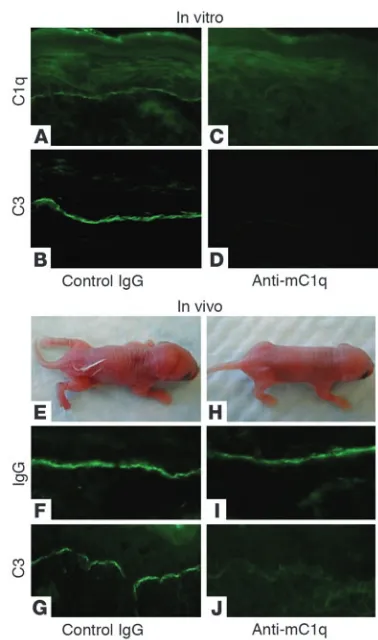
patho-genic IgG administration (Figure 1, A and I, and Table 1). Direct IF of perilesional skin of these mice showed in vivodeposition of pathogenic rabbit IgG (Figure 1, B and J) and murine C3 (Figure 1, C and K). H&E-stained skin sections from these mice revealed der-mal-epidermal separation with PMN infiltration (Figure 1, D and L). In contrast, C4–/– mice exhibited no blisters 12 hours after injection
with pathogenic IgG (Figure 1E). Direct IF of the skin of these mice showed in situdeposition of rabbit IgG (Figure 1F), but no murine C3 (Figure 1G), at the BMZ. Histologic examination of the skin of the C4–/– mice showed no signs of dermal-epidermal detachment
(Figure 1H). Indirect IF demonstrated that the titers of circulating rabbit anti-mBP180 IgG in the C4–/– mice were comparable to those
of the WT and Fb–/– mice (1:2,560 in 3 groups). These results
dem-onstrate that complement activation via the classical pathway but not the alternative pathway is essential for subepidermal blistering in experimental BP.
Inhibition of C1q abolishes experimental BP. Since C4–/– mice are
impaired in both the classical and lectin pathways, we then induced experimental BP in the absence of the classical pathway. If experi-mental BP depends on the classical pathway, then blocking C1q, the first component of the classical pathway, should block the skin disease. Our next set of experiments demonstrated this was the case. In vitro, anti-mC1q antibody blocked anti-mBP180–mediat-ed deposition of C1q and C3 at the BMZ (Figure 2, A–D). In vivo, WT mice (n =6) pretreated with anti-mC1q antibody followed by pathogenic anti-mBP180 IgG showed no complement activation
in the skin and developed no skin lesions (Figure 2, E–J, and Table 1). These data further implicated classical pathway activation as the major pathway in experimental BP.
[image:3.585.46.286.82.307.2]The alternative pathway plays an accessory role in experimental BP. To determine whether the alternative pathway augments the
Table 1
Role of the complement activation of different pathways in experimental BP
IgG injected Treatment n Mean disease activity
WT
R50 – 9 0.00 ± 0.00
R530 – 24 2.75 ± 0.10
R530 Anti-mC1q 8 0.10 ± 0.08A
R50 Compound 48/80 6 0.00 ± 0.00
C4–/–
R50 – 9 0.00 ± 0.00
R530 – 12 0.04 ± 0.04B
R50 Compound 48/80 6 0.00 ± 0.00 R530 Compound 48/80 6 3.00 ± 0.00
R530 PMNs 6 3.00 ± 0.00
R530 IL-8 6 3.00 ± 0.00
Fb–/–
R50 – 6 0.00 ± 0.00
R530 – 12 2.54 ± 0.10C
Neonatal WT, C4–/–, and Fb–/– mice were injected i.d. with control IgG
R50 or pathogenic rabbit anti-mBP180 IgG R530 alone (2.64 mg/g body weight) or were additionally pretreated with mC1q neutralizing anti-body (5 μg/g body weight), treated with compound 48/80 (1 μg/g body weight i.d.), treated with PMN chemoattractant IL-8 (50 ng/g body weight i.d.), or administered purified mouse PMNs (5 × 10⁵ cells in 50 μl PBS
i.d.) from WT mice 2 hours after IgG injection. Animals were examined 12 hours after IgG injection. Clinical disease severity (mean ± SEM) was scored as described in Methods and analyzed by paired Student’s t test. AP =0.00001; BP =0.0000001; CP =0.175 versus anti-mBP180
[image:3.585.308.530.453.633.2]IgG–injected WT mice. Three to four independent experiments were performed per group.
Figure 1
classical pathway in experimental BP, we injected WT, C4–/–, and
Fb–/– mice (n =12 per group) with a suboptimal dose (2.0 mg/g
body weight) of pathogenic antibody, with which the injected WT mice developed an average clinical disease score of 1.5 at 12 hours after injection. We also extended IgG incubation time beyond 12 hours and up to 48 hours. WT mice injected with the suboptimal dose of pathogenic antibody developed clinical disease scores of 1.5, 2.2, and 2.9 at 12, 24, and 48 hours, respectively (Figure 3A). The pathogenic IgG-injected Fb–/– mice developed clinical disease
scores of 1.25 (P =0.21), 1.75 (P =0.04), and 2.1 (P =0.007) at 12, 24, and 48 hours, respectively. The C4–/– mice showed no skin blisters
at any time point (P <0.001). These results suggest that the classi-cal pathway plays a major role in the early phase of subepidermal blistering and in the later phase the classical pathway in concert with the alternative pathway cause full-scale BP skin lesions.
To further confirm this conclusion, we extended the time course up to 14 days and compared the disease severity between WT and Fb–/– mice (n =5 per group). As expected, circulating levels of
pathogenic IgG in WT and Fb–/– mice decreased as the mice grew
bigger (Figure 3B). The WT group developed clinical disease scores of 2.5 ± 0.26, 1.6 ± 0.19, 0.6 ± 0.1, and 0 ± 0 on days 1, 3, 7, and 14, respectively, while the Fb–/– group developed clinical disease scores
of 1.83 ± 0.26 (P =0.04), 1.25 ± 0.17 (P =0.20), 0.58 ± 0.08 (P =0.90), and 0 ± 0 on days 1, 3, 7, and 14, respectively. Myeloperoxidase (MPO) activity assay revealed a significantly higher level of PMN infiltration in the WT group than in the Fb–/– group at 24 hours,
but similar levels of PMN infiltration were seen on days 3, 7, and 14 in the skin of the IgG-injected WT and Fb–/– mice (Figure 3C).
We also injected WT and Fb–/– mice with a higher dose of
patho-genic IgG (3 mg/g body weight) and observed no difference in clinical disease score or skin MPO activity between these 2 groups of mice at all time points (data not shown).
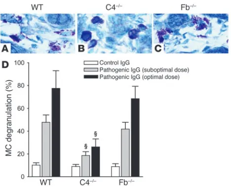
MC degranulation is impaired in C4–/– mice injected with pathogenic
anti-mBP180 antibodies. Complement activation generates comple-ment fragcomple-ments C3a and C5a, of which C5a can mediate PMN chemotaxis and both C3a and C5a can mediate MC degranulation (40). We then determined whether complement activation via the classical pathway was required for MC degranulation, PMN recruit-ment, or both by quantifying MC degranulation and PMN infiltra-tion. Neonatal WT, C4–/–, and Fb–/– mice were injected intradermally
(i.d.) with the suboptimal and optimal doses of pathogenic IgG and examined at 2, 12, 24, and 48 hours after injection. At 2 hours, when MC degranulation triggered by pathogenic IgG reaches its peak (38), toluidine blue staining revealed extensive MC degranulation in the dermis of WT and Fb–/– mice (Figure 4, A and C). In contrast,
C4–/– mice exhibited a minimal level of MC degranulation in the
dermis (Figure 4B). Quantification of MC degranulation revealed a significant reduction in the number of degranulating MCs in C4–/– mice compared with WT and Fb–/– mice (Figure 4D). WT and
Fb–/– mice showed similar levels of MC degranulation. Thus, MC
degranulation triggered by anti-mBP180 IgG mainly depends on complement activation of the classical pathway.
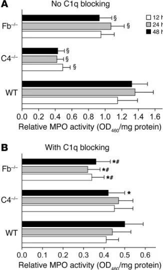
MPO activity assays revealed a significant difference in PMN infiltration at the IgG injection sites between WT and C4–/– mice
at all time points (Figure 5A). There was no difference statisti-cally between WT and Fb–/– mice at 12 hours (P = 0.057), but
significant differences were observed at 24 and 48 hours after IgG injection (Figure 5A). There was no difference in PMN infil-tration between anti-mC1q–treated WT and C4–/– mice at 12 and
24 hours, but there was a significant difference between these groups at 48 hours after injection (Figure 5B). However, anti-mC1q antibody–treated Fb–/– mice showed a drastic reduction in
PMN recruitment at all time points compared with anti-mC1q antibody–treated WT and C4–/– mice (Figure 5B). The degree of
PMN recruitment in the anti-mC1q–treated Fb–/– mice was
com-patible to that previously shown in the pathogenic IgG–injected C5–/– mice (39), suggesting that the lectin pathway does not
[image:4.585.66.255.79.399.2]par-ticipate in experimental BP. These results further substantiate the conclusion that the classical pathway plays a major role and the alternative pathway plays an accessory role in activating MCs, which in turn recruit PMNs to the dermal-epidermal junction, causing subepidermal blistering.
Figure 2
research article
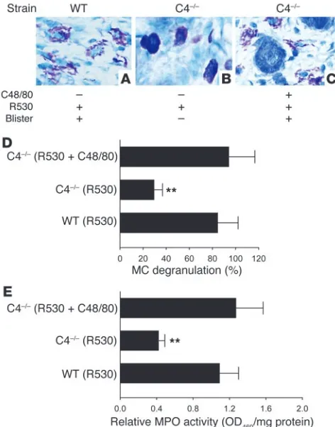
The Journal of Clinical Investigation http://www.jci.org Volume 116 Number 11 November 2006 2895 C4–/– mice pretreated with a MC degranulating agent become susceptible
to experimental BP. If the major role of complement activation in experimental BP is to activate MCs, then the requirement for the complement system for the subepidermal blistering in experimen-tal BP should be bypassed by artificial induction of MC degranula-tion. This is exactly what we found in the next set of experiments: C4–/– mice (n =6) injected locally with compound 48/80, a potent
MC degranulating agent, followed by pathogenic anti-mBP180 IgG developed clinical blisters to the same degree as the IgG-injected WT mice (Table 1). Like the diseased WT mice (Figure 6A), exten-sive MC degranulation took place in the dermis of the compound 48/80–treated C4–/– mice (Figure 6C). In contrast, C4–/– mice
with-out compound 48/80 treatment exhibited no skin lesions and minimal MC degranulation (Figure 6B). WT or C4–/– mice injected
with compound 48/80 alone as a control exhibited MC degranu-lation, but no skin lesions (Table 1). Quantification assays also revealed significantly elevated levels of MC degranulation (Figure 6D) and PMN infiltration (Figure 6E) in compound 48/80–treated C4–/– mice relative to C4–/– mice without compound 48/80
pre-treatment. These results further demonstrated that complement activation acts upstream of MC degranulation in the anti-mBP180 IgG–induced inflammatory cascade.
Pathogenic anti-mBP180 IgG induces subepidermal blistering in C4–/–
mice reconstituted with mouse PMNs or by i.d. injection of the PMN chemoattractant IL-8 Our findings indicate that a major function of complement activation in experimental BP is the activation of MCs. Since activated MCs degranulate, recruiting PMNs to the skin site in experimental BP, it is expected that artificially recruit-ing PMNs from the circulation to the dermis of C4–/– mice should
circumvent the requirement for both complement activation and
MC degranulation for disease phenotype. C4–/– mice were injected
with pathogenic IgG and then reconstituted with mouse PMN. The C4–/– mice reconstituted with 5 × 10⁵ PMNs (n =6) developed
subepidermal blisters 12 hours after IgG injection (Figure 7 and Table 1). In addition, C4–/– mice (n =6) given an i.d. injection of
IL-8 prior to, or coincident with, the treatment with anti-mBP180 IgG developed extensive blisters 12 hours later (Figure 7 and Table 1). Significantly higher levels of PMN infiltration were seen in the lesional skin of C4–/– mice with PMN reconstitution
com-pared with C4–/–mice without PMN reconstitution (Figure 7). The
mean MPO activity levels in skin extracts of C4–/– mice injected
with anti-mBP180 IgG plus IL-8 and anti-mBP180 plus PMN were 1.31 ± 0.29 and 1.43 ± 0.36 OD460/mg protein, respectively,
com-pared with 0.47 ± 0.04 OD460/mg protein in the skin from the
C4–/– mice injected with anti-mBP180 IgG alone (P < 0.01). Taken
together, these results clearly demonstrate that complement activation via the classical pathway mainly participates in PMN recruitment by directly activating MCs in experimental BP.
Discussion
The aim of this paper was to investigate the role of the comple-ment activation pathways in expericomple-mental BP. Mice genetically deficient in the classical pathway component C4 are resistant to experimental BP. Because C4–/– mice are impaired in both the
classical and lectin pathways, we induced experimental BP in WT mice pretreated with anti-mC1q antibody. Anti-mC1q antibody treatment completely abolished subepidermal blistering. Since BP is an autoantibody-mediated disorder, and activation of the classical pathway by antibody is initiated by C1q, it is likely that the attenuation of the pathology observed in the C4–/– mice in
experimental BP is due to the absence of the classical and not the lectin pathway. Mice deficient in the alternative pathway compo-nent Fb that were injected with pathogenic antibodies developed a delayed and less intense disease phenotype in our 0- to 14-day time course study. These results demonstrate that an intact alter-native pathway is necessary for the full development of the skin disease. Taken together, our data suggest that activation of the classical pathway is essential for disease development and that the alternative pathway acts in concert with the classical pathway in subepidermal blistering.
[image:5.585.57.273.76.394.2]We should point out that under the current experimental condi-tions the contribution of the alternative pathway to experimental BP might be underestimated and that the involvement of the lectin
Figure 3
pathway cannot be totally excluded. Given the fact that the IgG pas-sive transfer neonatal mouse model is limited to a single injection of pathogenic antibody and is relatively short term (0–14 days), it is important to develop an adult mouse model with on-going BP disease and to compare the disease severity in WT mice with that of mice deficient in different complement activation pathways.
Complement activation has long been implicated in the disease development of BP and HG based on the identification of com-ponents of the classical and alternative pathways in perilesional sites and blister fluids (21). Using the BP mouse model, we previ-ously demonstrated that complement activation is indeed a nec-essary step in the process of subepidermal blistering triggered by anti-mBP180 antibodies (37). In the present study, we provide the first direct evidence to our knowledge that subepidermal blister formation induced by pathogenic anti-mBP180 IgG requires both classical and alternative pathways. Our results suggest that local activation of the complement cascade is a key step in the formation of skin lesions in BP as well as HG patients. We should note that the complement pathway activated is determined by the isotype, subclass, and species of antibody used to elicit the lesion. In experi-mental BP, pathogenic rabbit anti-mBP180 IgG belongs to only one isotype and recognizes a single pathogenic epitope. In human BP, conversely, BP180-specific autoantibodies belong to IgG1, -2,
-3, and -4 and IgE, and these BP autoantibodies target multiple epitopes. It is possible that the complement pathway activated by pathogenic autoantibodies and the relative contribution of differ-ent pathways in human BP may not be the same as in experimen-tal BP. Future studies using a new mouse model of BP induced by anti-mBP180 autoantibodies will address which Ig isotype and subclass(es) are pathogenic and which complement pathway(s) are activated by the pathogenic antibody.
We have previously shown that anti-mBP180–mediated comple-ment activation is essential for PMN recruitcomple-ment to the cutane-ous BMZ (37, 38). One finding that demonstrated this link is that C5–/– mice injected i.d. with a PMN chemoattractant, either C5a or
IL-8, became susceptible to the pathogenic effects of anti-mBP180 IgG (39). These data were consistent with the hypothesis that com-plement activation via the classical pathway triggered by an anti-mBP180 immune complex results in the generation of C5a, which subsequently leads to dermal-epidermal detachment. Our present studies provide direct evidence to support this idea — an intact clas-sical pathway arm is required for subepidermal blister formation triggered by complement-fixing anti-mBP180 IgG. Thus, the major function of the complement system in experimental BP might well be to generate C5a, which would function in recruiting PMNs to the cutaneous BMZ. C5a could carry out this role directly (by func-tioning as a PMN chemoattractant itself), indirectly (by stimulat-ing local cells to release proinflammatory mediators such as IL-8), or through a combination of these 2 mechanisms (40). Recently, we have shown that in experimental BP, MC degranulation, which leads to PMN recruitment, mainly depends on complement activa-tion (38). Our present data further demonstrate that MC degranu-lation and subsequent PMN infiltration triggered by pathogenic anti-mBP180 antibodies was severely impaired in mice deficient in the classical pathway component C4. Taken together, our findings clearly reveal that the major function of complement activation via the classical pathway is to directly activate MCs, which in turn recruit PMNs. Several recent studies have shown that the alterna-tive pathway plays a key and, in most instances, obligate role in generating proinflammatory complement activation products in vivo (41). In experimental BP, the disease severity is significantly compromised without the alternative pathway. However, it is yet to be determined the exact role the alternative pathway plays in BP and how this pathway is activated.
In the K/BxN serum transfer model of inflammatory arthritis, the joint disease is triggered by anti–glucose-6-phosphate isom-erase (anti-GPI) antibodies and, like experimental BP, depends on PMNs and MCs (42–44). Unlike experimental BP, this arthritis mouse model requires the alternative pathway, and the classical pathway is entirely dispensable (45). The difference in the comple-ment pathway usage between these 2 animal models may be due to the difference in targeted antigens (BP180 versus GPI) and their presentation for antibody generation and binding. Alternatively, the isotype difference of pathogenic antibodies used may cause this discrepancy. In experimental BP, rabbit anti-mBP180 IgG mainly activates the classical pathway, while anti-GPI antibodies activate the alternative pathway, since the dominant isotype of the anti-GPI antibody in K/BxN mice is IgG1 and murine IgG1 is very poor at complement activation via the classical pathway (42).
[image:6.585.48.282.81.273.2]In summary, we have demonstrated that complement activation via the classical and alternative pathways plays a critical role in subepidermal blister formation in experimental BP. These findings may shed new light on the pathogenic mechanisms responsible
Figure 4
research article
The Journal of Clinical Investigation http://www.jci.org Volume 116 Number 11 November 2006 2897
for BP and HG and other subepidermal blistering diseases with an autoimmune response to BP180 such as mucous membrane pem-phigoid, pemphigoid gestationis, and lichen planus pemphigoides (9, 46, 47). These findings may lead to more effective management of these human autoimmune blistering diseases.
Methods
Laboratory animals. Breeding pairs of C57BL/6J WT mice and C57BL/6J C4–/– mice were purchased from The Jackson Laboratory. Fb–/– mice back-crossed 10 generations with C57BL/6J were generated as described previ-ously (48). The mice were maintained at the University of North Carolina at Chapel Hill Animal Center. Neonatal mice (24–36 hours old with body weights between 1.4 and 1.6 g) were used for passive transfer experiments. Animal care and animal experiments were approved by the Animal Care Committee at the University of North Carolina at Chapel Hill.
Preparation of pathogenic anti-mBP180 IgG. The preparation of recombinant mBP180 and the immunization of New Zealand White rabbits were per-formed as previously described (36, 49, 50). The pathogenicity of these IgG preparations was tested by passive transfer experiments as described below. A pathogenic anti-mBP180 IgG preparation (R530) and a nonpathogenic control IgG preparation (R50) were used in this study (39).
Induction of experimental BP and clinical evaluation of animals. Neonates were given on the back 1 i.d. injection of a sterile solution of IgG in PBS (50 μl IgG, 2.64 mg IgG/g body weight) as described previously (36). The site of the 50-μl injection, approximately 3–4 mm in diameter, was marked with ink. The skin at the IgG injection sites of the mice from the test and con-trol groups was examined at different time points after the IgG injection. For the 0- to 3-day time course experiments, neonatal mice were injected and placed in a temperature- and humidity-controlled incubator. To keep neonatal mice alive for longer than 3 days, the neonatal mice were returned to their cages immediately after injection. The extent of cutaneous disease was scored as follows: 0, no detectable skin disease; 1, mild erythematous reaction with no evidence of the epidermal detachment sign (elicited by
gentle friction of the mouse skin; when positive, this produced fine, per-sistent wrinkling of the epidermis); 2, intense erythema and epidermal detachment sign involving 10%–50% of the epidermis in localized areas; 3, intense erythema with frank epidermal detachment sign involving more than 50% of the epidermis in the injection site.
After clinical examination the animals were sacrificed, and skin and serum specimens were obtained. Each skin section (approximately 6 × 6 mm in size) was cut into 3 pieces from the central lines: one (approximately 3 × 3 mm) for routine histological examination by light microscopy (H&E staining) to localize the lesional site and PMN infiltration and toluidine blue staining to quantify MCs and MC degranulation, one (approximately 3 × 3 mm) for direct IF assays to detect rabbit IgG and mouse C3 deposition at the BMZ, and one (approximately 3 × 6 mm) for MPO enzymatic assay to quantify the PMN accumulation at the skin injection site as described below. The sera of injected animals were tested by indirect IF techniques to determine the titers of rabbit anti-mBP180 antibodies using mouse skin cryosections as the substrate. Direct and indirect IF studies were performed as previously described (36) using commercially available FITC-conjugated goat anti-rabbit IgG (Kirkegaard & Perry Laboratories Inc.). Monospecific goat anti-mC3 IgG was purchased from Cappel Laboratories.
Inhibition of C1q in vitro and in vivo. For in vitro C1q inhibition, neona-tal mouse skin sections were incubated with purified rabbit anti-mBP180 or control IgG for 1 hour, after which freshly prepared mouse serum was added as a complete complement source in the presence of purified rat anti-mC1q monoclonal antibody or rat isotype control IgG1κ. After 1 hour of incubation, in situdeposition of mouse C1q and mouse C3 at the BMZ were detected with FITC-conjugated rat anti-mC1q monoclonal antibody and FITC-conjugated monospecific goat anti-mC3 antibody, respectively. For in vivo C1q inhibition, neonatal C57BL/6J mice were injected i.d. with 5 μg purified rat anti-mC1q monoclonal antibody or isotype control rat IgG1κ. Two hours later, the mice were injected i.d. with pathogenic anti-mBP180 IgG (2.64 mg/g body weight) and then examined 12, 24, and 48 hours after pathogenic IgG injection as described above. Purified rat
anti-Figure 5
[image:7.585.77.240.85.355.2]mC1q monoclonal antibody and rat IgG1κ isotypic control were purchased from Cedarlane Laboratories Ltd.
Quantification of MCs and MC degranulation. Skin sections (approxi-mately 3 × 3 mm) of IgG-injected mice were fixed in 10% formalin. Par-affin sections (6 μm thick) were prepared and stained with toluidine blue and H&E. Dermal MCs were counted by 2 individuals in the labo-ratory in a blinded fashion and classified as degranulated (>10% of the granules exhibiting fusion or discharge) or normal in 5 random fields under a light microscope at ×400 magnification (38, 51). There was no significant difference between the 2 individual countings (for total MCs, 32.20 ± 6.52 versus 36.97 ± 11.55; P =0.082). MCs with complete degranulation that may be missed by toluidine blue staining were iden-tified by indirect IF with FITC-conjugated rat anti-mouse c-kit mono-clonal antibody (BD Biosciences). Results were expressed as percentage
of degranulated MC (number of degranulating MCs per total number of MCs in 5 random fields × 100%).
Quantification of PMN accumulation at antibody injection sites. Tissue MPO activity was used as an indicator of PMNs within skin samples of experi-mental animals as described elsewhere (52). We previously showed that clinical skin blistering is directly correlated with the number of infiltrating PMNs in the IgG injection site (39). The mouse skin samples (approximately 3 × 6 mm) were extracted by homogenization in 500 μl extraction buffer. MPO content was expressed as relative MPO activity (OD460/mg protein).
In vivo stimulation of MC degranulation. To stimulate MC degranulation, neonatal C4–/– mice were injected i.d. with compound 48/80 (1 μg/g body weight; Sigma-Aldrich), a MC degranulating agent. Skin MC degranula-tion was analyzed by toluidine blue staining, and skin PMN infiltradegranula-tion was quantified by MPO assay.
[image:8.585.45.281.83.384.2]Isolation and i.d. injection of PMNs Mouse PMNs were isolated from hepa-rinized blood by dextran sedimentation followed by separation on a den-sity gradient as described previously (53). PMN purity of the final cell preparation was consistently >96% as determined by cell-cytospin and LeukoStat staining (Fisher Scientific). The viability of the PMNs was >96% as determined by trypan blue exclusion. C4–/– mice were injected i.d. with pathogenic anti-mBP180 IgG (2.64 mg/g body weight in 50 μl PBS). Two
Figure 6
[image:8.585.336.508.585.741.2]Effect of MC degranulating agent on PMN infiltration and subepidermal blistering in pathogenic IgG–injected C4–/– mice. WT and C4–/– mice were injected with pathogenic anti-mBP180 R530 IgG (2.6 mg/g body weight) with or without pretreatment of the MC degranulating agent compound 48/80 (C48/80) and were examined 2 hours (for toluidine blue staining) and 12 hours (for clinical examination and MPO assay) after IgG injection. (A–C) Clinical examination and toluidine blue stain-ing of the mouse skin showed subepidermal blisters and MC degranu-lation in WT (A) and C4–/– mice with compound 48/80 pretreatment (C). (B) In contrast, C4–/– mice without compound 48/80 treatment exhibited no skin lesions and minimal MC degranulation. (D) MCs in the dermis of the skin were counted and classified as degranulated or normal. Total MCs in 5 random fields were counted (mean, 31.72 ± 8.41), and per-cent MC degranulation was calculated. As expected, MC degranulation was significantly reduced in C4–/– mice compared with WT mice. How-ever, a significant increase of MC degranulation was seen in the skin of compound 48/80–treated C4–/– mice. n =6 per group. (E) MPO activity assay revealed significantly higher levels of PMN infiltration in WT and C4–/– mice with compound 48/80 pretreatment than those without it. Tis-sue MPO activity at the injection sites was expressed as relative MPO activity. Three independent experiments were performed per group. n =6 per group. **P <0.01 versus WT and pretreated C4–/– mice.
Figure 7
research article
The Journal of Clinical Investigation http://www.jci.org Volume 116 Number 11 November 2006 2899 hours later, these mice were injected with 5 × 10⁵ PMNs i.d. at the same
site (54, 55). The animals were analyzed 12 hours after the IgG injections as described above.
IL-8 pretreatment of C4–/– mice. A single i.d. injection of recombinant human IL-8 (50 ng in 50 μl PBS; R&D Systems) or an equivalent amount of BSA was given to neonatal C4–/– mice, followed by i.d. injection of rabbit anti-mBP180 IgG (2.64 mg/g body weight in 50 μl PBS) 1 hour later (39). Control animals received an equivalent amount of normal rabbit IgG in place of the anti-mBP180 IgG. The animals were analyzed 12 hours after the IgG injections as described above.
Statistical analysis. Data were expressed as mean ± SEM and were analyzed using Student’s t test (SigmaPlot; Systat). A P value less than 0.05 was con-sidered significant.
Acknowledgments
We thank Matthew Fleming and Simon Warren for routine his-tology and Lowell Goldsmith for his critical reading of the man-uscript. This work was supported in part by U.S. Public Health Service NIH grants AI40768 and AI61430 (to Z. Liu), AR32599
and AR32081 (to L.A. Diaz), AR052109 (to N. Li), and AI25011 (to R.A. Wetsel). P.R. Schroder was a recipient of a Medical Scientist Training Program scholarship awarded by the Medical College of Wisconsin. K.C. Nelson received a research fellowship grant from the Holderness Foundation.
Received for publication January 17, 2003, and accepted in revised form August 15, 2006.
Address correspondence to: Zhi Liu, 3100 Thurston-Bowles, Department of Dermatology, University of North Carolina, Cha-pel Hill, North Carolina 27599, USA. Phone: (919) 966-0788; Fax: (919) 966-3898; E-mail: [email protected].
Pamela R. Schroeder’s present address is: Diabetes and Endocrine Center, Union Memorial Hospital, Baltimore, Maryland, USA. Kelly C. Nelson and Minglang Zhao contributed equally to this work.
1. Lever, W.F. 1953. Pemphigus. Medicine.32:1–123. 2. Shornick, J.K., Bangert, J.L., Freeman, R.G., and
Gilliam, J.N. 1983. Herpes gestationis: clinical and histological features of twenty-eight cases. J. Am. Acad. Dermatol. 8:214–224.
3. Jordon, R.E., et al. 1967. Basement zone antibodies in bullous pemphigoid. J. Am. Med. Assoc. 200:751–758. 4. Jordon, R.E. 1976. Complement activation in pem-phigus and bullous pemphigoid. J. Invest. Dermatol. 67:366–371.
5. Katz, S.I., Hertz, K.C., and Yaoita, H. 1976. Herpes gestationis. Immunopathology and characteriza-tion of the HG factor. J. Clin. Invest. 57:1434–1441. 6. Stanley, J.R., Hawley-Nelson, P., Yuspa, S.H.,
She-vach, E.M., and Katz, S.I. 1981. Characterization of bullous pemphigoid antigen: a unique basement membrane protein of stratified epithelia. Cell. 24:897–903.
7. Mutasim, D.F., et al. 1985. A pool of bullous pem-phigoid antigen(s) is intracellular and associated with the basal cell cytoskeleton-hemidesmosome complex. J. Invest. Dermatol. 84:47–53.
8. Labib, R.S., Anhalt, G.J., Patel, H.P., Mutasim, D.F., and Diaz, L.A. 1986. Molecular heterogeneity of bullous pemphigoid antigens as detected by immu-noblotting. J. Immunol. 136:1231–1235.
9. Morrison, L.H., Labib, R.S., Zone, J.J., Diaz, L.A., and Anhalt, G.J. 1988. Herpes gestationis auto-antibodies recognize a 180-kD human epidermal antigen. J. Clin. Invest. 81:2023–2026.
10. Tanaka, T., Parry, D.A.D., Klaus-Kovtun, V., Stein-ert, P.M., and Stanley, J.R. 1991. Comparison of molecularly cloned bullous pemphigoid antigen to desmoplakin I confirms that they define a new family of cell adhesion junction plaque proteins.
J. Biol. Chem. 266:12555–12559.
11. Sawamura, D., Li, K., Chu, M.-L., and Uitto, J. 1991. Human bullous pemphigoid antigen (BPAG1). Amino acid sequences deduced from cloned cDNAs predict biologically important peptide segments and protein domains. J. Biol. Chem. 266:17784–17790. 12. Diaz, L.A., et al. 1990. Isolation of a human
epi-dermal cDNA corresponding to the 180-kD auto-antigen recognized by bullous pemphigoid and herpes gestationis sera. Immunolocalization of this protein to the hemidesmosome. J. Clin. Invest. 86:1088–1094.
13. Giudice, G.J., Squiquera, H.L., Elias, P.M., and Diaz, L.A. 1991. Identification of two collagen domains within the bullous pemphigoid autoantigen, BP180. J. Clin. Invest. 87:734–738.
14. Giudice, G.J., Emery, D.J., and Diaz, L.A. 1992.
Cloning and primary structural analysis of the bullous pemphigoid autoantigen, BP-180. J. Invest. Dermatol. 99:243–250.
15. Hopkinson, S.B., Riddelle, K.S., and Jones, J.R.C. 1992. Cytoplasmic domain of the 180-kD bullous pemphigoid antigen, a hemidesmosomal compo-nent: molecular and cell biologic characterization.
J. Invest. Dermatol. 99:264–270.
16. Bedane, C., et al. 1997. Bullous pemphigoid and cicatricial pemphigoid autoantibodies react with ultrastructurally separable epitopes on the BP180 ectodomain: evidence that BP180 spans the lamina lucida. J. Invest. Dermatol. 108:901–907.
17. Hirako, Y., Usukura, J., Nishizawa, Y., and Owaribe, K. 1996. Demonstration of the molecular shape of BP180, a 180-kDa bullous pemphigoid antigen and its potential for trimer formation. J. Biol. Chem. 271:13739–13745.
18. Balding, S.D., Diaz, L.A., and Giudice, G.J. 1997. A recombinant form of the human BP180 ectodo-main forms a collagen-like homotrimeric complex.
Biochemistry. 36:8821–8830.
19. Giudice, G.J., et al. 1993. Bullous pemphigoid and herpes gestationis autoantibodies recognize a com-mon non-collagenous site on the BP180 ectodo-main. J. Immunol. 151:5742–5750.
20. Zillikens, D., et al. 1997. Tight clustering of extracellular BP180 epitopes recognized by bul-lous pemphigoid autoantibodies. J. Invest. Dermatol. 109:573–579.
21. Jordon, R.E., Kawana, S., and Fritz, K.A. 1985. Immunopathogenic mechanisms in pemphi-gus and bullous pemphigoid. J. Invest. Dermatol.
85(1 Suppl.):72s–78s.
22. Chorzelski, T.P., and Cormane, R.H. 1968. The presence of complement bound in vivo in the skin of patients with pemphigoid. Dermatologica. 137:134–138.
23. Provost, T.T., and Tomasi, T.B., Jr. 1973. Evidence for complement activation via the alternate path-way in skin diseases, I. Herpes gestationis, systemic lupus erythematosus, and bullous pemphigoid.
J. Clin. Invest. 52:1779–1787.
24. Jordon, R.E., Day, N.K., Sams, W.M., Jr., and Good, R.A. 1973. The complement system in bullous pem-phigoid. I. Complement and component levels in sera and blister fluids. J. Clin. Invest. 52:1207–1214. 25. Provost, T.T., and Tomasi, T.B., Jr. 1974. Immuno-pathology of bullous pemphigoid: basement mem-brane deposition of IgE, alternate pathway compo-nents and fibrin. Clin. Exp. Immunol. 18:193–200. 26. Jordon, R.E., Schroeter, A.L., Good, R.A., and Day,
N.K. 1975. The complement system in bullous pemphigoid. II. Immunofluorescent evidence for both classical and alternate pathway activation.
Clin. Immunol. Immunopathol. 3:307–314. 27. Jordon, R.E., Nordby, J.M., and Milstein, H. 1975.
The complement system in bullous pemphigoid. III. Fixation of C1q and C4 by pemphigoid anti-body. J. Lab. Clin. Med. 86:733–740.
28. Jordon, R.E. 1975. Complement activation in bul-lous skin diseases. J. Invest. Dermatol.65:162–169. 29. Jordon, R.E., Heine, K.G., Tappeiner, G., Bushkell,
L.L., and Provost, T.T. 1976. The immunopathol-ogy of herpes gestationis. Immunofluorescence studies and characterization of the “HP factor”.
J. Clin. Invest.57:1426–1433.
30. Diaz-Perez, J.L., and Jordon, R.E. 1976. The complement system in bullous pemphigoid. IV. Chemotactic activity in blister fluid. Clin. Immunol. Immunopathol. 5:360–370.
31. Carlo, J.R., Gammon, W.R., Sams, W.M., Jr., and Ruddy, S. 1979. Demonstration of the complement regulating protein, beta 1H, in skin biopsies from patients with bullous pemphigoid. J. Invest. Derma-tol.73:551–553.
32. Lee, C.W., and Jordon, R.E. 1980. The complement system in bullous pemphigoid: VII. Fixation of the regulatory protein beta 1H globulin by pemphi-goid antibody. J. Invest. Dermatol. 75:465–469. 33. Gammon, W.R., Ruddy, S., and Sams, W.M., Jr.
1981. Relationship of beta 1H globulin and cleav-age fragments of the third component of comple-ment in the skin of patients with bullous pemphi-goid and dermatitis herpetiformis. Clin. Immunol. Immunopathol.20:21–30.
34. Dahl, M.V., Falk, R.J., Carpenter, R., and Michael, A.F. 1984. Deposition of the membrane attack complex of complement in bullous pemphigoid.
J. Invest. Dermatol. 82:132–135.
35. Jordon, R.E., Xia, P., and Geoghegan, W.D. 1992. Bullous pemphigoid autoantibodies reactive with intracellular basal keratinocyte antigens: studies of subclass distribution and complement activation.
J. Clin. Immunol. 12:163–169.
36. Liu, Z., et al. 1993. A passive transfer model of the organ-specific autoimmune disease, bullous pem-phigoid, using antibodies generated against the hemidesmosomal antigen, BP180. J. Clin. Invest. 92:2480–2488.
37. Liu, Z., et al. 1995. The role of complement in experimental bullous pemphigoid. J. Clin. Invest. 95:1539–1544.
on neutrophil recruitment in experimental bul-lous pemphigoid. J. Clin. Invest.108:1151–1158.
doi:10.1172/JCI200111494.
39. Liu, Z., et al. 1997. A major role for neutrophils in experimental bullous pemphigoid. J. Clin. Invest. 100:1256–1263.
40. Muller-Eberhard, H.J. 1988. Molecular organiza-tion of the complement system. Annu. Rev. Biochem.
57:321–347.
41. Thurman, J.M., and Holers, V.M. 2006. The cen-tral role of the alternative complement pathway in human disease. J. Immunol. 176:1305–1310. 42. Kouskoff, V., et al. 1996. Organ-specific disease
pro-voked by systemic autoimmunity. Cell.87:811–822. 43. Wipke, B.T., and Allen, P.M. 2001. Essential role of
neutrophils in the initiation and progression of a murine model of rheumatoid arthritis. J. Immunol. 167:1601–1608.
44. Lee, D.M., et al. 2002. Mast cells: a cellular link between autoantibodies and inflammatory arthri-tis. Science.297:1689–1692.
45. Ji, H., et al. 2002. Arthritis critically dependent on innate immune system players. Immunity.
16:157–168.
46. Bernard, P., et al. 1992. The major cicatricial pemphi-goid antigen is a 180-kD protein that shows immu-nologic cross-reactivities with the bullous pemphi-goid antigen. J. Invest. Dermatol.99:174–179. 47. Tamada, Y., et al. 1999. Lichen planus
pemphi-goides: identification of 180 kD hemidesmosome antigen. J. Am. Acad. Dermatol.32:883–887. 48. Matsumoto, M., et al. 1997. Abrogation of the
alternative complement pathway by targeted dele-tion of murine factor B. Proc. Natl. Acad. Sci. U. S. A. 94:8720–8725.
49. Li, K., Tamai, K., Tan, E.M.L., and Uitto, J. 1993. Cloning of type XVII collagen. Complementary and genomic DNA sequences of mouse 180-Kilodalton bullous pemphigoid antigen (BPAG2) predict an interrupted collagenous domain, a transmembrane segment, and unusual features in the 5′-end of the gene and the 3′-untranslated region of the mRNA.
J. Biol. Chem. 268:8825–8834.
50. Liu, Z., Diaz, L.A., Haas, A.L., and Giudice, G.J. 1992. cDNA cloning of a novel human ubiquitin carrier protein. An antigenic domain specifically
recognized by endemic pemphigus foliaceus auto-antibodies is encoded in a secondary reading frame of this human epidermal transcript. J. Biol. Chem. 267:15829–15835.
51. Wershil, B.K., Wang, Z., Jordon, J.R., and Galli, S.J. 1991. Recruitment of neutrophils during IgE-dependent cutaneous late phase responses in the mouse is mast cell dependent: partial inhibition of the reaction with antiserum against tumor necrosis factor-α. J. Clin. Invest. 87:446–453.
52. Bradley, P.P., Priebat, D.A., Christensen, R.D., and Rothstein, G. 1982. Measurement of cutaneous inflammation: estimation of neutrophil content with an enzyme marker. J. Invest. Dermatol. 78:206–209. 53. Metcalf, J.A., Gallin, J.I., Nauseef, W.M., and Root,
R.K. 1986. Laboratory manual of neutrophil function.
Raven Press. New York, New York, USA. 2–10. 54. Liu, Z., et al. 1998. Gelatinase B-deficient mice
are resistant to experimental BP. J. Exp. Med. 188:475–482.