Copyright © 1998, American Society for Microbiology. All Rights Reserved.
Characterization of La Crosse Virus RNA in Autopsied
Central Nervous System Tissues
LAURA J. CHANDLER,
1† MONICA K. BORUCKI,
1DAWN K. DOBIE,
1‡ LEONARD P. WASIELOSKI,
1§
WAYNE H. THOMPSON,
2CAMERON B. GUNDERSEN,
3KAY CASE,
3ANDBARRY J. BEATY
1*
Arthropod-borne and Infectious Diseases Laboratory, Department of Microbiology, College of Veterinary Medicine and
Biomedical Sciences, Colorado State University, Fort Collins, Colorado 80523
1; Gundersen Lutheran Medical
Center, La Crosse, Wisconsin 54601
3; and Department of Preventive Medicine, University of Wisconsin,
Madison, Wisconsin 53706
2Received 16 March 1998/Returned for modification 30 June 1998/Accepted 20 August 1998
A reverse transcription-PCR (RT-PCR) technique was used to detect La Crosse (LAC) virus RNA in the
central nervous system (CNS) tissues of two patients who died of LAC encephalitis in 1960 and 1978. Viral RNA
was readily detected by RT-PCR although the tissues had been stored frozen for up to 37 years. LAC virus was
detected in the cerebral cortex but not in other CNS tissues. RT-PCR allowed detection of replicative forms of
the virus, indicating that the virus was actively replicating in the specific CNS tissues. The small (S) RNA
segments of the viruses from the CNS samples were demonstrated to be genetically similar by single-strand
conformation polymorphism analyses. These S RNA segments were then sequenced; only two base changes
were demonstrated between the 1960 and the 1978 samples, suggesting that LAC virus is genetically stable in
areas of endemicity. The RT-PCR analyses of analyte directly from CNS tissues allows study of the virus
without passage in cell culture.
La Crosse (LAC) virus is the causative agent of La Crosse
encephalitis, a vector-borne disease of children in the United
States. LAC virus is a member of the family Bunyaviridae, in
the genus Bunyavirus (5). LAC virus was first identified in 1965,
when it was isolated from the brain of a child who had died of
meningoencephalitis in 1960 (30). At that time it was
recog-nized as a California serogroup virus but distinct from
Califor-nia encephalitis virus, the prototype virus of the serogroup. It
was subsequently named LAC virus.
LAC virus is distributed throughout the eastern half of the
United States (5, 6). Human infections with LAC virus occur
frequently (11); however, less than 1.5% of LAC virus
infec-tions are clinically apparent (11, 31). Nonetheless, LAC
en-cephalitis is the most commonly reported cause of pediatric
arboviral encephalitis in the United States (5, 10).
Approxi-mately 75 to 100 cases are reported each year (10, 12, 31).
Although the virus is quite widely distributed in the United
States, most (
.
90%) of the reported cases of LAC encephalitis
occur in Minnesota, Wisconsin, Iowa, Ohio, Illinois, and
Indi-ana (1, 12, 15, 31). It is not known whether the differences in
disease incidence are due to differences in viruses (virulence
phenotypes), recognition and reporting of disease, or other
factors. Genetic analyses of LAC virus isolates from different
geographic areas will be required to determine whether
phe-notypic differences between viruses account for differences in
disease incidence (3, 16).
LAC encephalitis is currently diagnosed by classical
serolog-ical techniques such as immunoglobulin G and
immunoglobu-lin M detection (2). Recently, reverse transcription-PCR
(RT-PCR) techniques have been used to detect California
serogroup viruses, including LAC virus (4, 7, 9, 17, 33, 34).
RT-PCR techniques are sensitive and specific when used for
the detection of these viruses, but they have not been used for
diagnosis of California serogroup virus infections from post
mortem tissues.
In this paper, we report the development and use of a LAC
virus-specific RT-PCR technique to detect LAC virus RNA
species in autopsied central nervous system (CNS) tissues of
patients who died from LAC encephalitis in 1960 and 1978.
The LAC virus RT-PCR technique also identified the specific
tissues in which the virus was replicating and allowed us to
genetically characterize the viruses without passage of the virus
in mice or cell culture. The viruses were then genetically
com-pared by single-strand conformation polymorphism (SSCP)
and sequence analyses.
MATERIALS AND METHODS
Virus stocks.Prototype LAC virus was obtained from the Yale Arbovirus Research Unit, New Haven, Conn. This virus was originally isolated from the brain of a patient who died of encephalitis in 1960 (30). The virus had been passaged three times in suckling mice and six times in BHK-21 cells. Stock virus was prepared by standard techniques (2).
Human CNS tissues.Human CNS tissues had been obtained at autopsy from two patients who died of LAC encephalitis. One of the samples was from a patient who died in 1960 and was the sample from which the prototype virus was originally isolated in 1965 (30). The second CNS sample was from a patient who died in 1978 (A-78-134). The CNS samples from patient A-78-134 had previously been separated into discrete tissues: medulla, temporal lobe, cerebral cortex, spinal cord, cerebellum, and basal ganglion. Each was stored separately. All CNS materials had been stored at270°C.
Extraction of total cellular RNA from infected BHK-21 cells or from CNS tissue.For positive controls, prototype LAC virus was inoculated into BHK-21 cells and RNA was extracted 24 h postinfection. For negative controls, total BHK-21 cellular RNA was extracted from noninfected cells. Approximately 107
BHK-21 cells were washed with sterile phosphate-buffered saline (PBS) and then pelleted and subjected to the RNA extraction procedure. For RNA extraction from the CNS tissues, six to eight small pieces of tissue (approximately 100 mg each) were cut from the sample with a sterile scalpel blade.
Total cellular RNA was extracted from BHK-21 cells or from CNS tissue using
* Corresponding author. Mailing address: Department of
Microbi-ology, Colorado State University, Fort Collins, CO 80523. Phone:
(970) 491-2988. Fax: (970) 491-1815. E-mail: [email protected]
.edu.
† Present address: Center for Tropical Diseases, Department of
Pathology, University of Texas Medical Branch, Galveston, TX
77555-0609.
‡ Present address: The Neurosciences Institute, San Diego, CA 92121.
§ Present address: U.S. Army Medical Research Institute of
Infec-tious Diseases, Fort Detrick, MD 21702-5011.
3332
on May 15, 2020 by guest
http://jcm.asm.org/
RT-PCR.For RT synthesis of first-strand cDNA, total cellular RNA (approx-imately 0.5mg of RNA in 5ml) was mixed with 15 pmol of the appropriate primer. Samples were incubated at 70°C for 10 min to denature secondary structure in the RNA and then cooled to 20°C for primer annealing. Following priming, reverse transcription was performed by adding 4ml of 53buffer, 2ml of 0.1 mM dithiothreitol, 4ml of deoxynucleoside triphosphate mix (equal parts of each nucleotide at 10 mM), and 1.5 U of Superscript II reverse transcriptase (Gibco-BRL). Synthesis of cDNA was performed by incubating at 40°C for 1 h, and then the RT reaction was terminated by heating the tubes to 95°C for 10 min. For PCR, 5ml of cDNA was removed from each reaction mixture and added to 40ml of 13PCR buffer containing 200 pmol of the appropriate primers. The solution was overlaid with 50ml of mineral oil (Perkin-Elmer), and samples were heated to 80°C. While samples were held at 80°C, 10ml of 13PCR buffer containing 1.5 U of Taq DNA polymerase (Promega, Madison, Wis.) was added to each tube. The samples were then thermocycled as follows: 92°C for 1 min, 56°C for 1 min, and 72°C for 2 min for 25 cycles and a final extension step at 70°C for 7 min. After amplification, samples were held at 4°C for 12 to 16 h. PCR products were analyzed by electrophoresis in 1.5% agarose.
As a positive control for the PCR, a plasmid containing a full-length cDNA copy of the LAC small (S) RNA segment (20) was used. Reaction tubes con-taining water in place of RT reaction mixtures were included as negative controls for the PCR.
SSCP analysis of PCR products.SSCP analysis was performed as previously described for LAC virus (4). Immediately following the PCR, 4ml of PCR product was removed and mixed with 1.5ml of SSCP loading buffer (10 mM NaOH, 95% formamide, 0.05% bromophenol blue, and 0.05% xylene cyanol). The DNA was denatured by heating at 95°C for 2 min and then quickly cooled in ice to allow intrastrand reannealing of the single strands. Samples were immediately loaded onto a 12% polyacrylamide gel and electrophoresed in 13 TBE buffer (8.9 mM Tris, 8.9 mM boric acid, 0.2 mM EDTA [pH 8.0]) for 18 h at 6 mA and 4°C. Following electrophoresis, the gel was fixed for 1 h in 10% acetic acid and stained for 30 min at room temperature in 0.15% (wt/vol) silver nitrate–0.055% (vol/vol) 37% formaldehyde in distilled H2O. Following staining,
the bands were visualized by developing the gel in a 3% sodium carbonate– 0.0002% sodium thiosulfate–0.15% formaldehyde solution. Development was stopped with 10% acetic acid, and the gel was rinsed in water and allowed to dry overnight.
Sequence analyses.PCR products were cloned into the plasmid vector pCR2.1 with a TA cloning kit (Invitrogen, San Diego, Calif.). The insert was amplified by
and expected product sizes are listed in Table 1. Nucleotide sequences were aligned by using SEQAID II (22) and CLUSTAL W version 1.6 (29).
Virus isolation from CNS tissue.Virus isolation from the A-78-134 cortex tissue was attempted by following standard procedures (2). Small pieces of tissue (approximately 100 mg each) were removed aseptically and homogenized in sterile PBS containing 10% fetal bovine serum. The homogenate was centrifuged (4,0003g for 10 min at 4°C), and the supernatant was inoculated intracranially
into 2-day-old suckling mice (approximately 0.025 ml per mouse). Three different samples were prepared, and each was inoculated into one litter of mice. A total of 32 suckling mice were inoculated. Virus isolation from the 1960 CNS sample was not attempted.
Confirmation of virus identity. Fluorescent-antibody testing and RT-PCR were used to confirm the identity of the virus isolated from the CNS tissues. Virus isolated in the suckling mouse brain was diluted 1:100 and inoculated onto Vero cells. Virus was allowed to replicate for 24 h, at which time the cells were fixed in cold (40°C) acetone for 20 min. A LAC virus-specific monoclonal anti-body (807-09) was obtained from Francisco Gonzalez- Scarano, University of Pennsylvania (8). Antibody was used at a working dilution of 1:500. The indirect fluorescent-antibody test was performed as described previously (2), using fluo-rescein isothiocyanate-conjugated goat anti-mouse immunoglobulin as the de-tector antibody. For identification of the isolated virus by RT-PCR, RNA was extracted from the mouse-passaged material. This RNA was subjected to RT-PCR with the LAC virus-specific primers. Following amplification, the material was sequenced as described above.
Nucleotide sequence accession numbers.The sequences of the 668-bp S seg-ment fragseg-ments from the 1960 and 1978 viruses have been submitted to Gen-Bank and given accession no. AFO25478 and AFO25479, respectively.
RESULTS
Detection of LAC virus RNA in CNS extracts.
LAC virus
RNA was detected by RT-PCR in the CNS tissues of patients
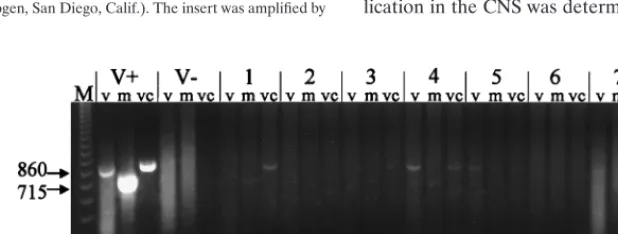
who had died in 1960 and 1978 (Fig. 1 and 2, respectively). In
addition to genomic RNA, mRNA and vcRNA were detected,
indicating that the virus had replicated in the CNS of the
patients. For patient A-78-134, the exact location of viral
rep-lication in the CNS was determined. RNA was extracted from
FIG. 1. RT-PCR detection of LAC virus RNA in CNS tissues of a patient from 1960. Total cellular RNA extracted from CNS tissues was subjected to RT-PCR with primers specific for LAC virus genomic and replicative forms of RNA. V1and V2are control reaction mixtures containing RNA extracted from BHK-21 cells inoculated with LAC virus (V1) or from noninfected BHK-21 cells (V2). Lane groups 1 through 7 contain seven different tissue samples from CNS of patient from 1960. Each group contains material from RT-PCR using primers specific for viral genomic (v), mRNA, or vcRNA. RT-PCRs were performed as described in the text. M, molecular weight markers (123-bp DNA ladder). Numbers on the left are base pairs.
on May 15, 2020 by guest
[image:2.612.145.454.565.682.2]six different areas of the brain; viral RNA was detected only in
the cerebral cortex tissue (Fig. 2). When RNA extracted from
LAC virus-infected BHK-21 cells was subjected to RT-PCR,
specific bands of either 715 or 860 bp were obtained (Table 1;
Fig. 1). No bands were obtained when RNA extracted from
noninfected cells was subjected to RT-PCR (Fig. 1).
Analysis of genetic heterogeneity by using SSCP.
SSCP was
used to investigate genetic heterogeneity between the
proto-type LAC virus and the viral RNA detected in the CNS tissues.
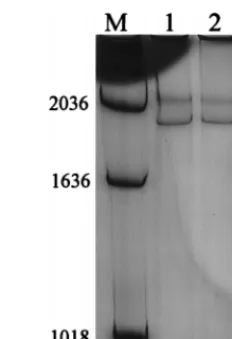
SSCP analysis was performed only on the PCR products
ob-tained with the primer that amplified genomic RNA. All three
viruses yielded the same SSCP pattern (Fig. 3). Thus, the
prototype laboratory virus that had been isolated 30 years
earlier from the original brain specimen assayed in these
ex-periments and had been passaged in the laboratory many times
did not differ substantially from the virus in the 1960 CNS
tissue or from the virus in the 1978 CNS tissue. These results
indicated that although the encephalitis cases occurred 18
years and approximately 50 miles apart, the viruses causing the
infections were genetically similar.
Sequence analysis.
To investigate nucleotide sequence
dif-ferences between the 1960 virus and the 1978 virus from the
CNS tissues, a 668-bp fragment of the S segment was
se-quenced. The laboratory strain of prototype LAC virus was
also sequenced and compared to GenBank sequences of
pro-totype LAC virus S segment (accession no. K00108) to
deter-mine if nucleotide changes occurred during laboratory passage.
Analysis was performed only on the PCR products obtained by
using the primer that amplified genomic RNA. Nucleotide
sequences from the prototype LAC virus (those obtained from
GenBank and from sequencing of laboratory prototype LAC
virus) were identical to those obtained from the 1960 CNS
tissues. Two base changes were seen in the virus from the 1978
case compared to the 1960 sample. One occurred at position
180, resulting in an amino acid change from serine to leucine
in the N protein and a change from histidine to tyrosine in the
NSs protein. A second base change occurred at position 481,
but this did not result in an amino acid change in the N protein
and did not affect the coding of the NSs protein, which
termi-nates at base 377. Thus, although the two encephalitis cases
occurred 18 years and 50 miles apart, the viruses were
genet-ically similar.
Virus isolation and identification.
A total of 32 suckling
mice were inoculated with the 1978 CNS homogenate, and 5
mice showed signs of encephalitis at day 3 or 4 postinoculation.
The brains of these mice were removed aseptically, pooled,
and homogenized in PBS–10% fetal bovine serum. The
ho-mogenate was centrifuged, and the supernatant was aliquoted.
This virus was called passage 1. The mouse-passaged material
was propagated in Vero cells, and identity of the virus isolate
was confirmed as LAC virus by using monoclonal antibody
(data not shown). RNA was extracted from a portion of the
first-passage material, and RT-PCR revealed a specific band,
confirming the identity of the virus as LAC virus (data not
shown). The mouse passage of the virus was also sequenced;
no nucleotide changes were detected.
DISCUSSION
LAC viral RNA was detected by RT-PCR in CNS tissues
from two children who died of encephalitis in 1960 and in 1978.
Although the CNS tissues had been stored frozen at
2
70°C for
37 and 19 years, respectively, viral RNA was readily detected in
both by using RT-PCR. In addition, LAC virus was isolated
from the cerebral cortex samples of patient A-78-134. RT-PCR
may be a more sensitive technique for detection of virus in
CNS tissues than virus isolation, and it is certainly more rapid.
RT-PCR revealed that LAC virus analyte was restricted to the
cerebral cortex. It is possible that other brain tissues were
infected but that extraction of analyte from these tissues did
not yield amplifiable LAC RNA. However, all samples did
contain abundant RNA as determined by spectrophotometry
(data not shown).
[image:3.612.62.282.67.293.2]Thus, the RT-PCR technique has potential as a tool for
diagnosis of LAC virus infections. Because this disease can
FIG. 2. RT-PCR detection of LAC virus RNA in CNS tissues of a patient [image:3.612.369.485.68.238.2]from 1978. Total cellular RNA was extracted from the medulla (M), cerebral cortex (CO), cerebellum (CE), spinal cord (SC), basal ganglion (BG), or tem-poral lobe (T), each containing two sets of tissues subjected to RT-PCR with primers specific for viral genomic, mRNA, or vcRNA as described for Fig. 1. V1 and V2are control reaction mixtures containing RNA extracted from BHK-21 cells inoculated with LAC virus (V1) or from noninfected BHK-21 cells (V2). P and W, plasmid (positive) and water (negative) PCR controls. m, molecular weight markers (123-bp DNA ladder). Numbers in white are base pairs.
FIG. 3. SSCP analysis of PCR products obtained from control RNA (lane 1), the 1960 CNS sample (lane 2), and the 1978 CNS sample (lane 3). M, molecular weight markers. The PCRs were performed with the LNF and LNR primer set (product size5715 bp), which detects genomic RNA. Numbers on the left are base pairs.
on May 15, 2020 by guest
http://jcm.asm.org/
circulating to investigate the genetic basis of emergence,
viru-lence changes, or other important epidemiologic data (27).
Our studies illustrate the potential application of molecular
techniques to the study of LAC viral epidemiology. By using
direct detection of LAC virus RNA in the CNS tissues, rapid
genetic analysis without passage of the virus is possible. This
precludes genetic variation that may occur with
bioamplifica-tion.
The SSCP analyses of the LAC viral S segment RNA
re-vealed that although the two encephalitis cases occurred 18
years and 50 miles apart, the viruses were genetically similar.
Sequence analyses confirmed the SSCP results and revealed
only two nucleotide changes between the two isolates in the
668-bp fragment. Interestingly, one amino acid change
oc-curred in the conserved overlapping reading frame of the S
segment that codes for the N and NSs proteins.
The similarity of the virus sequences was somewhat
surpris-ing, since the cases were separated temporally and spatially. A
similar lack of heterogeneity in the M RNA segment of the
same LAC virus isolates was recently demonstrated (14);
in-deed, these investigators speculated that fatal LAC
encepha-litis may result from infections with a limited range of virus
genotypes. The genome of Ebola virus, which also has a
neg-ative-sense RNA genome, has also been shown to exhibit
re-markable genomic stability over time and space (28). Genetic
stability of other RNA viruses, such as measles virus, has
also been demonstrated (23–26). The epidemiological
sig-nificance of LAC virus genomic stability remains to be
de-termined.
ACKNOWLEDGMENTS
This work was supported by Public Health Service grant AI 19688
from the National Institutes of Health.
The expert technical assistance of Bernard Kalfayan and William
Sweeney is acknowledged.
REFERENCES
1. Beaty, B. J., and C. H. Calisher. 1991. Bunyaviridae—natural history. Curr. Top. Microbiol. Immunol. 169:27–78.
2. Beaty, B. J., C. H. Calisher, and R. E. Shope. 1995. Arboviruses, p. 189–212.
In N. J. Schmidt and R. W. Emmons (ed.), Diagnostic procedures for viral,
rickettsial and chlamydial infections, 7th ed. American Public Health Asso-ciation, Washington, D.C.
3. Beaty, B. J., D. W. Trent, and J. T. Roehrig. 1988. Virus variation and evolution: mechanisms and epidemiological significance, p. 59–85. In T. P. Monath (ed.), The arboviruses: epidemiology and ecology, vol. I. CRC Press, Boca Raton, Fla.
4. Black, W. C., D. L. Vanlandingham, W. P. Sweeney, L. P. Wasieloski, C. H.
Calisher, and B. J. Beaty.1995. Typing of LaCrosse, snowshoe hare, and Tahyna viruses by analyses of single-strand conformation polymorphisms of the small RNA segments. J. Clin. Microbiol. 33:3179–3182.
5. Calisher, C. H. 1994. Medically important arboviruses of the United States
Tsai. 1994. Bunyavirus diseases, p. 439–454. In R. R. McKendall and W. G. Stroop (ed.), Handbook of neurovirology. Marcel Dekker, Inc., New York, N.Y.
13. Hosoya, M., K. Honzumi, and H. Suzuki. 1997. Detection of enterovirus by polymerase chain reaction and culture in cerebrospinal fluid of children with transient neurologic complications associated with acute febrile illness. J. In-fect. Dis. 175:700–703.
14. Huang, C., W. H. Thompson, N. Karabatsos, L. Grady, and W. P. Campbell. 1997. Evidence that fatal human infections with La Crosse virus may be associated with a narrow range of genotypes. Virus Res. 48:143–148. 15. Kappus, K. D., T. P. Monath, R. M. Kaminski, and C. H. Calisher. 1983.
Reported encephalitis associated with California serogroup virus infections in the United States, 1963–1981. A.R. Liss, New York, N.Y.
16. Klimas, R., W. H. Thompson, C. H. Calisher, G. G. Clark, P. R. Grimstad,
and D. H. L. Bishop.1981. Genotypic varieties of La Crosse virus isolated from different geographic regions of the continental United States and evi-dence for a naturally occurring intertypic recombinant La Crosse virus. Am. J. Epidemiol. 114:112–131.
17. Kuno, G., C. J. Mitchell, G.-J. J. Chang, and G. C. Smith. 1996. Detecting bunyaviruses of the Bunyamwera and California serogroups by a PCR tech-nique. J. Clin. Microbiol. 34:1184–1188.
18. McJunkin, J. E., R. Khan, E. C. de los Reyes, D. L. Parsons, L. L. Minnich,
R. G. Ashley, and T. F. Tsai.1997. Treatment of severe La Crosse enceph-alitis with intravenous ribavirin following diagnosis by brain biopsy. Pediat-rics 99:261–267.
19. Mitchell, P. S., M. J. Espy, T. F. Smith, D. R. Toal, P. N. Rys, E. F. Berbari,
D. R. Osmon, and D. H. Persing. 1997. Laboratory diagnosis of central nervous system infections with herpes simplex virus by PCR performed with cerebrospinal fluid specimens. J. Clin. Microbiol. 35:2873–2877.
20. Powers, A. M., K. E. Olson, S. Higgs, J. O. Carlson, and B. J. Beaty. 1994. Intracellular immunization of mosquito cells to La Crosse virus using a recombinant Sindbis virus vector. Virus Res. 32:57–67.
21. Read, S. J., K. J. M. Jeffery, and C. R. M. Bangham. 1997. Aseptic meningitis and encephalitis: the role of PCR in the diagnostic laboratory. J. Clin. Microbiol. 35:691–696.
22. Rhoads, D. D., and D. J. Roufa. 1991. SEQAID II user’s manual, version 3.81. Kansas State University, Manhattan.
23. Rima, B. K., J. A. P. Earle, K. Baczko, P. A. Rota, and W. J. Bellini. 1995. Measles virus strain variations. Curr. Top. Microbiol. Immunol. 191:65– 83.
24. Rima, B. K., J. A. P. Earle, R. P. Yeo, L. Herlihy, K. Baczko, V. ter Meulen,
J. Carabana, M. Caballero, M. L. Celma, and R. Fernandez-Munoz.1995. Temporal and geographical distribution of measles virus genotypes. J. Gen. Virol. 76:1173–1180.
25. Rota, J. S., J. L. Heath, P. A. Rota, G. E. King, M. L. Celma, J. Carabana,
R. Fernandez-Munoz, D. Brown, L. Jin, and W. J. Bellini.1996. Molecular epidemiology of measles virus: identification of pathways of transmission and implications for measles elimination. J. Infect. Dis. 173:32–37.
26. Rota, J. S., P. A. Rota, S. B. Redd, S. C. Redd, S. Pattamadilok, and W. J.
Bellini.1997. Genetic analysis of measles viruses isolated in the United States, 1995–1996. J. Infect. Dis. 177:204–208.
27. Sable, C. A., and G. L. Mandell. 1996. The role of molecular techniques in the understanding of emerging infections. Mol. Med. Today 2(3):120– 128.
28. Sanchez, A., S. G. Trappier, B. W. J. Mahy, C. J. Peters, and S. T. Nichol. 1996. The virion glycoproteins of Ebola viruses are encoded in two reading frames and are expressed through transcriptional editing. Proc. Natl. Acad. Sci. USA 93:3602–3607.
29. Thompson, J. D., D. G. Higgins, and T. J. Gibson. 1994. CLUSTAL W: improving the sensitivity of progressive multiple sequence alignment through sequence weighting, position-specific gap penalties and weight matrix choice. Nucleic Acids Res. 22:4673–4680.
on May 15, 2020 by guest
30. Thompson, W. H., B. Kalfayan, and R. O. Anslow. 1965. Isolation of Cali-fornia encephalitis group virus from a fatal human illness. Am. J. Epidemiol.
81:245–253.
31. Tsai, T. F. 1991. Arboviral infections in the United States. Infect. Dis. Clin. N. Am. 5:73–102.
32. Tyler, K. L. 1994. Polymerase chain reaction and the diagnosis of viral central nervous system diseases. Ann. Neurol. 36:809–811.
33. Vodkin, M. H., T. Streit, C. J. Mitchell, G. L. McLaughlin, and R. J. Novak. 1994. PCR-based detection of arboviral RNA from mosquitoes homogenized in detergent. BioTechniques 17:114–116.
34. Wasieloski, L. P., A. Rayms-Keller, L. A. Curtis, C. D. Blair, and B. J. Beaty. 1994. Reverse transcription-PCR detection of La Crosse virus in mosquitoes and comparison with enzyme immunoassay and virus isolation. J. Clin. Mi-crobiol. 32:2076–2080.