UCSF
UC San Francisco Electronic Theses and Dissertations
Title
Examining Self-directed Multicellular Organization and Morphogenesis of Human Induced Pluripotent Stem Cells
Permalink
https://escholarship.org/uc/item/3f16d7r1
Author
Libby, Ashley Rachel Greeley
Publication Date
2019
by
Submitted in partial satisfaction of the requirements for degree of
in
in the
GRADUATE DIVISION
of the
UNIVERSITY OF CALIFORNIA, SAN FRANCISCO
Approved:
______________________________________________________________________________
Chair
______________________________________________________________________________
______________________________________________________________________________
______________________________________________________________________________
Copyright 2019
By
Dedicated to the Carl Family.
ACKNOWLEDGEMENTS
I have had the unique opportunity to pursue my PhD in a truly supportive environment without which I would not have made it through the rollercoaster of science, emotion, and at times mind numbing work that it took to complete my degree. As such, I have a long list of mentors, fellows, trainees, friends and family to thank for my success.
First, I would like to thank the UCSF Developmental Stem Cell Biology Program as a whole. I moved to California with no contacts in the area and the program and the people within it immediately because my first source of refuge, friendships, and learning. Specifically, my class (Bonnie, Akela, Hanna, Daniel, and Buddy) provided unlimited support early on, staying late at night in the RMB boardroom completing our genetics homework, eating at Kiki’s Sushi every week, and collectively trying to figure out what Tregs were at our first BMS retreat. Beyond my class
the DSCB community is one of both support and adventure, from tubbing trips, to house parties, to camping in Yosemite, I have loved it all, and I will miss the time I got to spend with my fellow students.
I would like to thank my co-advisors Dr. Bruce Conklin and Dr. Todd McDevitt, for their unmatched support of my work over the past 5 years. At the start of my PhD, Bruce accepted me into his lab with the recommendation that I check out Todd’s lab because he thought I would love the collaboration. I cannot thank Bruce enough for not only recognizing my interests, but also providing the push to seek out scientific advisors in areas outside of my expertise. In tangent, I must also thank Todd for never questioning my sudden appearance in his lab and welcoming me with open arms despite at first feeling out of place as a biologist in a lab full of bioengineers. Because of Todd’s unwavering trust, I had the opportunity to start the early development working group in the lab, recruit graduate students to join the “dark side” of developmental biology, and pursue a project that veered away from the lab’s expertise. Bruce and Todd made a perfect co-mentor match, where Bruce loved to think of big grandiose picture agendas, Todd was willing to painstakingly go over the nitty gritty details of my experiments, fellowship, and presentations.
Further, Todd set up collaborations (Demarcus Briers and Calin Belta) that I never would have dreamed of and forced me to step outside of my comfort zone. Over the past five years, under the mentorship of Bruce and Todd, I have learned many key lessons on how to present my science, write clearly, manage my time, and never forget the big picture goals of the science we do in lab. I am truly thankful to have been trainned by Bruce and Todd.
I would like to thank my committee members Dr. Susan Fisher and Dr. Zev Gartner for their intellectual support of my thesis project and future career. Susan and Zev have provided key advice on how to focus my experiments and develop my project. Furthermore, they have been excellent advisors in my future career path, providing seemingly endless letters of recommendation for my seemingly endless fellowships.
I have found throughout my PhD that environment is one of the most important things to the success of a graduate career, and I have had a fantastic one in the Conklin and McDevitt labs and at the Gladstone as a whole. From the beginning of my PhD, I had an insane number of senior graduate students and outside mentors to shape my writing and line of scientific questioning. Specifically, I must thank McDevitt lab senior members (Jenna Wilson, Josh Zimmermann, Tracy Hookway, and Jessie Butts) for covering all my proposals in red ink, painfully teaching me how to write with “strong” language. I must thank fellow graduate students Bonnie Cole and Ariel Kauss for being in my quals boat and making sure it didn’t sink. I want to thank Dr. Polin So in the Conklin lab for answering every question, reading every document, and providing endless amounts of support and advice. Polin has played an unprecedented roll in my development as a developmental biologist and has always been a firm point of reference for the type of scientist I aim to become.
I would like to thank my collaborators Drs. Calin Belta, Iman Haghighi, and Demarcus Briers. Specifically, Demarcus for spending hours every week on skype calls with me as we perfected the ins and outs of our computational model. It is because of this collaboration that I
in the future. Despite being across the country from one another, only ever meeting in person three times, and Demarcus and Iman graduating, we made it work. Thank you for teaching me how to be a good collaborator. It is a vital skill that I will take with me and it was an honor to learn from Demarcus.
As my PhD continued the McDevitt lab (McMurder) was a continued resource of support and fun far beyond just academic science. I know intricate details of baking pie crust (David Joy), crafting the perfectly toasted crème brulé (Mike Kang), and endless recipes for a variety of Indian dishes (Vaishaali Natarajan). While my stomach was full, Oriane Matthys ensured that I always worked it off between spin class or swimming, and Ana Silva made sure that I always had a song to hum even if I couldn’t dance like her. Further, everyone’s enthusiasm for Halloween (my favorite holiday) has painted my PhD with so many happy moments, even if I will never forgive Ivana Vasic for skipping our Halloween party.
I would like to thank all of the interns (Jocelyn Cervantes), research associates (Nik Mendoza-Camacho and Martina (Marty) Krakora) and graduate students (Ivana Vasic, Jasmine King, Emily Bulger, Nick Elder) that for whatever reason Todd thought it was a good idea to let me train. Jocelyn Cervantes, my first intern, whose time in lab was marked by sticky notes strewn across my desk, taught me how to teach science and manage two people’s time. Nik with his magic cloning hands and Marty with her crazy coding skills taught me that a little bit of sass can go a long way. All the graduate students that I have helped train in lab, have taught me the necessity of peripheral vision as they hovered by my desk with their latest problem. They constantly kept me on my toes with infinite questions ranging from “where is X reagent?” to “Do we really believe the French Flag Model?” to “Ashley, why is my differentiation breaking?”. I am truly sorry that I will not get to see the continuation of their development as scientists, but it has been an honor training them to this point. Overall, mentorship and helping others become excited about science has been one of the highlights of my PhD and solidified my drive to become a professor in the future.
I would like to specifically thank the early development team in the McDevitt lab (David Joy and Ivana Vasic) who took the leap of faith and joined me in a new section of the lab. Technically, David was the first student I ever trained, which for anyone that knows David now, it seems laughable that he was ever at intro level in anything. It has been an incredible experience working with David, who in return for cell culture knowledge taught me how to code, grown my appreciation for dinosaur t-shirts, and tackled every side project Todd threw at us, all while complaining about how we need more robots in lab. He is the most talented person I know, and I can’t express the gratitude I have for his constant support in lab. I would not have a PhD without him. I also want to thank Ivana, who completed our early development family. Anyone that meets Ivana will immediately be taken in by her bubbly presence. She forced David and I to leave behind our introverted nature as every day we were dragged into the conference room to once again argue over the details of embryonic development. Her frustration with the limits of our knowledge and drive to overcome them has been one of the highlights of working with her. All in all, there are no words that truly capture how important working in this team has been to me over the course of my PhD, and I am truly grateful to both David and Ivana.
Finally, I have to thank those who have kept me going outside of lab. I have relied on weekly DND sessions (Tawny, Hanna, Nate, Bonnie, Steph, Rebecca, Dan, and Natanya) to both keep my imagination alive and give me something to look forward to at the end of the day. I want to thank Black Hound Equestrian for giving me a place to sneak off to with fuzzy animals to pat. Particularly, my trainer Kate Jutagir and fellow riders Katie and Mareva, with whom I spent every Saturday morning riding for the past 5 years. I want to thank my family, who have supported me from across the country, providing endless emotional support even if they had no idea what I was doing. I want to thank the Kirby family for taking me in every Thanksgiving and providing an adoptive family away from home. I need to thank my high school friends Lynn Pincus, Elizabeth Aspinwall, and Marissa Edwards for always cheering me on and cheering me up. And finally, and
have not only been my roommates but have also truly become my family. They have welcomed me home every day and lived through the highs and lows of this degree with me. We have gone on endless adventures in the Bay Area (Mt. Tamalpais, Jane bread, Cool Patch Pumpkins, and Kirby Cove), California (Yosemite, Big Sur, and San Diego), and beyond (Hawaii, Japan (coming soon)). They have given me refuge, reminded me to laugh, and encouraged me to reach for the stars. I will always consider them my Ohana.
Contributions
The texts of Chapter 2, 3, and 4 are reprints of the material as it appears in “Spatiotemporal mosaic self-patterning of pluripotent stem cells using CRISPR interference”, “Automated Design of Pluripotent Stem Cell Self-Organization”, and “Axial Elongation of Caudalized Human PSC Organoids Mimics Neural Tube Development
”
. The co-author listed in these publications directed and supervised the research that forms the basis of this dissertation/thesis.Examining Self-directed Multicellular Organization and
Morphogenesis of Human Induced Pluripotent Stem Cells
Ashley Rachel Greeley Libby
Abstract
Embryonic morphogenesis is a critical determinant of tissue generation and function, yet many of the mechanisms that regulate morphogenic processes remain elusive due to the complex and dynamic nature of multicellular interactions and the limited tools to manipulate these systems at single cell resolution. Similarly, morphogenesis of human induced pluripotent stem-cell (hiPSC) derived organoids proceeds largely through self-organized pattern formation that crudely mimics organogenesis. The ability to control specific morphogenic processes would greatly enhance our ability to create bona vide human tissue and organ structures; however, robustly directing organoid morphogenesis requires development of novel control approaches. The four studies described in this dissertation developed systems to study pattern emergence and lineage fate specifications in hiPSC 2D and 3D cultures. First, the effects of mechanical regulation on population emergence and lineage fate specification was interrogated by mosaic knockdown of two mechanical regulators associated with cortical tension and cell-cell adhesion: Rho-associated kinase-1 (ROCK1) and E-cadherin (CDH1). Mosaic knockdown induced symmetry breaking (a pre-requisite of morphogenesis), which resulted in differential patterns of cell sorting and multicellular organization without disrupting pluripotency state. These results describe the spatiotemporal dynamics of multicellular self-organization that occur with the emergence of heterotypic cell populations within pluripotency allowing for population segregation before lineage fate commitment. Second, using machine learning, experimental parameters were optimized to yield pre-specified spatial patterns before successful validation in vitro with hiPSCs. Furthermore, asymmetric pre-patterning of iPSCs differentially impacted initial germ layer specification within differentiating colonies. This study demonstrated demonstrate that parameter optimization
achieved by automated machine learning could be used to efficiently predict and control hiPSC self-organization. Third, the regulation of CDH1 on organization and lineage fate was examined in 3D gastruloid cultures, where both microenvironment and CDH1 knockdown triggered the emergence of extra embryonic lineages. This study indicated that microenvironment is essential for specific lineage emergence in the early embryo. Fourth in the final study, population emergence was examined in neuronal organoid cultures that displayed axial extensions similar to neural tube formation and extension. Extending aggregates were dependent on the emergence of a neuromesodermal progenitor population alongside neuronal populations. This model of neural tube extension provides a platform to probe how disruption of population emergence affects morphological phenotypes. Overall, these studies provided avenues to elucidate the underlaying multicellular interactions that enable the development of complex patterning events within the developing human embryo on multiple scales and across multiple developmental periods.
TABLE OF CONTENTS
Chapter 1 : Introduction 1
1.1 Overview 1
1.2 Symmetry Breaking in Early Development 4
1.2.1 Compaction 4
1.2.2 Gastrulation and Axis Formation 5
1.2.3 Spinal Cord Development 6
1.3 Stem Cell Modeling of Embryonic Development 8
1.3.1 Tri-lineage Monolayer Differentiations 9
1.3.2 Organoids 10
1.3.3 CRISPRi as a Tool to Induce Asymmetry 11
1.4 Mechanical Regulation of Patterning 13
1.4.1 Cell-cell Adhesion Regulation of Morphogenesis 13 1.4.2 Cortical Tension Regulation of Morphogenesis 17 1.5 Computational Modeling of Cellular Behaviors 19
1.5.1 Agent Based Models 20
1.5.2 Cellular Potts Model 21
1.5.3 Pattern Classification, Machine Learning, and Optimized Pattern Prediction 22
1.6 Concluding Remarks 23
1.7 Bibliography 25
Chapter 2 : Spatiotemporal mosaic self-patterning of pluripotent stem cells using
CRISPR interference 45
2.1 Introduction 45
2.2 Materials and Methods 46
2.2.1 Human iPSC line generation and culture 46
2.2.3 Western blot 49
2.2.4 RNA isolation and qPCR 49
2.2.5 Atomic Force Microscopy 50
2.2.6 Time-lapse imaging 50
2.2.7 Immunofluorescence staining 51
2.2.8 EDU Incorporation Assay 51
2.2.9 FLOW Analysis 52
2.2.10 FACS 52
2.2.11 Fluidigm 96.96 Array 52
2.2.12 Differentiations 55
2.2.13 Computational Image Analysis 56
2.2.14 Statistics 56
2.3 Results 57
2.3.1 CRISPRi KD in human iPSCs modulates epithelial morphology 57 2.3.2 Mosaic CRISPRi silencing results in multicellular organization 61 2.3.3 Mosaic human iPSC colonies retain a pluripotent phenotype 66 2.3.4 Mixed populations of KD cells display transient gene expression changes in
coordination with emergence of patterns 68
2.3.5 Mixed populations allow for restricted germ lineage emergence 73
2.4 Discussion 74
2.5 Conclusion 81
2.6 Bibliography 82
Chapter 3 : Automated Design of Pluripotent Stem Cell Self-Organization 87
3.1 Introduction 87
3.2.2 Generation of CRISPRi knockdown iPSC lines 90
3.2.3 Mixed Colony Generation 92
3.2.4 Immunofluorescence Staining and Imaging 92
3.2.5 Protein Quantification 93
3.2.6 mRNA quantification 93
3.2.7 Time Lapse Imaging 95
3.2.8 Comparison of in vitro and in silico Spatial Patterns 95
3.2.9 BMP4 differentiations 98
3.2.10 Cellular Potts Model Environment 98
3.2.11 Cellular Potts Model Dynamics 100
3.2.12 Physical Units and Other Cellular Phenomena of Cellular Potts Model 103
3.2.13 Model Fitting to Empirical Data 103
3.2.14 TSSL Scoring and Pattern Optimization 110
3.2.15 Quantification and Statistical Analysis 118
3.2.16 Software 118
3.2.17 Data Availability 119
3.3 Results 119
3.3.1 Pattern Synthesis: in silico prediction and automated discovery of spatial
behaviors 119
3.3.2 Data-Driven Computational Model of Human iPSC Self-Organization 120 3.3.3 Formulating Parameters for Design Automation 124
3.3.4 Quantitative Pattern Classification 126
3.3.5 Automated Discovery of Pattern Producing Conditions 127 3.3.6 In Silico Model Accurately Predicts In Vitro Experimental Validation 131 3.3.7 Colony Organization Impacts Patterns of iPSC Differentiation 137
3.5 Conclusion 141
3.6 Bibliography 142
Chapter 4 : Loss of E-cadherin promotes extraembryonic fates accompanying
multilineage differentiation 148
4.1 Introduction 148
4.2 Materials and Methods 149
4.2.1 Human Induced Pluripotent Stem Cell Culture 149
4.2.2 Encapsulated Mixed Aggregate Generation 150
4.2.3 Un-encapsulated Mixed Aggregate Generation 150
4.2.4 Immunofluorescence Staining and Imaging 151
4.2.5 Whole Mount Lightsheet Imaging 151
4.2.6 Single Cell RNA Sequencing Sample and Library Preparation 152 4.2.7 Genome Annotation, RNA-seq Read Mapping, and Estimation of Gene and
Isoform Expression 153
4.2.8 Data Acquisition, Processing, and Merging
153
4.2.9 Cluster Analysis 154
4.3 Results 154
4.3.1 Loss of CDH1 Promotes Protrusion Morphology when Encapsulated 154 4.3.2 Unencapsulated and Encapsulated CDH1(-) Aggregates Display Differing
Morphologies 155
4.3.3 Emergence of Extraembryonic-like Cell Fates in Encapsulated Aggregates 158 4.3.4 Human iPSC Aggregates Have Similar Transcriptomes to Pre-implantation
Human Embryos 159
4.4 Discussion 161
4.6 Bibliography 164 Chapter 5: Axial Elongation of Caudalized Human PSC Organoids Mimics
Neural Tube Development 170
5.1 Introduction 170
5.2 Materials and Methods 171
5.2.1 Human Induced Pluripotent Stem Cell Line Generation and Culture 171
5.2.2 Organoid Differentiation 172
5.2.3 Organoid Elongation Imaging and Quantification 173 5.2.4 Real Time Quantitative Polymerase Chain Reaction 175 5.2.5 Histology, Immunocytochemistry and Imaging 176
5.2.6 Whole Mount Lightsheet Imaging 177
5.2.7 Single Cell RNA Sequencing Sample and Library Preparation 177 5.2.8 Genome Annotation, RNA-seq read mapping, and Estimation of Gene and
Isoform Expression 178
5.2.9 Cluster Analysis 179
5.2.10 RNAScope 179
5.2.12 Statistical Analysis 180
5.3 Results 180
5.3.1 Emergence of Axial Extensions from Neuronal Organoids 180 5.3.2 Wnt Mediated Increase in Extensions and SOX2(+)T(+) Progenitor
Populations 185
5.3.3 Regionalized HOX Gene Expression in Extending Aggregates 185 5.3.4 Manipulation of Endogenous BMP Signaling by CRISPR Interference 189
5.4 Discussion 190
5.6 Bibliography 192
Chapter 6: Future Considerations 197
6.1 CRISPRi Colony Pattern Formation Screens 198
6.2 Expanding CRISPRi Perturbations in the Cellular Potts Model 199 6.3 Interrogating Hippo Signaling in Pluripotent Stem Cell Transitions 201 6.4 Examining Neuromesodermal Progenitor Fate Regulation 202
6.5 Conclusions 204
LIST OF FIGURES
Figure 2.1: Maintenance of normal human iPSC karyotypes 48
Figure 2.2: CRISPRi of ROCK1 and CDH1 modulate physical properties of the cell 59 Figure 2.3: Protein knockdown time course for ROCK1 and CDH1 60
Figure 2.4: Morphology of ROCK1 knockdown human iPSCs 60
Figure 2.5: Maintenance of Tight junctions in knockdown human iPSCs 61 Figure 2.6: Cell-autonomous pattern emergence in mixed population colonies 63 Figure 2.7: Pattern emergence of variable ratios of mixed populations 64
Figure 2.8: Cell population change over time 65
Figure 2.9: Maintenance of nuclear pluripotency markers and epithelial phenotype 67 Figure 2.10: Maintenance of cell junction localized β-catenin in knockdown cells 68 Figure 2.11: Pluripotent and early germ layer gene expression in knockdown cells 69 Figure 2.12: Transient gene expression changes in mixed populations 71 Figure 2.13: Gene expression changes in WT-GFP cells mixed with CRISPRi cells 72 Figure 2.14: Germ lineage differentiations of mixed colonies 75 Figure 2.15: Inducible pattern emergence through the KD of molecules that affect
human iPSC physical properties 76
Figure 3.1: Overview of Synthesis of Spatial Patterns 89
Figure 3.2: Karyotype of CRISPRi Gen2 CDH1 hiPS Cell line 91 Figure 3.3: Knockdown of CDH1 quantified by immunofluorescence imaging 94 Figure 3.4: Characterizing in vitro morphological metrics 96
Figure 3.5: Segmentation workflow 97
Figure 3.6: CRISPRi cell line mRNA knockdowns 109
Figure 3.7: Pairwise experiments to characterize dynamic changes in spatiotemporal
Figure 3.8: Quantitative Pattern Classifier with TSSL 128
Figure 3.9: TSSL increase with pattern formation 131
Figure 3.10: Computational synthesis of de novo spatial patterns and in vitro validation 134 Figure 3.11: In silico vs. in vitro comparative metrics of generated target patterns 135 Figure 3.12: Titration of gene knockdown mixing ratios in vitro and in silico 136
Figure 3.13: Differentiation of Bullseye colonies 139
Figure 4.1: Encapsulated 50 cell human iPSC aggregate morphologic changes 156 Figure 4.2: Unencapsulated human iPSC 50 cell aggregate morphologies 157 Figure 4.3: Single cell sequencing analysis of encapsulated and unencapsulated
aggregates 160
Figure 4.4: Lineage trajectories of 50 cell aggregates 161
Figure 4.5: Comparison of in vitro aggregate transcriptome with preimplantation human
embryos 162
Figure 5.1: Emergence of axial extensions from neuronal organoids 182
Figure 5.2: Aggregate extension phenotypes 183
Figure 5.3: Wnt mediated increase in extensions and SOX2(+)T(+) progenitor
population 184
Figure 5.4: Progenitor and HOX gene expression in extending and non-extending
aggregates 187
Figure 5.5: Gene expression analysis of extending and non-extending aggregates by
single cell RNA sequencing 188
LIST OF TABLES
Table 2.1: CRISPRi gRNA sequences 47
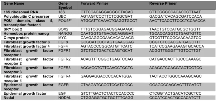
Table 2.2: qPCR primer sequences 53
Table 2.3: Fluidigm primer sequences 53
Table 3.1: List of design parameters that map to experimental perturbations 104 Table 3.2: Model fitting parameters for the Space vs. Time, Protein Expression vs.
Time, and the Protein Expression vs. Space characterizations 104
Table 5.1: CRISPRi guides 172
Table 5.2: qPCR primers 175
Table 5.3: Antibodies 177
Chapter 1: Introduction
1.1 Overview
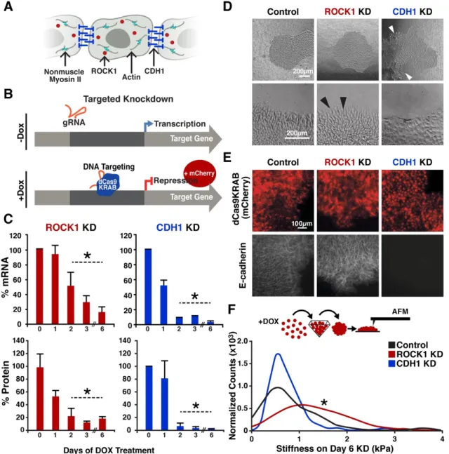
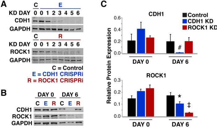
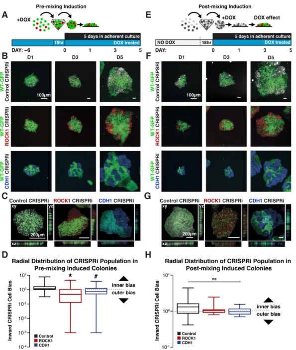
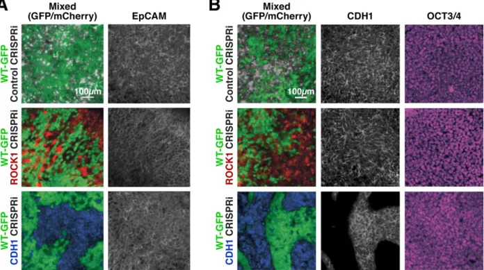
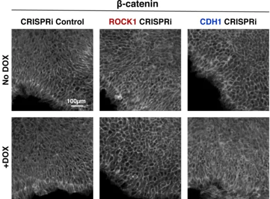
The goal of this dissertation was to interrogate mechanisms behind the symmetry breaking events that regulate population emergence and organization within the developing embryo. Symmetry breaking is an overarching term used in developmental biology to describe instances in the development of complex organisms when organizational events occur that divide what was once a homogeneous population into separate heterogeneous populations. The emergence of heterogeneity can take the form of changes in gene expression, changes in cell shape, physical movement into patterns, etc. However, often developmental symmetry breaking events involve a wide variety of complex genetic, behavioral, and morphologic changes at once, making it difficult to interrogate the cause of the initial emergent heterogeneity. Therefore, this dissertation outlines multiple methods in the form of four studies to interrogate aspects of symmetry breaking events using human induced pluripotent stem cells (iPSCs) as a model for early embryonic development. The goal of the first study presented in this dissertation was to develop a system in human iPSCs that would enable robust control over symmetry breaking events. Thus, an inducible CRISPR interference (CRISPRi) system was used in mixed populations of human iPSCs where only a portion of the colony contained the CRISPRi machinery. Then, with CRISPRi induction, a relatively homogeneous human iPSC population would undergo controlled symmetry breaking as only a subpopulation experienced genetic knockdown. This platform was used to interrogate mechanical regulators of population organization: the cell-cell adhesion molecule E-cadherin (CDH1) and the tension regulator Rho-associated coiled-coil kinase (ROCK1). Interestingly, mosaic knockdown triggered population organizations within human iPSC colonies without disrupting pluripotency. When patterned colonies were then differentiated, cells lacking CDH1
preferentially differentiated to the mesendoderm when in mixed colonies, while cells lacking ROCK1 were able to differentiate to all germ lineages equally. This study provided a unique method to control symmetry breaking events and demonstrated the importance of adhesion driven symmetry breaking in the regulation of lineage fate decisions.
With the development of a system to genetically control sub-population emergence, the goal of the second study was to apply the behaviors observed in the symmetry breaking in vitro studies to a computational model capable of predicting novel patterning events and organizations of human iPSCs within colonies. Combining a Cellular Potts-based computational model and a pattern recognition framework, a computational system to predict specific experimental parameters was created to derive patterning events upon induction of symmetry breaking. Executing the in silico derived experimental set up in vitro resulted in multicellular population organization that remarkably reflected the in silico predictions in both frequency and extent of pattern formation. Furthermore, differentiation of patterned human iPSC colonies resulted in divergent patterned cell fate commitment upon morphogen exposure, indicating that directed multicellular organization impacts lineage co-emergence. This study demonstrated that morphogenic dynamics could be predicted in silico to accurately manipulate human iPSCs in vitro to produce desired morphogenetic events, a critical first step towards robustly engineering more complex tissues.
After establishing control over symmetry breaking events and subsequent human iPSC organization in monolayer culture, the goal of the third study was to interrogate the regulation of symmetry breaking through changes in adhesions in a 3D space to better mimic multi-dimensional organization of embryonic development. Specifically, compaction of the embryo from the morula to the early blastocyst and subsequent gastrulation involves a wide variety of multi-dementional population organizations and migrations that are difficult to recapitulate in monolayer cultures. Therefore, the previously described mosaic knockdown of CDH1 was initiated in 50 cell human iPSC 3D aggregates encapsulated in an alginate hydrogel. With mosaic CDH1 knockdown
population segregation observed and further complete knockdown of CDH1 triggered emergent morphologies reminiscent of embryonic stages. Transcriptome analysis of encapsulated and unencapsulated aggregates revealed performed emergence of all three embryonic germ lineages without exposure to external morphogens. Moreover, the presence of an extraembryonic-like population in only the encapsulated aggregates indicated that micro-environment may play a role in generating cues for lineage emergence. Overall, this study demonstrates an organoid culture system mimicking aspects of both morphogenic arrangements and cellular lineage fates that arise in the early embryo.
Moving beyond the initial symmetry breaking events in the embryo such as compaction and gastrulation, the final study used the three dimensional culture techniques from the third study to interrogate more complex symmetry breaking in the form of body axis extension. An in vitro organoid model of axial extension was developed from a previously reported human iPSC interneuron differentiation protocol. The resultant organoids recapitulated a large number of morphologic and gene expression features found in in vivo axial elongation, forming internal tube-like epithelial compartments. Additionally, these organoids contained cells simultaneously expressing brachyury(T) and SOX2 suggesting the emergence of neuromesodermal progenitors (NMPs), which in vivo contribute to both the closing neural tube and paraxial mesoderm. Elongations increased in a dose dependent manner with the addition of the small molecule Wnt agonist CHIR, recapitulating the in vivo role of Wnt signaling in caudalization and NMP emergence. Single cell RNA sequencing revealed increased MIXL1(+) paraxial mesoderm populations in extending aggregates. HOX gene expression was regionalized in extending aggregates, with hindbrain (HOXB1) expression at the base, while brachial (HOXC6) and thoracic (HOXB9) were expressed in the extensions. To explore dorsoventral polarity in the organoids, knockdowns of BMP inhibitors Noggin and Chordin were generated using CRISPR interference, yielding increased expression of dorsal neural fates in inhibited lines due to loss of endogenous
system. Further, it demonstrates the power of human iPSC organoid models to elucidate many of the underlaying multicellular interactions that drive symmetry breaking enabling the development of complex human tissues.
This dissertation provides the first description of controlled symmetry breaking events in human iPSC culture, a quantitative computational model capable of predicting human iPSC patterning events, and a description of multiple unique organoid systems that highlight morphogenic symmetry breaking events. Furthermore, it interrogates mechanisms by which adhesions regulate lineage emergence in early embryogenesis, enabling the future study of the cumulative mechanisms by which robust morphogenesis is controlled. The following introduction provides the necessary background on topics critical to this work including, symmetry breaking in early embryonic development, stem cell modeling of embryonic development, mechanical regulation of patterning, and computational modeling of cellular dynamics.
1.2 Symmetry Breaking in Early Embryonic Development
Symmetry breaking in biological systems occurs with the emergence of a heterogeneous subpopulation, most commonly undergoing changes in cellular polarity, gene expression, replication, or cellular morphology. In embryonic development, symmetry breaking events characterize the repeated emergence of different cell fates and dictate the creation of the complex structures that comprise embryonic tissues. The following sections describe key events in early mammalian development that rely on symmetry breaking events and subsequent organization to regulate embryo morphogenesis as it transitions between stages.
1.2.1 Compaction
One of the earliest symmetry breaking events in the developing embryo occurs at compaction where a subpopulation of cells in the morula becomes transcriptionally distinct,
leading to morphogenic reorganization to form the blastocyst. The blastocyst is divided into two structurally distinct tissues: an outer layer of polarized trophectoderm cells which surrounds a cavity called the blastocoel, and inner layer of non-polar cells called the inner cell mass (Gueth-Hallonet and Maro 1992). The emergence of the trophectoderm and distinction from the inner cell mass depends on the co-expression of the transcription factors CDX2 and GATA3 (Ralston et al. 2010, 2; Niwa et al. 2005; Strumpf et al. 2005, 2) regulated by the farther upstream transcription factor TEAD4 where embryos lacking TEAD4 fail to produce trophectoderm or develop the blastocoel cavities (Nishioka et al. 2008; Yagi et al. 2007).
In preimplantation mouse embryos, changes in TEAD4 signaling are largely regulated by the Hippo pathway where nuclear accumulation of Yes-associated protein 1 (YAP1) directs TEAD4’s localization within the genome, regulating its transcriptional activity (Hirate et al. 2012; Nishioka et al. 2009). Interestingly, changes in YAP1 nuclear accumulation are linked to changes in cell-cell adhesions and subsequent cellular polarity as the embryo grows in size and cell number (Hirate et al. 2013; Mihajlović and Bruce 2016). Furthermore, YAP1 nuclear accumulation has been shown to be mechanically responsive; changes in ECM stiffness as well as tension across cellular junctions can drive nuclear localization of YAP1 (Benham-Pyle, Pruitt, and Nelson 2015; Dupont et al. 2011), suggesting that the physical rearrangement of cells during compaction may contribute to trophoblast fate specification. However, the exact relationship between changing adhesions and trophoblast specification is unclear.
1.2.2 Gastrulation and Axis Formation
The next symmetry breaking event that occurs in development is formation of the epiblast and the primitive endoderm, followed by gastrulation of the late blastocyst, which also relies on changes in polarity and adhesion (Montero and Heisenberg 2004). The late blastocyst is composed of two compartments: the epiblast, which will form the embryo proper, and the primitive
from a relatively homogeneous population of epithelial pluripotent cells to a spatially organized, multicellular tissue of distinct progenitor cells. This occurs when cells within the epithelial layer begin to express mesenchymal transcription factors like SNAIL and Brachyury, delaminate from the epithelial layer, and invade into the surrounding tissue forming the primitive streak (Tada, Concha, and Heisenberg 2002; Montero and Heisenberg 2004; Tam and Behringer 1997). The delamination of cells not only delineates a change in fate to different mesodermal populations, but it is also accompanied by changes in adhesion as cells change from epithelial to mesenchymal phenotypes and migrate across the embryo. With movements associated with gastrulation comes the formation of the body axis where the primitive streak delineates the dorsal side of the body forming the dorsal-ventral axis as well as establishing the anterior-posterior axis with its direction of extension and inherently the left-right body axis (Beddington and Robertson 1999). However, the specific dynamics of these processes are not well established due to gastrulation occurring post implantation in many model mammalian systems making its direct examination difficult.
1.2.3 Spinal Cord Development
As the embryo continues to develop past gastrulation, multiple tissues arise and begin to morphologically shape the embryo, leading to anisotropic morphologies. For example, the spine, a crucial structure that both enables physical support as well as protection of essential neural projections that connect the body to the brain, begins to develop at gastrulation. Dysregulation of the key processes leading to its formation results in several congenital abnormalities (Kaplan, Spivak, and Bendo 2005). The generation of the spine patterns and elongates the anterior-posterior body axis and specifies the tissues that contribute to the spinal cord (Steventon et al. 2016; Wilson, Olivera-Martinez, and Storey 2009; Schiffmann 2006; Yamaguchi 2001). Although neural tube formation and axial extension have long been studied in model organisms such as chick and amphibians, it is complicated to study the multi-cellular interactions that drive this axial extension at high spatiotemporal resolution in mammalian embryos because it occurs
post-implantation (Viebahn 1999; Beddington and Robertson 1999). The following subsection outlines our current understanding on how neural tube patterning is established.
The process of neural tube formation, or neurulation, is characterized by thickening and flattening of the neural plate in response to signals from the notochord, followed by folding inward ventrally and finally pinching off at the top of the fold to close the tube (J. L. Smith and Schoenwolf 1989; Tam and Behringer 1997). Coincident with neural tube closure is extension of the anterior-posterior axis as the entire embryo breaks symmetry and elongates. Concomitant with elongation is the establishment of different progenitor zones within the neural tube creating both ventral-dorsal and rostral-caudal patterning of the neural tube. These patterning events rely on a combination of morphogen gradients across the embryo that establish the proper signaling milieu to determine progenitor fate based on dorsal-ventral or rostral-caudal location within the developing neural tube. Dorsal-ventral gradients differences in sonic hedgehog (Shh) released ventrally from the notochord and bone morphogenic proteins (BMP) released dorsally from the roof plate (Ericson et al. 1997; Liem et al. 1995; McMahon et al. 1998). Patterning the rostral-caudal axis of the embryo is the Wingless-type MMTV integration site family (Wnts), retinoic acid (RA) released from the neighboring somites, and fibroblast growth factors (FGFs), which dictate convergent extension, progenitor proliferation, and have been shown to regulate HOX gene profiles (Janssens et al. 2010; Carpenter 2002; Corral and Storey 2004; del Corral et al. 2003; Bel-Vialar, Itasaki, and Krumlauf 2002).
As the neural tube continues to extend, an additional axial pool of progenitors, called neuromesodermal progenitors (NMPs), contributes to both the neural tube and the surrounding paraxial mesoderm that eventually becomes that somites (Henrique et al. 2015). This axial stem cell pool of NMPs and are marked by high expression of the transcription factors SOX2 and Brachyury (Henrique et al. 2015; Gouti et al. 2014) and reside in the caudal lateral epiblast just below the closing neural tube. Interestingly, this population of cells is hypothesized to regulate
cord and vertebrae. However, due to NMP location in the post implantation embryo making visualization difficult, the dynamics of NMP symmetry breaking that regulate the production of two different tissues are largely unknown, leaving multiple unanswered questions about their maintenance, differentiation, patterning, and subsequent regulation over axis extension.
Overall, spinal cord development provides multiple examples of symmetry breaking. First, morphogenic symmetry breaking occurs with physical elongation of the embryonic axis through neural tube closure and extension. Second, symmetry breaking is observed in coordinated population emergence and patterning of neuronal subtypes specific to location within the developing neural tube. Finally, the maintained progenitor pools like NMPs continuously demonstrate regulation over population emergence as the embryo continues to develop providing a constant source of new tissue through symmetry breaking lineage fate decisions.
1.3 Stem Cell Modeling of Embryonic Development
The repeated relevance of symmetry breaking events in the development of the early embryo and the health implications of their dysregulation in human congenital diseases (congenital heart disease, spinal malformations, etc.) highlights the importance of mechanistically understanding these basic processes across embryonic development (Kaplan, Spivak, and Bendo 2005; Srivastava 2000). However, the dynamics of development have typically been difficult to interrogate in vivo due to the physical restrictions, optical opacity, and complex signaling milieu inherent to the developing embryo. Furthermore, human specific embryonic processes are largely unknown as using human embryos to study morphogenic events presents ethical dilemmas. Therefore, to study symmetry breaking events and morphogenesis, it is essential to establish an in vitro human system that promotes the coincident development of analogous heterogeneous populations.
Human pluripotent stem cells (hPSCs) provide an unlimited source of cells that can mimic developmental differentiation processes and maintain the ability to self-organize into tissue-like structures, such as optic cups, gut organoids, or stratified cortical tissues (Eiraku et al. 2008; 2011; Spence et al. 2010). Human pluripotent stem cells are split into two main categories: embryonic stem cells (ESCs) and induced pluripotent stem cells (iPSCs). ESCs are isolated from the inner cell mass of embryos and maintain a cellular state of pluripotency, meaning that they have the ability to both renew themselves and differentiate to all three germ lineages (Trounson 2001). Similarly, iPSCs, which are cells reprogramed from fibroblasts by activating the pluripotent gene regulatory network, demonstrate the ability to both renew and differentiate to all three germ lineages (Yamanaka 2012; Takahashi et al. 2007). However, despite the inherent ability of both ESCs and iPSCs to form multiple cell types in 2D culture or in 3D organoids, stem cell differentiations are intrinsically variable (Bredenoord, Clevers, and Knoblich 2017). Historically, differentiations that mimic symmetry breaking at gastrulation, allowing for the emergence of all three lineages, have been in the form of embryoid bodies. These models are inherently random, difficult to robustly reproduce, and do not recapitulate body plan formation during embryonic development (Kurosawa 2007; Carpenedo, Sargent, and McDevitt 2007). As a result of this variability and the lack of alternative human models that faithfully promote asymmetric emergence, many of the mechanisms that control and coordinate human morphogenesis remain undefined. Therefore, new approaches to reliably control the emergence and organization of multiple cell types would greatly advance tissue modeling and organ developmental studies.
1.3.1 Tri-lineage monolayer differentiations
Monolayer stem cell differentiations have been developed for a wide variety of cell types and range in their purity and reproducibility (Butts et al. 2017; Lian et al. 2012; Spence et al. 2010; Gouti et al. 2014). Often, these differentiations rely on the use of either morphogens or small
cell lineage fate decisions to the desired cell type. However, even with external signals directing cell fate, differentiations in monolayer culture often produce a heterogenous mixture of cells that remain largely unorganized, despite the emergence of multiple cell types. These differentiations do not reflect the organized tissue patterning observed within the developing embryo. To address this, recent techniques have been developed that allow for both multi-population emergence as well as organization of multiple cell types in the form of tri-lineage differentiations which mimic aspects of both population emergence and patterning as in the early embryo.
Patterned tri-lineage differentiations can be achieved by stimulating monolayer cultures of PSCs on micropatterned surfaces that restrict colony boundaries (Warmflash et al. 2014). When morphogens present at gastrulation (BMPs, Wnts, Activin) are added to cultures with such boundary conditions, radial patterns of specification emerge: first an outer ring of CDX2(+) cells forms, followed by an inner ring of brachyury(+) cells, and a center of SOX2(+); marking an extraembryonic-like population, a mesendoderm population and a ectoderm population, respectively (Warmflash et al. 2014; Tewary et al. 2017; James et al. 2005; Britton et al. 2019). These studies demonstrate that PSCs have an intrinsic ability to undergo symmetry breaking and self-organize when presented with boundary conditions. However, despite the ability to generate patterns of cells, these systems only produce radial organization events, leaving many unanswered questions about how a radially symmetrical embryo at the morula or blastocyst stage can robustly generate anisotropic patterning events such as the primitive streak or axial elongation.
1.3.2 Organoids
An additional example of symmetry breaking and patterning in vitro, is the creation of organoid models where PSC differentiations are conducted in three dimensional environments, yielding multiple cell types that spatially organize into structures reminiscent of embryonic tissues (Bredenoord, Clevers, and Knoblich 2017). Such organoid models have been established for
multiple organ systems such as gut, brain, and liver (Lancaster and Knoblich 2014; Clevers 2016). In these systems, PSCs or progenitors are exposed to signaling cues which drive lineage fates toward specific tissues; a signaling milieu which is reinforced by endogenous signaling between cells. A combination of these exogeneous and endogenous signals allows for the emergence of the many cell types involved in that tissue’s structure and function. Organoids offer potential in both therapeutic research and basic scientific discovery as they can be used to recapitulate elements of development and disease, offering opportunities that range from mechanistic interrogation of biological processes to drug testing. However, a major limitations of organoid models is the wide variety of phenotypes that are generated from a single differentiation. Structural organization within organoids systems is difficult to both control and repeat (Bredenoord, Clevers, and Knoblich 2017). This lack of precision is highly contrast to the robustness of embryonic development and organogenesis where generation of body plan and tissue structure is tightly regulated. Therefore, engineered platforms that control symmetry breaking and organization would facilitate the mechanistic study of the biological processes that regulate morphogenesis.
1.3.3 CRISPRi as a Tool to Induce Asymmetry
A hallmark of symmetry breaking is the asymmetric co-emergence of distinct cell populations that then can self-organize to form developmental patterns, multicellular structures, and ultimately, functional tissues and organs (Bronner 2016; Lancaster and Knoblich 2014; Sasai 2013). Controlling cellular heterogeneity in vitro is often achieved by independent differentiation of hPSCs followed by re-combination of distinct cell types, which fails to mimic parallel cell-type emergence (Matthys, Hookway, and McDevitt 2016). Attempts to engineer in vitro systems that yield controlled emergence of spatial organization often rely on extrinsic physical restriction of cells to direct subsequent multicellular pattern formation (Warmflash et al. 2014; Hsiao et al.
regions, but artificially restrict cell behaviors by limiting the degrees of freedom in which morphogenic phenomena can occur. Additionally, current tools to interrogate gene function, such as genetic knockouts or siRNA (Boettcher and McManus 2015), cannot selectively perturb gene expression of subpopulations of cells in situ, which is required to generate controlled asymmetry analogous to embryonic morphogenesis.
Several of these limitations can be addressed with inducible CRISPR interference (CRISPRi) systems in mammalian cells (Larson et al. 2013; Mandegar et al. 2016; Gilbert et al. 2014). CRISPRi uses the gene editing tools of CRISPR (Qi et al. 2013; Jinek et al. 2013; Cong et al. 2013) but prevents double stranded breaks by using a catalytically dead Cas9 (dCas9). With additional fusion of the dCas9 to a Krüppel associated box (KRAB) domain allows for gene repression by guide RNA mediated binding of the dCas9-KRAB to the transcription start site of the gene or interest, preventing polymerase binding and subsequent transcription (Larson et al. 2013). Furthermore, inducible CRISPRi systems enable temporal regulation over specific genetic targets with limited off-target effects (Boettcher and McManus 2015). Thus, with temporal knockdown constraints, precisely-controlled biological systems can be engineered to induce well-defined genetic perturbation at explicit times and within well-defined populations of cells, mimicking developmental symmetry-breaking events. Moreover, applying such a technology to stem cell populations will facilitate mechanistic study of symmetry breaking in a wide variety of tissues as the same PSC line containing a CRISPRi system can be used in trilineage differentiations or in multiple types of organoids systems, allowing for robust genetic control over population emergence. Overall, the advent of CRISPR technologies presents multiple avenues to regulate population emergence in vitro which not only engineers control over heterogenous systems, but also provide methods by which to robustly interrogate morphogenic processes dependent of population emergence and symmetry breaking.
1.4 Mechanical Regulation of Patterning
Symmetry breaking events can occur not only at the gene expression level, but also at the physical morphogenic level. Morphogenic asymmetries arise from reorganization of cells due to local changes in mechanical tissue stiffness and cell adhesions that facilitate physical organization of developing embryos (Krieg et al. 2008; Maître et al. 2012; Heisenberg and Bellaïche 2013). Mechanical rearrangement is necessary for many aspects of morphogenesis, including cell polarity, collective movement, multicellular organization, and organ size regulation (Arboleda-Estudillo et al. 2010; Maître 2017). Differential adhesion (Foty and Steinberg 2004; 2005) and cortical tension (Essen 1997; Krieg et al. 2008) are critical determinants of mechanically-driven cell sorting, in which both processes are known to contribute to tissue organization (Lecuit and Lenne 2007). In cortical tension-dominated sorting, variable actin cytoskeleton-generated cortex tension stimulates sorting of individual cells, whereas differential adhesion sorting promotes segregation of cell populations due to intercellular homophilic adhesions. In this thesis specific molecules regulating either cell-cell adhesion or cortical tension are manipulated to introduce physical asymmetries within populations of iPSCs. However, although the regulation of cellular organization by physically forces has been highly studied, how changing adhesions and cortex tension regulate the coordination of lineage fate decisions with emergence of patterns is not completely understood.
1.4.1 Cell-cell Adhesion Regulation of Morphogenesis
The regulation of changing adhesions is required for many developmental morphogenic processes including mesoderm formation, neural crest formation, heart field migration, and heart septa formation (Heasman et al. 1994; Montero and Heisenberg 2004; Radice et al. 1997; Xu, Baribault, and Adamson 1998). These distinct morphogenic events that rely on epithelial to mesenchymal transitions (EMT) are often characterized by subpopulation organization,
delamination from an epithelial layer, and subsequent migration across the embryo (C.A. Burdsal, C.H. Damsky, and R.A. Pedersen 1993); all of which rely on changing intercellular adhesion. Consequently, the regulation of changing intercellular contacts in coordination with lineage transitions is essential for robust morphogenic processes. Despite much research into how changing intercellular adhesion molecules regulate cell population movement, there has been little work mechanistically examining connections between changing intercellular adhesions and lineage fate decisions.
An important family of intercellular adhesion molecules involved in morphogenesis are cadherins. Cadherins are single pass Ca2+-dependent transmembrane glycoproteins that mediate
cell-cell adhesions. E-cadherin (CDH1), a type I classical cadherin, is widely associated with early developmental morphogenesis(Ringwald et al. 1987). Unsurprisingly, the reduction of CDH1 levels is essential to EMT and its regulation of subsequent morphogeneic processes (Heasman et al. 1994; Przybyla, Lakins, and Weaver 2016). Consequently, CDH1 has been implicated in the control of downstream lineage fate decisions that help to regulate morphogenesis in a range of animal models (Przybyla, Lakins, and Weaver 2016; Li et al. 2010; C.A. Burdsal, C.H. Damsky, and R.A. Pedersen 1993). For example, prevention of its down regulation in chick embryos results in wide spread developmental defects including lack mesenchymal mesoderm and neural crest formation(Nieto et al. 1994). Additionally, in vivo over-expression of CDH1 in Xenopus laevis prevents the induction of the mesoderm lineage during gastrulation(Heasman et al. 1994). Furthermore, changes in CDH1 contacts coordinate with in vitro stem cell lineage decisions. These studies suggest that CDH1 may play a larger role in lineage specification during morphogenesis, in addition to regulating the self-organization and migration of cell populations.
Overlap between CDH1 control over adhesion and potential control over lineage specification, lies with the intracellular domain of the CDH1 complex. The intracellular CDH1 complex involves multiple scaffolding proteins: β-catenin, α-catenin, γ-catenin (plakoglobin) and p120-catenin. These proteins serve as mediators that attach the cytoplasmic domain of CDH1 to
the actin cytoskeleton, allowing for physical anchorage and stabilization of intercellular adhesions (Leckband and Rooij 2014; Steinberg and McNutt 1999; Huber et al. 2001). Interestingly the CDH1 complex proteins have been characterized to have multiple interactions within the cell outside of their role in adhesion within the CDH1 complex (A. L. Smith et al. 2012; Munemitsu et al. 1995; Przybyla, Lakins, and Weaver 2016; Heuberger and Birchmeier 2010). In particular, the protein β-catenin has the ability to translocate to the nucleus and drive transcriptional regulation. Within the nucleus, canonical β-catenin transcriptional regulation is key to the Wnt signaling(Dale 1998). Wnt ligand binding disrupts the β-catenin destruction complex from degrading cytoplasmic β-catenin, allowing for β-catenin accumulation, phosphorylation, and eventual translocation to the nucleus. In the nucleus, β-catenin interacts with LEF/TCF transcription factors, promoting the transcription of downstream targets of the Wnt pathway essential to lineage specification(Dale 1998). Consequently, the inhibition of β-catenin prevents mesoderm induction in Xenopus embryos(Heasman et al. 1994). However, the TCF transcription factors involved in the Wnt pathway are not the only interactors reported for β-catenin within the nucleus. In fact, a large body of scientific literature indicates the overlap of the Hippo, TGFβ, and Wnt signaling pathways by varying transcription factor interactions with β-catenin(B. Zhou et al. 2012; Benham-Pyle, Pruitt, and Nelson 2015; Dale 1998; Attisano and Wrana 2013). β-catenin can interact with the YAP/TAZ transcription factor complex as a part of the Hippo pathway as well as with SMAD2/3 nuclear complex involved in the TGFβ signaling pathway(B. Zhou et al. 2012; Benham-Pyle, Pruitt, and Nelson 2015). This poses additional intrigue into the regulation of lineage fate decisions by β-catenin because the YAP/TAZ pathway has been shown to help establish and maintain pluripotent expansion of stem cells (Ohgushi, Minaguchi, and Sasai 2015). And the Activin and Nodal pathways, a sub-set of the TGFβ family, are associated with continued maintenance of the transcriptional pluripotency network in human stem cells (James et al. 2005).
have been shown to result in nuclear localization of β-catenin, and conversely canonical-Wnt signaling activates Snail and Slug, transcription factors that down-regulate CDH1 expression(Heuberger and Birchmeier 2010; Cano et al. 2000; B. P. Zhou et al. 2004; Bolós et al. 2003; Benham-Pyle, Pruitt, and Nelson 2015). Mechanical strain via CDH1 contacts results in nuclear localized Yap1 and β-catenin, which have been shown to complex and regulate epithelial cell cycle entry and consequently tissue size (Benham-Pyle, Pruitt, and Nelson 2015, 1; Heallen et al. 2011). Moreover, activation of the TGFβ/Activin/Nodal pathway in human embryonic stem cells can be induced by mechanical strain and contributes to prevention of differentiation (Saha et al. 2008; James et al. 2005; Saha et al. 2006). The combination of these findings provides a strong scientific premise that CDH1’s mechanical activity at the plasma membrane may regulate how β-catenin interacts with these downstream pathways during differentiation. The changing adhesions in EMT mediated by reduction CDH1 contacts may result in the release of membrane-bound β-catenin that could activate and amplify the canonical-Wnt pathway as well as interact with the Activin/Nodal and YAP/TAZ pathways to help facilitate a transition out of pluripotency (Przybyla, Lakins, and Weaver 2016). Consequently, in vivo mesodermal defects with CDH1 overexpression, similar to β-catenin depletion, may be due to the recruitment of β-catenin to over expressed CDH1 junctions, reducing the cytoplasmic and nuclear pool of signaling-competent β-catenin (Heuberger and Birchmeier 2010). In fact, in pathological situations where CDH1 is down-regulated, β-catenin once held at CDH1 junctions can translocate to the nucleus, resulting in EMT (Kam and Quaranta 2009; Gottardi, Wong, and Gumbiner 2001). Despite the evidence pointing toward a transcriptional regulation of lineage fate via CDH1 regulated β-catenin availability and downstream signaling, there has been little mechanistic interrogation of how changing adhesions associated with developmental morphogenesis and EMT play a larger role in lineage specification. More over the vast majority of our understanding of CDH1 comes from the study of animal models, which have different transcriptional networks in the control of pluripotency maintenance and lineage transitions (Kattman et al. 2011; Dalton 2013; Takashima et al. 2014),
making conclusions about human CDH1 developmental mechanisms difficult to solidify. As a result, fundamental questions about how CDH1 coordinates human morphogenic events and lineage fate have yet to be answered and have led to contradictory literature on CDH1’s role in human pluripotency maintenance.
1.4.2 Cortical Tension Regulation of Morphogenesis
As the embryo begins to form complex tissue shapes, in parallel with the changing adhesions that regulate processes such as EMT, changes in tensile forces also help to shape epithelial tissues within the early embryo. In particular, the tension across the actin cytoskeleton and adhesions effectively couple cells within an epithelial sheet (Brodland 2002; Hočevar Brezavšček et al. 2012). This tension can generate a wide range of morphogenic changes where the mechanically steady state of the tissue and thus its subsequent shape are dictated by generation of force across the tissues surface or the cortex or each individual cell (Lecuit and Lenne 2007; Lecuit and Yap 2015; Brodland 2002).
Individual cells generate tension through the activation of the actin cytoskeleton contractile machinery. Specifically, non-muscle myosin II motors are actin binding proteins that localize to organized bundles of actin filaments at the cortex of cells. Here they act as linkers between actin filaments and can drive constriction of the cellular cortex by movement along parallel actin filaments (Vicente-Manzanares et al. 2009; Henn and Cruz 2005). Interestingly, the asymmetric distribution of actin filaments and myosin motors drives several processes in early developmental morphogenesis, such as inner cell partitioning of the early embryo (Samarage et al. 2015; Ducibella and Anderson 1975), gastrulation (Krieg et al. 2008), axis extension (Tahinci and Symes 2003; Butler et al. 2009; Dawes-Hoang et al. 2005), and neural plate folding (Rolo, Skoglund, and Keller 2009).
phosphatase (Vicente-Manzanares et al. 2009; Somlyo and Somlyo 2000). Myosin phosphatase itself is regulated by both fluctuations in intracellular calcium as well as Rho family GTPases and downstream Rho kinases where RhoA activates Rho-associated coiled-coil containing protein kinase (ROCK1) which then in turn activates myosin phosphatase through phosphorylation. This complicated signaling cascade allows for multiple cellular signaling mechanisms to regulate myosin activation and subsequent generation of tension. For example, the wnt planar cell polarity pathway activates actin cytoskeleton reorganization via activation of RhoA, initiating the signaling cascade that results in contractility (Tada, Concha, and Heisenberg 2002; Matthews et al. 2008; Kim and Han 2005).
Beyond cortical tension’s roles in cellular and tissue organization, the changing of tension has also been implicated in lineage fate decisions within the embryo as well as PSC differentiations. At gastrulation, the embryo generate three separate germ lineages which have been shown to display differences in cortical tension both in vivo as well as in in vitro culture (Krieg et al. 2008; Sliogeryte et al. 2014). Organ stratification of the epidermis has been shown to be reinforced by changes in cortical tension that lead to differences in proliferation and differentiation as cells move upward through the stratified tissue (Miroshnikova et al. 2018). Furthermore, uneven distribution of myosin leads to asymmetric cell division in multiple progenitors as they undergo differentiation (Ou et al. 2010; Lechler and Fuchs 2005), suggesting that symmetry breaking in tension generation at the individual cell level can also regulate lineage fate decisions. In addition, the reduction of asymmetries in cortex tension is associated with stabilization of cellular fates. For example, ROCK inhibition and thus reduction in tension generation is often used in human pluripotent cell culture and has been implicated in pluripotency maintenance (Ohgushi, Minaguchi, and Sasai 2015; McBeath et al. 2004). However, how changes in cortex tension might mechanistically regulate cell fate as well as changes in morphology is not well understood.
1.5 Computational Modeling of Cellular Behaviors
Computational modeling offers a method to systematically study the regulation of symmetry breaking events and morphogenesis without the limitations presented by in vitro or in vivo experimentation. Specifically, patterns generated by biological systems during morphogenesis often resemble solutions to mathematical models such as non-linear reaction diffusion systems from chemistry or magnetic spin model systems from solid state physics (Heisenberg 2017; Turing 1952; Marcon and Sharpe 2012; Graner and Glazier 1992). As a result, the interplay between in silico mathematical modeling of biological systems and in vivo and in vitro experiments interrogating such models has shaped our understanding of developmental morphogenesis. However, such experiments are limited by the ability to measure characteristic properties of the biological system and perturb those properties in a spatio-temporally controlled way. Where computational models can control all aspects of an experiment, often both in vivo and in vitro systems have limited manipulations that can be performed to test a hypothesis, limiting the ability to match a computational model with a biological one with high fidelity.
For example, although computational approaches can test general principles of biology in silico, it is often difficult to directly map these models to specific in vitro mechanisms and perturbations, making it challenging to systematically synthesize experimentally tractable perturbations in silico that can be accurately reproduced in vitro. Previously, several groups (M. Molitoris et al. 2016; Tewary et al. 2017; Warmflash et al. 2014) have induced radial organization of differentiated germ layers by restricting hPSC colonies to micropatterned islands, or have used molecular engineering of cell surface and/or substrate properties to extrinsically control cell location and subsequent multicellular patterning in vitro (M. Molitoris et al. 2016; Hsiao et al. 2009; Chandra Ravi A. et al. 2005; L. MacKay, Sood, and Kumar 2014; Toda et al. 2018). However, the resulting patterns that arise spontaneously afford limited control of precise multicellular organization or override the intrinsic mechanisms that regulate cell-mediated
morphogenic assembly. Theoretical in silico frameworks have been developed to computationally model multicellular organization (Bartocci et al. 2016; Briers et al. 2016; Sharpe 2017) and automate the design of non-spatial cellular logic (Nielsen et al. 2016). However, despite the independent development of these in vitro and in silico frameworks for multicellular patterning, the ability of the in silico framework to predict a set of manipulations to generate de novo multicellular organization in vitro has yet to be fully demonstrated. In Chapter 3 of this dissertation, we discuss the development of an extended Cellular Potts computational model that successfully relates in silico predictions of pattern formation with attainable in vitro outputs through the use of machine learning and mathematical optimization.
1.5.1 Agent Based Models
Agent based models (ABMs) offer a unique opportunity to computationally model collective cellular behaviors while maintaining individual cell identity. ABMs consist of agents that maintain autonomous decision making processes in reaction to a given set of inputs (Bonabeau 2002). By representing cells as individual agents, ABMs allow for discrete models of cell behavior to environmental stimuli where individual cells make cell state decisions (Van Liedekerke et al. 2015; Thorne et al. 2007). ABMs are incredibly dynamic and can incorporate environment, time, and interactions between agents, making them particularly efficient at representing spatial patterning as a result of multiple interactions (Glen, Kemp, and Voit 2019). Additionally, ABMs are often designed to relate measurable metrics of cellular behavior directly to the established rules for decision making in the model, allowing for simulations that go beyond theory and are based on biological measurements (Thorne et al. 2007; Glen, Kemp, and Voit 2019). Furthermore, because ABMs allow for individual cellular behaviors to be established, they allow for the examination of emergent properties at the tissue level that result from distinct single cell decisions.
1.5.2 Cellular Potts Model
There are several established ABM frameworks that can be used to model symmetry breaking and cellular population emergence such as vertex models (Fletcher et al. 2014), spring force models (Güdükbay, Özgüç, and Tokad 1997), or the Cellular Potts model (Voss-Böhme 2012; Marée, Grieneisen, and Hogeweg 2007). The large differences between these ABM frameworks are based on their ability to incorporate the movement of cells, where the spring force models do not allow for cell movement (Gusev 2004; Güdükbay, Özgüç, and Tokad 1997), vertex based models allow for epithelial sheet movement (Hočevar Brezavšček et al. 2012), and Cellular Potts models allow for dynamic cellular movements (Scianna, Preziosi, and Wolf 2012; Szabó and Merks 2013). Because, early mammalian development relies on a wide variety of cellular movements in coincidence with fate emergence (Montero and Heisenberg 2004), the Cellular Potts model provides a rich, computationally-efficient framework to interrogate multicellular morphogenic patterning as a result of symmetry breaking events at the single cellular level.
The Cellular Potts Model (CPM) is an extension of Isling models (Yang 1952) and represents the spatial environment of cells grown in a monolayer using a 2D square lattice grid (Marée, Grieneisen, and Hogeweg 2007). Each square region in the grid (i.e. a lattice site) represents a partial region of a cell’s membrane or the medium surrounding a cell. Therefore, cells are represented in the model as combinations of connecting lattice sites. A cell ID is assigned to each lattice site to identify the region of a cell that occupies a lattice site. For example, 100 connected lattice sites each having a cell ID equal to 1 represent a single cell. Complex behaviors such as preferential cell-cell adhesions, cortical tension, and cell migration, are achieved by probabilistically copying lattice sites to adjacent regions, which in the CPM is known as a copy attempt. Each copy attempt is accepted with a probability related to a Hamiltonian function which represents the sum of competing forces influencing intracellular behaviors and cell interactions with the environment. For example, conservation of cell area, locally polarized cell migration,