Biogeography of a human oral microbiome at the
micron scale
Jessica L. Mark Welcha,b,1, Blair J. Rossettia,b, Christopher W. Riekenb, Floyd E. Dewhirsta,c, and Gary G. Borisya,b,1
aThe Forsyth Institute, Cambridge, MA 02142;bMarine Biological Laboratory, Woods Hole, MA 02543; andcHarvard School of Dental Medicine, Boston, MA 02115
Contributed by Gary G. Borisy, December 24, 2015 (sent for review November 10, 2015; reviewed by Patricia I. Diaz, Michael A. Fischbach, and John J. Mekalanos)
The spatial organization of complex natural microbiomes is critical to understanding the interactions of the individual taxa that com-prise a community. Although the revolution in DNA sequencing has provided an abundance of genomic-level information, the biogeog-raphy of microbiomes is almost entirely uncharted at the micron scale. Using spectral imaging fluorescence in situ hybridization as guided by metagenomic sequence analysis, we have discovered a distinctive, multigenus consortium in the microbiome of supragingival dental plaque. The consortium consists of a radially arranged, nine-taxon structure organized around cells of filamentous corynebacteria. The consortium ranges in size from a few tens to a few hundreds of microns in radius and is spatially differentiated. Within the structure, individual taxa are localized at the micron scale in ways suggestive of their functional niche in the consortium. For example, anaerobic taxa tend to be in the interior, whereas facultative or obligate aerobes tend to be at the periphery of the consortium. Consumers and producers of certain metabolites, such as lactate, tend to be near each other. Based on our observations and the literature, we propose a model for plaque microbiome development and maintenance consistent with known metabolic, adherence, and environmental considerations. The consortium illustrates how complex structural organization can emerge from the micron-scale interactions of its constituent organisms. The understanding that plaque community organization is an emergent phenomenon offers a perspective that is general in nature and applicable to other microbiomes.
biofilm
|
imaging|
microscopy|
microbial ecologyB
iogeography—the study of the distribution of organisms across the globe—seeks to recognize patterns in the spatial distri-bution of organisms and discover the forces that underlie those patterns. Bacteria are micron-sized, and many of the forces and factors that underlie their distributional patterns operate at mi-cron scales and are qualitatively different from the large-scale factors, such as climate, that drive traditional biogeography. To frame the analysis of microbial distribution patterns at the scale that microbes themselves experience, we introduce the concept of micron-scale biogeography: the study of the distribution of mi-crobes relative to micron-scale features of their environment. These features include the host or inanimate surfaces on which the microbes reside as well as local gradients of nutrients and oxygen. Key components of the micron-scale environment, particularly in biofilms and other densely populated habitats, are the microbes themselves, serving as substrates for attachment of other mi-crobes, creating spatial structure, and acting as point sources for diffusible metabolites.Micron-scale biogeography is critical to understanding the physiology and ecology of the community as well as its systems biology and its effects on human health and disease. Close proximity or physical contact between two microbes can sub-stantially alter their physiology, for example conferring on an anaerobe the ability to survive in an aerobic environment (1) or dramatically altering the range of metabolites produced com-pared with those produced by the same organism in isolation (2–5). Thus, a mechanistic understanding of the physiology of key players depends on knowing the identity of the neighbors with which they
commonly interact. When the microbiota is host-associated, its physiology and ecology become intimately connected with those of the host at both micron scales and host scale and are capable of critically influencing the promotion of health or the progression toward disease. Thus, it is necessary to know not only who is next to who but also, who is next to what.
Dental plaque is a human microbiome community with study that dates back to the initial observations of Leeuwenhoek over 300 years ago (6). Modern studies have analyzed taxon–taxon associations through pairwise binding interactions between members of different oral microbial species. These interactions, termed“coadhesion”or“coaggregation,”have been described in an extensive body of literature (7, 8) and form the basis for an influential model describing the structure and development of dental plaque as an ecological succession (9). This model begins with the salivary pellicle coating the teeth and the initial attach-ment of Streptococcusspp. andActinomyces spp. to the pellicle. These attached microbes then serve as a substrate for the binding of a variety of other colonizers, includingFusobacterium nucleatum, which functions as a bridge between the early colonizers and the late-colonizing pathogens by virtue of its capacity to bind physically to both sets of microbes. This model synthesizes in vitro and in vivo observations to make testable predictions about the spatial struc-ture of mastruc-ture dental plaque, but a direct test of the model by high-resolution imaging has not previously been undertaken.
The study of microbial communities has been revolutionized by metagenomic and metatranscriptomic approaches, which have
Significance
The physiology and ecology of complex microbial communities are strongly dependent on the immediate surroundings of each microbe, including the identity of neighboring microbes; however, information on the micron-scale organization of microbiomes is largely lacking. Using sequencing data com-bined with spectral fluorescence imaging, we have discovered a multigenus, highly organized microbial consortium in human dental plaque. The spatial structure of the consortium reveals unanticipated interactions and provides a framework for un-derstanding the organization, metabolism, and systems biology of the microbiome and ultimately, its effect on the health of the human host. Our synthesis of high-throughput sequencing data with spatial and structural information shows the informative value of microbial biogeography at the micron scale.
Author contributions: J.L.M.W., F.E.D., and G.G.B. designed research; J.L.M.W., B.J.R., C.W.R., and G.G.B. performed research; J.L.M.W., B.J.R., and F.E.D. contributed new reagents/ analytic tools; J.L.M.W., B.J.R., C.W.R., F.E.D., and G.G.B. analyzed data; and J.L.M.W. and G.G.B. wrote the paper.
Reviewers: P.I.D., University of Connecticut Health Center; M.A.F., University of California, San Francisco; and J.J.M., Harvard Medical School.
The authors declare no conflict of interest.
1To whom correspondence may be addressed. Email: [email protected] or
This article contains supporting information online atwww.pnas.org/lookup/suppl/doi:10. 1073/pnas.1522149113/-/DCSupplemental.
MIC
ROBIOLOGY
PNAS
methods that revealed the complexity have the drawback that the sample must be homogenized for nucleic acid extraction, thereby destroying any spatial structure at the micron scale that might have existed. The absence of detailed spatial information represents a fundamental gap in knowledge that precludes a full understanding of the assembly and interactions of complex microbial communities. The integration of spatial information with high-throughput sequencing data by direct visualization of spatial structure opens an entirely different window into understanding community structure. Fluorescence in situ hybridization (FISH) targeting rRNA (11, 12) can be used to identify nearly any microbe, but because of technical limitations, it is generally used to differentiate only two or three microbial types simultaneously. The resulting images reveal distinctive distributions of individual organisms (13– 15) but not the overall structure of the community. However, fluorescence spectral imaging allows the differentiation of many fluorophores and creates an opportunity to take a systems-level view of the spatial structure of the microbiota (16), simultaneously imaging and identifying all members of a complex microbial consortium. Here, we analyze sequencing data from the Human Microbiome Project (HMP) to identify the major bacterial taxa likely to be important in the structure and function of supra-gingival plaque, and by imaging the spatial organization of these most abundant taxa, we describe a complex, spatially organized, multigenus consortium. This synthesis of high-throughput se-quencing data with spatial and structural information may serve as a case study in microbial biogeography at the micron scale.
Results
Identification of Bacterial Taxa Important in Supragingival Plaque.
The Human Oral Microbiome Database (HOMD) (17) contains 707 entries at the species level. This enormous diversity poses an enormous challenge for efforts to sort out the spatial and struc-tural relationships of the taxa. In an attempt to reduce the com-plexity to manageable proportions, we sought guidance from the 16S rRNA gene sequencing data generated by the HMP (18). We previously applied an information theory approach to analysis of the oral microbiome at the single-nucleotide level, resulting in high-resolution sequence groups termed oligotypes (19). The oligo-types were assigned to HOMD species and analyzed for each of nine oral habitats defined by the HMP. This analysis showed that most species of oral bacteria are habitat specialists and that the complexity can be reduced simply by considering only the bacteria resident in the oral habitat of interest. In the following discussion, we consider plaque to mean specifically the biofilm that forms on teeth as opposed to other oral substrates, such as gums or tongue, and we focus on the microbiota resident in plaque above the gum line, supragingival plaque.
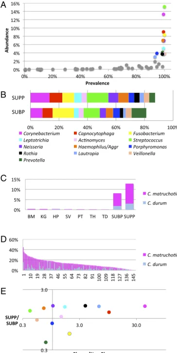
As an initial basis for identifying key taxa in supragingival plaque, we assessed the abundance and prevalence of the oligo-types grouped by genus. This analysis readily identified a group of bacterial genera that were both abundant and prevalent (Fig. 1A). Of 57 genera detected in supragingival plaque (SUPP), most had both low abundance and low prevalence. In contrast, 13 genera had at least 3% mean abundance and were also highly prevalent, each being detected in more than 90% of SUPP samples. Collectively, these 13 genera accounted for 85% of the sequencing data from SUPP. The same 13 genera also accounted for more than 80% of the subgingival plaque (SUBP) data (Fig. 1B), indicating a close relationship between the two plaque sites. Because of their abun-dance and prevalence, these taxa are likely to form both the spatial and the metabolic framework of the healthy plaque microbiome.
Taxa that are present primarily or exclusively in one site may provide clues to the distinctive features of the habitat and the role that those taxa contribute to the site. Habitat analysis of the oral microbiome suggested that one genus,Corynebacterium, in particular was strikingly specific to supragingival and subgingival
and on six of eight oral surfaces sampled (the tongue, buccal mucosa, keratinized gingivae, hard palate, tonsils, and throat) but made up 8% of the bacterial community in SUBP and more than 12% in SUPP (Fig. 1C) (19, 20). The HOMD recognizes six oral species within the genusCorynebacterium. However, of these six, only two,Corynebacterium matruchotiiandCorynebacterium durum, were present at significant levels in plaque. AlthoughC. matruchotiiwas the dominant species in most individuals,C. durumwas domi-nant in some (Fig. 1D). Taking the two species together, the ge-nus not only had a high mean abundance, but also, it was consistently abundant, with a relative abundance of 3% or more in 90% of the individuals. The abundance and prevalence of Co-rynebacteriumsuggest that it plays an important role in the plaque community, whereas its plaque specificity suggests that it occupies a niche that is dependent on properties of the tooth surface and/or the gingival crevice.
Habitat analysis of 12 other abundant plaque genera (Fig. 1E) showed large differences in their degree of specificity to plaque but only modest differences in their relative abundance in SUPP compared with SUBP. Genera with strong plaque specificity, in addition toCorynebacterium, includedCapnocytophaga, which was 10-fold more abundant in plaque than at nonplaque sites, as well asLautropia andRothia. By contrast, genera such as Strepto-coccus occupied a broad range of habitats. Despite being the single most abundant genus in SUPP, Streptococcus was sub-stantially more abundant at nonplaque sites than in plaque on average. This wide-ranging habitat preference likely reflects the capacity ofStreptococcusto be an efficient colonizer of multiple oral surfaces. Additional genera with broad habitat range in the mouth includeHaemophilusandVeillonella. Supragingival plaque is often characterized as being composed primarily of Gram-positive aerobes, whereas Gram-negative anaerobes come to dominate subgingival plaque, particularly in individuals affected by peri-odontitis (21). However, habitat analysis of genera shows that the similarities between the two plaques are more striking than their differences in the healthy individuals sampled by the HMP. Most of the abundant genera are enriched in SUPP compared with SUBP by a small and relatively constant factor of∼1.3–1.6. Some genera (Actinomyces,Porphyromonas, andVeillonella) are equally abundant. A few predominantly anaerobic genera, notably Prevotella and Fusobacterium, are more abundant in SUBP but only by a factor of ∼2. Thus, the overall similarity of distribution suggests a close connection between these two spatially adjacent communities.
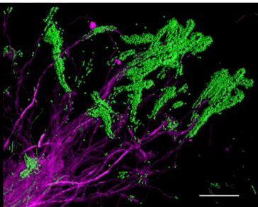
Plaque Microbiota Is Organized into Highly Structured, Multigenus Consortia.Bacteria are micron-sized and live in a chemical and structural environment with micron-scale heterogeneity. There-fore, an understanding of the micron-scale spatial organization of bacterial communities is necessary for understanding how these communities function. A striking degree of spatial organization can become visible, even with simple procedures, when plaque samples are prepared gently to retain their structure. For example, when plaque was spread out on a slide, conventional FISH with two probes revealed clumps ofCorynebacterium, with long filaments that were coated at their tips by brightly staining cocci (Fig. 2). Associations between cocci and filaments in plaque were docu-mented over 40 years ago and have been called“corncobs”(22, 23). However, the arrangement in dissected plaque, which we vi-sualized with FISH, is striking in that the filaments are clearly continuous from the base of the clump to the tips, but the cocci are restricted to the tips or distal ends of the filaments (Fig. 2). This spatial arrangement suggests a role forCorynebacteriumas a foundational taxon that structures the environment in a way that creates a microenvironment favorable to the growth of the cocci. Why the cocci are restricted to the distal ends is a key question, the answer to which requires more complete information about the surrounding structure.
We used two complementary methods designed to preserve and visualize the spatial structure of the plaque community: whole-mount preparations and methacrylate embedding. Whole mounts permitted the imaging of entire 3D structures, including long filaments, but at the expense of slight spatial distortion resulting from compression. Embedding and sectioning preserved micron-scale spatial relationships more accurately but at the expense of loss of 3D continuity. Regardless of the preparation method, we de-tected similar microbial consortia in all samples. For a systems-level analysis of the spatial organization of these samples, we used Combinatorial Labeling and Spectral Imaging FISH (CLASI-FISH) (16) to differentiate up to 15 taxa simultaneously. In our previous proof of concept of CLASI-FISH, we labeled plaque that was partially dispersed to single-cell thickness (16), so that spectral sig-natures created by binary combinations of fluorophores could be read unambiguously. For this study, we wished to analyze more intact 3D structures, in which multiple cells may lie on top of one another, even in a single optical plane of focus. In such samples, overlapping cells with different binary signals in the same pixel could generate ambiguity in taxon identification. To avoid this ambiguity, we used a simplified labeling strategy, in which a single fluorophore served as the spectral signature to identify each taxon, and as many as 10 distinct fluorophores were used simultaneously. The combination of sequence analysis with imaging allowed an assessment of spatial organization that was taxonomically both wide-reaching and refined. We used FISH probes with broad coverage, using probes for four phyla, two classes, three families, and 15 genera (Table S1). More specificity was provided by HMP sequencing data, which showed that, for most plaque genera, a small number of species was dominant. Of the 13 most abundant genera, one genus was represented by only a single major plaque species (Lautropia mirabilis), and six were represented primarily by two or three major species or small clusters of species (Table S2). Collectively, the probes that we used targeted 96–98% of the cells in a healthy supragingival plaque microbiome as judged by rRNA tag sequencing data from the HMP (Table S2). Among these probes, 2 family- and 11 genus-level probes covered 88% of the sequencing data and are shown in Figs. 2–8 andFigs. S1–S4. When describing imaging results in the following section, we will use the taxon name as shorthand for cells in the image that are reactive with the taxon-specific probe, but it should be kept in mind that these organisms are likely to be members of the species shown in Table S2. The generaHaemophilusandAggregatibacterare phylogenetically inter-twined in the family Pasteurellaceae and targeted by probe Pas111, which we refer to as Haemophilus/Aggregatibacter. The genera Neisseria, Kingella, and Eikenella are likewise intertwined in the family Neisseriaceae and targeted by probe Nei1030, which we refer to asNeisseriaceae.
We detected in plaque a complex microbial consortium char-acterized by the presence of a mass ofCorynebacteriumfilaments withStreptococcusat the periphery. We refer to this structure as a “hedgehog”because of its spiny, radially oriented filaments. We identified nine taxa as regular participants in hedgehog structures: Corynebacterium,Streptococcus,Porphyromonas, Haemophilus/ Aggregatibacter,Neisseriaceae,Fusobacterium,Leptotrichia, Capno-cytophaga, andActinomyces. Other genera were detected rarely or inconsistently in the hedgehog structures. To visualize the regular constituents of the consortium simultaneously, we constructed a probe set consisting of 10 probes: the 9 probes targeting these taxa plus the universal probe Eub338 reactive with essentially all bac-teria. Each of these 10 probes consisted of a unique oligonucleo-tide conjugated to a unique fluorophore (Table S3). To validate
A
B
C
D
E
Fig. 1. Metagenomic sequence analysis points toCorynebacteriumas a key taxon in supragingival plaque. (A) Prevalence abundance plot for supragingival plaque. (B) Cumulative abundance of genera in both supra- and subgingival plaque. Genera with greater than 3% abundance in SUPP, mean across 148 subjects, are indicated by colored dots inAand bar segments inB;Balso shows the abundance of these genera in SUBP. Data are from the HMP (18) V3–V5 region of 16S rRNA, analyzed by oligotyping (19), and grouped by genus. (C)Corynebacteriumis far more abundant in plaque than in other oral sites. Mean abundances ofC. matruchotiiandC. durumare shown for each oral site analyzed by oligotyping (19). BM, buccal mucosa; HP, hard palate; KG, keratinized gingiva; PT, palatine tonsils; SV, saliva; TD, tongue dorsum; TH, throat. (D) Cory-nebacteriumis a major component of most plaque samples. Relative abundance of Corynebacterium in the HMP SUPP samples from 148 individuals (19). C. matruchotiiis usually more abundant, butC. durumdominates some samples. (E) Habitat analysis identifies genera that are strongly characteristic of SUPP. The plaque to nonplaque ratio measures the relative abundance of each genus in two plaque sites compared with seven nonplaque sites sampled by the HMP [calcu-lated as (mean SUBP+mean SUPP)/(mean BM+mean KG+mean HP+mean SV+mean PT+mean TH+mean TD)]. This ratio identifiesCorynebacteriumand
Capnocytophagaas the taxa most preferentially abundant in plaque. The SUPP to SUBP ratio identifies these genera as relatively more abundant in SUPP than in SUBP. Colors inEare the same as those inAandB.
MIC
ROBIOLOGY
PNAS
the probes for specificity, we applied the 10-probe set to pure cultures, which we hybridized and imaged under the same condi-tions as natural plaque samples. All probes reacted strongly with the target taxon and insignificantly with the nontarget taxa (Fig. S1).
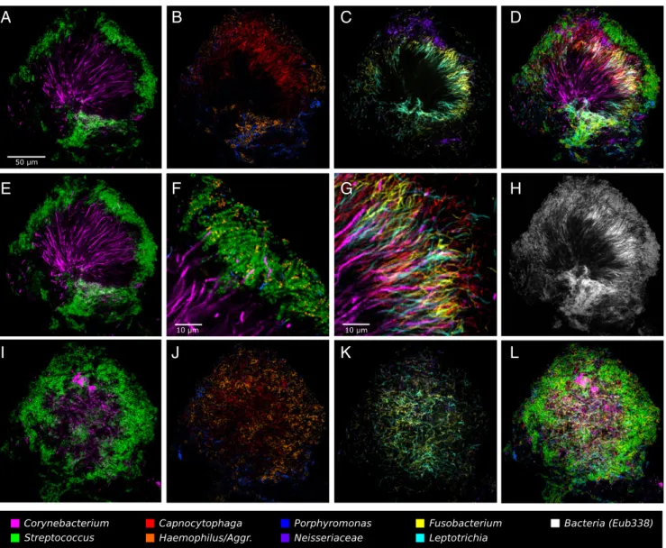
This 10-probe set revealed large, organized hedgehog structures with a generally consistent composition and spatial arrangement (Fig. 3). The fluorescence signal from each of the probes was ac-quired with a spectral, confocal microscope, was differentiated using a linear unmixing algorithm (Materials and Methods), and is presented in false color, with combinations of probes shown superimposed as detailed in Fig. 3. Fig. 3A–Dand F–H shows a single focal plane near the middle of the structure. Co-rynebacteriumfilaments radiate outward from near the center of the image. The coccoidStreptococcuscells are arranged around the distal tips of theCorynebacteriumfilaments (Fig. 3A). Also located at the periphery of the structure, in the same region as the Strep-tococcus, are cells of Haemophilus/Aggregatibacter and Porphyr-omonas(Fig. 3B).Capnocytophagaoccupies a wide band just inside the periphery (Fig. 3B). Also occupying this band but forming a more complete ring or annulus between the periphery and the base areFusobacteriumandLeptotrichia(Fig. 3C).Neisseriaceaeforms clusters in and near the periphery (Fig. 3C).Actinomyces, which was represented by only a small number of cells in this particular structure, tended to be located near the base. All taxa are shown superimposed in Fig. 3D.
The spatial arrangement ofCorynebacteriumrelative to other taxa in the structure is detailed in Fig. 3E–G. Long filaments that move in and out of the plane of focus can be only partially captured in a single optical section (∼1-μm thickness). To visu-alize the continuity of these filaments, we generated a maximum intensity projection of three adjacent optical sections (Fig. 3E), which shows single filaments that are continuous for more than 50μm and reach from the core to the periphery of the structure. Some filaments remain visible after they enter the region that contains Streptococcus, whereas others apparently disappear when they enter this zone. A detail of the periphery (Fig. 3F) shows that the corncob structures are composed of a filamentous core (sometimes visualized as Corynebacterium but frequently not stained) surrounded primarily by Streptococcus but also by
gatibacter, both of which are in close contact withStreptococcus cells. On their way to this corncob region in the periphery, the Corynebacterium filaments traverse the annulus that is densely populated with elongated rods of Fusobacterium, Leptotrichia, and Capnocytophaga, with cells of all four taxa oriented in roughly the same direction (Fig. 3G).
Completing the overview of the structure, a comparison of the fluorescent signal from the universal probe (Fig. 3H) to the overlay of nine specific probes (Fig. 3D) shows that the taxon-specific probes identify nearly all of the cells in the structure. A second focal plane near the exterior of the structure (Fig. 3I–L) shows a view of the outer shell composed primarily of corncobs. Toward the center of the image, the edge of theFusobacterium–Leptotrichiaannulus can be seen in end-on view (Fig. 3K). In summary, the plaque hedgehog is a radially organized, multigenus consortium with a framework composed primarily ofCorynebacterium, a multitaxon filament-rich annulus, and a periphery of corncob structures.
Corncobs are defined morphologically as structures in which coccoid cells,“kernels,”surround a central filament. Our CLASI-FISH results revealed that the kernels were of different taxo-nomic types and could be either single or double layer (Fig. 4). Single-layer corncobs had kernels of eitherStreptococcusor Por-phyromonas; double-layer corncobs consisted of a combination of Streptococcusas the inner layer andHaemophilus/Aggregatibacter as the outer layer. The most common corncob had a single layer ofStreptococcuskernels surrounded by a partial or complete layer ofHaemophilus/Aggregatibacter.Porphyromonaskernels could be colinear withStreptococcus around the same filament, or could form entire corncobs of their own, but in either case were always organized in a single layer. In contrast, cells ofHaemophilus/ Aggregatibacterwere never observed to form their own corncobs with Corynebacteriumfilaments. When present, they were always found adjacent to Streptococcus cells. The Haemophilus/Aggregatibacter– Streptococcusassociation was evidently specific, becauseHaemophilus/ Aggregatibacterwas not found adjacent to cells ofPorphyromonas or other taxa in the absence ofStreptococcus. Overall, the close spatial proximity of multiple taxa in corncobs suggests the pos-sibility of significant competitive, exploitative, or mutualistic in-teractions among these taxa.
In a substantial fraction of corncob structures, weak or no fluorescence signal was detected from the central filament in the region where the kernels were present. Lack of hybridization to the central filament was particularly frequent in whole-mount preparations (Fig. 4A). In embedded and sectioned preparations, the central filament was more consistently visualized (Fig. 4B). Higher magnification images of longitudinal and cross-section views (Fig. 4C) illustrate the visualization or lack thereof of the central filament. However, in all cases in which the central fila-ment was clearly labeled, it hybridized with theCorynebacterium probe, even in cases in which cells of other taxa, including Fuso-bacterium, Leptotrichia, and Capnocytophaga, were present in abundance immediately adjacent to the corncobs (Fig. S2). This observation indicates that, rather than binding promiscuously to any available filamentous substrate, the cocci are engaged in a highly specific interaction withCorynebacterium.
The filaments or elongated rods inhabiting plaque hedgehogs were striking in both their density and their spatial organization. Both Fusobacterium and Leptotrichia showed elongated mor-phology and were dispersed thinly at the periphery of the hedgehog but reached very high densities in the region immediately proximal to the periphery (Fig. 3C), a region that we call the filament-rich annulus.Capnocytophaga, likewise, reached high densities in the annulus but also extended into regions of the consortium that were rich inNeisseriaceae(Fig. 3BandD). In whole-mount prepara-tions, many cells in the filament-rich annulus overlapped in images in which all taxon channels were superimposed (Fig. 3D). This overlap was likely caused, in part, by compression of the 3D
Fig. 2. Corncob structures formed byCorynebacteriumand cocci in plaque. Corynebacteriumcells (magenta) are visible as long filaments, with cocci (green) bound to the tips of the filaments. Partially disrupted plaque was hybridized with a probe for Corynebacterium and a universal bacterial probe. Image was acquired using a Zeiss AxioImager 63×Plan-Apochromat 1.4 N.A. objective and Apotome structured illumination. (Scale bar: 20μm.)
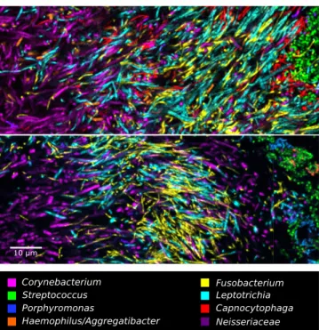
structure in whole-mount preparations, so that the cells were more densely packed than would occur in uncompressed material. In plaque embedded in methacrylate and sectioned, the compression was eliminated, and the images showed cells that were tightly packed but clearly resolved and distinct from one another (Fig. 5). Notably, these images showed that bacteria do not form large single-taxon clusters within hedgehogs. Instead, cells of at least four different taxa were intermingled at micron scales. These images show that the local environment of a cell in hedgehog consortia includes cells of several other taxa, even when we define local to mean within a radius of as little as 5–10μm.
By contrast, the localization of Actinomyces relative to Co-rynebacteriumwas characterized more by patchy clusters than by intermingling.Actinomycescells were generally detected in clumps
within the base of the hedgehog or adjacent to hedgehogs, as shown in Figs. 6 and 7. The presence ofActinomycesnear the base of a hedgehog is suggestive of the possibility thatCorynebacterium finds its attachment site in plaque not directly on the tooth but on a preexisting biofilm containingActinomyces.
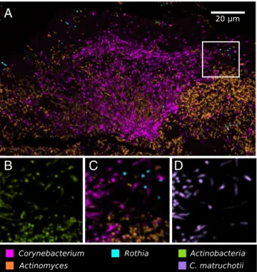
Application of a nested probe set allowed identification of the frameworkCorynebacteriumtaxon to the species level. As shown in Fig. 7 andFig. S3, only three genera,Corynebacterium, Actino-myces, andRothia, comprised virtually all of theActinobacteria present in plaque, and the speciesC. matruchotiicomprised nearly all of theCorynebacteriumin the hedgehog structure.
Hedgehog structures showed near-universal prevalence among individuals, but the fraction of plaque consisting of hedgehogs was highly variable from sample to sample, even within a single
A
B
C
D
E
F
G
H
I
J
K
L
Fig. 3. A hedgehog structure in plaque showing spatial organization of the plaque microbiome. Plaque was hybridized with a set of 10 probes each labeled with a different fluorophore. Each panel shows the superposition of several of these individual fluorophore channels.A–DandF–Hshow a single focal plane near the center of the structure, with two to three fluorophore channels shown in each ofA–Cand all nine specific probes superimposed inD. (E) Maximum intensity projection of three planes, representing a total of∼2μm of thickness, to visualize the continuity ofCorynebacteriumfilaments from the center toward the edge of the structure.Fis a detailed view of corncob structures.Gis a detailed view of mixed filaments.Hshows the fluorophore channel corresponding to the universal bacterial probe, showing that the specific probes (D) identify most of the cells that hybridize to the universal probe.I–Lshow a second focal plane near the periphery of the structure. Fluorophore channels shown correspond to the following genera in the figure: (A,E, andI) Co-rynebacteriumandStreptococcus; (BandJ)Capnocytophaga,Porphyromonas, andHaemophilus/Aggregatibacter; (CandK)Fusobacterium,Leptotrichia, and Neisseriaceae; (D and L) all nine specific probes; (F) Corynebacterium, Streptococcus, Porphyromonas, and Haemophilus/Aggregatibacter; (G) Co-rynebacterium,Fusobacterium,Leptotrichia, andCapnocytophaga; and (H) Bacteria. The plaque sample was fixed in 2% (wt/vol) paraformaldehyde, stored in 50% (vol/vol) ethanol, and spread onto the slide in 50% (vol/vol) ethanol in preparation for FISH. MIC
ROBIOLOGY
PNAS
individual. We detected hedgehogs in every individual who was sampled on multiple occasions and in∼80% of individuals sampled only once. The most exposed surface sampled, the tooth surface on the buccal side, yielded hedgehogs; so did plaque from the gingival margin. Some samples contained multiple hedgehog structures adjacent to one another (Fig. S4). Other samples lacked hedgehogs but contained other consortia. For example, clusters of Lautropia formed the center of a structure that also contained Streptococcus,Haemophilus/Aggregatibacter, andVeillonella and was reminiscent of a cauliflower (Fig. 8). Most samples contained a mixture of hedgehogs and other consortia. Because of this extensive variability and the time-intensive nature of spectral imaging analysis, higher-throughput imaging methods will be required to conduct a comprehensive analysis of spatial, temporal, and individual variation in the abundance of hedgehogs and other consortia in plaque.
In summary, we have discovered distinctive, multigenus con-sortia in dental plaque, with each taxon localized in a precise and well-defined spatial zone. The precision and reproducibility of
reflects a finely tuned interaction among the cells comprising oral microbial communities.
Discussion
Organization of Hedgehog Structures.The spatial organization of hedgehog consortia provides a framework for understanding the community structure and metabolism of the plaque microbiome. A modest number of abundant taxa makes up the clear majority of the cells in the structure, and these taxa are arranged in an organized spatial framework, within which each microbe oc-cupies a characteristic position. Based on the literature, we in-terpret the metabolic, adhesive, and environmental drivers of plaque spatial structure as diagrammed in Fig. 9.
The radial organization of hedgehogs, built on a framework of Corynebacterium, suggests that Corynebacterium is the founda-tion taxon of the consortium: it structures the environment, thereby creating habitat for other organisms and nucleating a plaque-characteristic consortium. Consistent with this view, our habitat analysis showed thatCorynebacteriumwas the genus most characteristic of plaque. The physical environment of plaque is distinctive from all other oral habitats because of the tooth surface itself: the tooth represents a solid surface permanently exposed in the mouth, whereas all other oral surfaces are cov-ered in epithelial cell layers that frequently shed. Our model suggests thatCorynebacterium proliferates in plaque and struc-tures the plaque environment because it has adopted a strategy of filamentous growth outward from the tooth, anchored in a base cemented to that permanent, exposed surface. By embed-ding itself in a biofilm matrix attached to the tooth, Co-rynebacterium could anchor the entire structure and create a protected reservoir from which it can regrow after its removal by abrasion or oral hygiene procedures, thus accounting for how
A
B
C
Fig. 4. Complex corncob structures in SUPP. (AandB) Clusters of corncobs at the perimeter of hedgehog structures. (A) Whole mount of plaque hybridized with probes forCorynebacterium,Fusobacterium,Streptococcus, Porphyr-omonas, and Haemophilus/Aggregatibacter. (B) Methacrylate-embedded section hybridized with probes forCorynebacterium,Streptococcus,Porphyromonas, andHaemophilus/Aggregatibacter. (C) Gallery of representative images showing types of corncobs frequently observed. (Scale bar:C, 5μm.)
Fig. 5. Filaments and rods of several genera intermingle at micron scales in an annulus of the hedgehog structure. The two images shown are from methacrylate-embedded, sectioned plaque from two different donors. Both samples were hybridized with probes forCorynebacterium,Fusobacterium, Leptotrichia, Streptococcus, Porphyromonas, Haemophilus/Aggregatibacter, andNeisseriaceae; the probe set inUpperalso included a probe for Capno-cytophaga.
this organism maintains a high relative abundance and a prom-inent position specifically in the microbiota associated with the tooth surface, i.e., dental plaque.
Other taxa involved in the hedgehog are stratified into distinct zones within the structure, so that different sets of taxa are characteristic of the perimeter, the filament-rich annulus, and the base. A consequence of this stratification is to reduce the number of taxa likely to be the major drivers of community me-tabolism in each of the zones. Of the nine taxa that we show to be abundant participants in hedgehogs, the three that are most likely to be key participants in the aerobic metabolism at the perimeter are Streptococcus,Haemophilus/Aggregatibacter, andPorphyromonas. In addition,Neisseriaceaemay contribute, although it is less regular in its presence at the perimeter. Key participants in metabolism of the filament-rich annulus are likely to be the three taxa that proliferate most specifically and abundantly there:Fusobacterium, Leptotrichia, andCapnocytophaga. The base is likely dominated by the metabolism ofCorynebacteriumitself, modulated by other taxa, such asActinomyces, when they are present. Imaging the spatial organization of the structure focuses attention on small sets of taxa that are highly likely to interact with one another in the healthy human mouth, and can serve to guide studies of metabolic interaction in the most promising direction.
The selection and spatial stratification of taxa in the hedgehog are likely driven by environmental and biochemical gradients. Plaque is bathed in a flow of saliva that presents an oxidizing environment, but microbes at the surface of a biofilm can rapidly consume the O2, resulting in a hypoxic environment within 10–20μm
of the surface (24).Streptococcus, a facultative aerobe, can thrive in an oxygen-rich environment, consistent with its location at the periphery of the consortium. In agreement with our lo-calization of the Streptococcus-containing corncobs to the pe-riphery of the hedgehog, earlier electron micrographs of the plaque colonizing teeth and enamel crowns clearly show the lo-calization of corncob structures at the periphery of plaque, on the side away from the enamel surface (23, 25), although these earlier studies were unable to assign a taxonomic identity to the participants in the corncob structures. The location of Strepto-coccusat the periphery, in turn, drives biochemical gradients. Streptococci consume sugars and oxygen and generate lactate, acetate, CO2, and hydrogen peroxide (26, 27). Both lactate and
hydrogen peroxide can be inhibitory to susceptible microbes but not to other participants in corncobs: hydrogen peroxide is de-toxified by catalase-producing members of the commensal microbiota, including Corynebacteriumand Aggregatibacter, and lactate is used preferentially as a growth substrate by Aggregati-bacter (28). Capnocytophaga requires CO2 for growth, and its
abundance just inside the corncob shell suggests that it is making use of a CO2-rich environment generated byStreptococcus.
FusobacteriumandLeptotrichiaare generally considered anaer-obes, but strains of both have recently been found to grow efficiently as microaerophiles as well (1, 29–31), and their pro-liferation in the annulus proximal to the corncob shell suggests a low-oxygen environment there. In summary, the precise localiza-tion of taxa within this complex structure is consistent with the modulation of the chemical environment that we would expect to be carried out by Streptococcus but likely includes other, as yet unknown metabolic interactions among neighboring cells. In con-trast, the localization of Porphyromonasin the outer shell of the hedgehog structure, in a presumably aerobic environment, is un-expected. Both of the widely studied species of Porphyromonas, Porphyromonas gingivalis and Porphyromonas endodontalis, are strict anaerobes. However, those two species are rare in the plaque of healthy individuals, whereas two additional species, Porphyr-omonas catoniaeandPorphyromonas pasteri(previously Porphyr-omonassp. HOT 279), are abundant (19). It is likely that the Porphyromonasspp. that we detect in hedgehogs are represen-tatives of these less well-known taxa and are at least moderately aero-tolerant.
Within each of the strata in the hedgehog, cells of each taxon are arranged not as single-taxon microcolonies but in close proximity to cells of other genera. Corncob structures, for ex-ample, are characterized by direct physical contact between the
A
B
C
D
Fig. 7. Nested probing for species-level identification ofCorynebacterium. Methacrylate-embedded, sectioned plaque was hybridized with a nested probe set targeting cells at the taxonomic levels of phylum, genus, and species. (A) Low-magnification image shows the three major oral genera of phylum Actinobacteria:Corynebacterium,Actinomyces, andRothia. High-magnification views show (B) all Actinobacteria, (C) the three genera, and (D)C. matruchotii.
Fig. 6. Localization ofActinomyceswithin hedgehogs, in patches within the base region of hedgehogs, and adjacent to them.
MIC
ROBIOLOGY
PNAS
central Corynebacteriumfilament and the surrounding Strepto-coccus(23). We detectedPorphyromonascells sometimes forming separate corncobs, but also present in mixed corncobs immediately adjacent toStreptococcus. This arrangement raises the question of whether the Porphyromonas cells are in direct competition withStreptococcus, for example competing for attachment sites on theCorynebacteriumfilament that allow the attached cell to be bathed in the surrounding nutrient-rich saliva. Alternatively, it is conceivable thatStreptococcusandPorphyromonasmay facilitate each other’s attachment in the corncob and thereby gain some cross-feeding metabolic advantage. The positioning of Haemo-philus/Aggregatibacter in corncobs, by contrast, was not directly adjacent to the central filament in the absence of Streptococcus. This arrangement could arise simply from the binding properties of the cells, if they find attachment sites only onStreptococcus, or alternatively could reflect a metabolic need for close proximity to Streptococcus. Whether each taxon–taxon interaction in corncobs is primarily mutualistic, commensal, or parasitic is a subject for future research, as is the degree to which the relationships might change depending on local conditions, and the degree to which active competition shapes the composition and spatial organiza-tion of the assemblage. Similar quesorganiza-tions arise for the cells in the filament-dense annulus and the hedgehog base.
From the point of view of microenvironments inhabited by the component cells of the hedgehog, one taxon stands out as being part of multiple communities: a single filament ofCorynebacteriummay experience several distinctive microenvironments along its length. The proximal part of the filament inhabits the hedgehog base, which is largely dominated by Corynebacterium but may contain Actino-mycesand is also populated by a scattering of other cells. Farther along its length, theCorynebacteriumfilament traverses the filament-rich annulus, where its neighbors consist ofFusobacterium, Lepto-trichia, andCapnocytophaga. At its distal end, theCorynebacterium filament is encased in a corncob shell of Streptococcus and Por-phyromonas, with frequently abundantHaemophilus/Aggregatibacter andNeisseriaceae. These different environments may alter the local physiology ofCorynebacterium, even within a single filament.
The prevalence of hedgehog structures alters our understanding of the dynamics of colonization of oral surfaces and the succes-sional development of plaque. Clean enamel, glass, or hydroxy-apatite surfaces in the mouth are initially colonized by a mixed community in whichStreptococcusandActinomycesare prominent (32–34). Previous models of development and succession in plaque, after initial colonization, assign a central role to Fuso-bacteriumspp. in physically linking early and late colonizers (9, 35) or creating the conditions necessary for colonization of plaque by pathogens (1, 36, 37). Whether these models were meant to de-scribe interactions in supragingival as opposed to subgingival plaque is not entirely clear; the work on initial colonization gen-erally used substrates mounted supragingivally in the mouth, whereas the pathogens in the climax community were subgingival anaerobes. The genus Corynebacterium is conspicuously absent from the early microbiota colonizing enamel and from these models but is one of the more abundant plaque taxa detected in cultivation-independent analyses based on sequencing of rRNA genes (18, 20, 38). These cultivation-independent analyses repre-sent neither the very earliest stages in colonization nor the highly mature and complex subgingival biofilm associated with perio-dontitis, but instead represent ordinary daily plaque accumulation sampled from healthy subjects. The results that we present here, using HMP sequencing data and samples of ordinary plaque from healthy volunteers, showCorynebacteriumas the taxon that pro-vides a physical link to each of the other taxa in the hedgehog structure. Our results do not suggest a central role forFusobacterium
Fig. 8. A cauliflower structure in plaque composed ofLautropia, Strepto-coccus,Haemophilus/Aggregatibacter, andVeillonella. Scattered cells of Pre-votella,Rothia, andCapnocytophagaare also visible.
Fig. 9. Summary hypothesis for interpretation of hedgehog structures. Corynebacteriumfilaments bind to an existing biofilm containing Strepto-coccusandActinomyces. At the distal tips of theCorynebacteriumfilaments, corncob structures form in which the filaments are surrounded by cocci, includingStreptococcusandPorphyromonas, in direct contact with the Co-rynebacteriumfilament as well asHaemophilus/Aggregatibacterin contact withStreptococcus. Clusters ofNeisseriaceaealso occupy the periphery of the hedgehog. TheStreptococcuscells create a microenvironment rich in CO2, lac-tate, and acelac-tate, containing peroxide, and low in oxygen. Elongated filaments ofFusobacterium andLeptotrichiaproliferate in this low-oxygen, high-CO2 environment in an annulus just proximal to the corncob-containing peripheral shell of the hedgehog. The CO2-requiringCapnocytophagaalso proliferates abundantly in and around this annulus. The base of the hedgehog is dominated byCorynebacteriumfilaments and thinly populated by addi-tional rods, filaments, and/or cocci.
in physically connecting members of the consortium. Although it may contribute to consortium organization,Fusobacteriumis only one of four filamentous taxa in hedgehogs, and it is neither the most abundant nor the most spatially extensive. Bioinformatic analysis suggests that the genus Fusobacterium as a whole is substantially more abundant in subgingival than in supragingival plaque (20). Our FISH probe detects the entire Fusobacteriumgenus, but within the genus, onlyF. nucleatumhas high abundance in plaque. Furthermore, several distinct subspecies ofF. nucleatum are recognized in the HOMD, and in HMP se-quencing data, these subspecies showed distinctive habitat prefer-ences: some for subgingival and some for supragingival plaque (19). We conclude that theFusobacteriumin hedgehogs is likely one of the supragingival-abundant subspecies ofF. nucleatum.
As a slowly growing taxon that is not prominent or even fre-quently represented among initial colonizers of enamel, how does Corynebacteriumand the hedgehog structures that it apparently organizes come to be so abundant in plaque? It seems likely that Corynebacteriumfinds attachment sites on the preexisting biofilm consisting ofStreptococcusandActinomyces, which are among the early colonizers (33) and can be found near the base of hedgehog structures. EM examination of the microbial community colo-nizing removable enamel chips worn inside the mouth showed scattered filamentous cells oriented perpendicularly to the pri-marily coccus-covered surface at 24 hours and a mixed com-munity of abundant filamentous organisms by 48 hours (39), suggesting that colonization with Corynebacterium may take place around the 24-hour stage in plaque development. In the subgingival crevice or within the dental calculus that precipitates around these filamentous aggregates (40), the attachment sites ofCorynebacterium would be protected, and when distal por-tions of the structure are lost, by abrasion or oral hygiene procedures similar to our sampling methods, the filaments could rapidly regrow from these reservoirs.
Understanding Microbiomes: Genes Vs. Organisms.The imaging ap-proach cuts through the overwhelming complexity of detail in mi-crobial communities and allows common patterns to shine through. With deep sequencing, it has become clear that many taxa in the oral microbiota are shared across individuals but are abundant in some samples and almost vanishingly rare in others (19, 38). These differences in abundance may result from real differences be-tween individuals, fluctuations within a single individual over time, or a combination of the two. Whatever its cause, the lack of a consistently abundant microbial“core”has led to the idea that perhaps it is not organisms but genes and functions that are conserved within the microbiome, distributed across a variety of organisms whose identities are irrelevant. The discovery of hedgehog consortia argues against this idea, at least for some microbiomes. The consistency of the composition and structure of the hedgehog across many individuals suggests that organisms are highly relevant to understanding the roles, organization, and dynamics of the members of the consortium. We suggest that it is neither necessary nor desirable to disregard taxonomy and reduce the microbiome to a collection of genes or metabolites unmoored from their source organism. Instead, we suggest the converse, that an understanding of the ecology and physiology of the organisms in the consortium will provide an organizing principle for understanding and interpreting metagenomic and metatranscriptomic data.
The consortium that we have described is composed of organ-isms of not only different species but nine different genera clas-sified into eight families in five phyla. Thus, it is a consortium not of close relatives but of highly disparate organisms. The intimate association of widely diverged taxa may be a general property of spatially organized microbial consortia. Darwin (41) was the first to recognize that the“struggle for existence”was most severe be-tween closely allied forms and that“divergence of character”would
support the greatest amount of life. In his words,“the advantages of diversification of structure, with the accompanying differences of habit and constitution, determine that the inhabitants, which thus jostle each other most closely, shall, as a general rule, belong to what we call different genera and orders”(41). Although our im-aging studies to date have mostly been restricted to genus-level analysis, we suggest that the members of each genus are not in-terchangeable with one another. Future, more taxonomically refined analyses will reveal which representatives of each genus participate in hedgehog structures. The likely identity of these representatives can be inferred from sequencing data and is generally small in number, as noted above for genus Corynebacteriumwith only two major oral species.
Potential Significance of Plaque Spatial Structure for Health and Disease and for Modeling Using Synthetic Communities.The sources of both spatial and temporal heterogeneity in the plaque community and their significance are a rich field for additional investigation. The existence of heterogeneity is clearly visible in the sequencing data, and its functional significance is suggested by morphological studies showing, for example, that primarily filamentous and pri-marily coccoid communities coexist side by side in supragingival plaque, and that the filamentous communities are more likely to form calculus (40). Would plaque rich in hedgehog structures, compared with other alternate consortia, make the preservation of health or transition to disease more or less likely? Critical topics for future study include whether there are features of host biology, behavior, or immune system that encourage the development of the hedgehog consortium over other microbial assemblages in plaque and what, if any, role hedgehog structures play in the maintenance of oral health or progression toward disease. The ability to create realistic in vitro models of the plaque biofilm is critical for advancing the testing of antimicrobial or microbe-modulating compounds as well as exploring biochemical and metabolic interactions among taxa. Plaque as currently recon-stituted in flow cells or other in vitro systems bears little re-semblance to the structures that we see in plaque harvested from the mouth. Improving these reconstituted models, with success judged by their ability to recapitulate the spatial structure of natural plaque, will open up a vast area of mechanistic inquiry. In summary, the biogeography of a microbial community on the micron scale reveals a spatial organization critical to un-derstanding the physiology, ecology, and functional significance of the community. Whether the community in question is host-associated or environmental, these insights are generalizable to the study of any microbiome.
Materials and Methods
Sample Collection, Fixation, and Storage.We collected supragingival plaque from 22 healthy volunteers, each of whom had given informed consent. For 10 volunteers, oral health was confirmed by clinical examination; the rest were self-reported as both orally and systemically healthy. Volunteers refrained from oral hygiene for 12–48 h before sample collection. Plaque was collected using toothpicks to scrape visible plaque from the gingival margin or tooth surface, or using floss to collect plaque from throughout the mouth. Samples were prepared in three ways. (i) Plaque was applied directly to slides and air-dried, and the samples were fixed directly on the slide, washed, dehydrated through an ethanol series, and subjected to FISH. (ii) Plaque was fixed in paraformaldehyde, stored in 50% (vol/vol) ethanol, spread onto slides in 50% (vol/vol) ethanol, and air-dried immediately be-fore FISH. (iii) Plaque was fixed in paraformaldehyde followed by immediate embedding in methacrylate resin, which was later sectioned, and the sec-tions were applied to slides and then subjected to FISH directly on the slide. Samples were handled gently with a minimum of agitation so as to preserve as much spatial structure as possible. Fixation was carried out in 2% (wt/vol) paraformaldehyde in PBS or 10 mM Tris (pH 7.5) for at least 1.5 h on ice. For samples not fixed directly on the slides, sample was allowed to sediment by gravity, and the supernatant was removed by pipetting. Samples were washed in 1×PBS or 10 mM Tris (pH 7.5) for 15 min and again sedimented by gravity, and the supernatant was removed. Samples were resuspended in
MIC
ROBIOLOGY
PNAS
at−20 °C, or they were immediately dehydrated through an ethanol series, washed with acetone, and embedded in Technovit 8100 methacrylate resin (EMSdiasum.com). Embedded blocks were stored at room temperature and sectioned dry using a triangular tungsten carbide knife (Delaware Diamond Knives, Inc.).
Sequencing and Sequence Analysis.Sequence analysis was carried out on a previously published oligotyping analysis (19) using publicly available data from the HMP (18); details of sample collection, sequencing, and human subjects research approvals are contained in the original publications.
Spectral Imaging FISH, Image Acquisition, and Analysis.Fluorophore-labeled oligonucleotides were purchased fromThermoFisher.comorwww.Biomers.net. FISH was carried out using standard protocols (42) on sections or whole-mount samples mounted onto UltraStick Slides (Thermo Scientific). Hybridization solution [900 mM NaCl, 20 mM Tris, pH 7.5, 0.01% SDS, 20% (vol/vol) formamide, each probe at a final concentration of 2 nM] was applied to samples and
amide. Slides were then washed in wash buffer (215 mM NaCl, 20 mM Tris, pH 7.5, 5 mM EDTA) at 48 °C for 15 min, dipped in cold water, air-dried, mounted in ProLong Gold Antifade Solution (ThermoFisher), covered with a #1.5 coverslip, and allowed to cure in the dark overnight. Slides were imaged using a Zeiss LSM 780 Confocal Microscope with a 40×1.4 N.A. Plan-Apochromat objective. Each field of view was imaged using sequential excitation with the 633-, 594-, 561-, 514-, 488-, and 405-nm laser lines. Linear unmixing was performed using the Zeiss ZEN software using reference spectra acquired from cultured cells hybrid-ized as above with the Eub338 probe labeled with the appropriate fluorophore. Unmixed images were assembled and false-colored using Fiji (43).
ACKNOWLEDGMENTS. We thank Alex Valm for probe design; Katherine Lemon and Matthew Ramsey for helpful discussions; and Jake Casper, Louie Kerr, Carissa McKinney, Janina Schuhmann, Braden Tierney, Steven Wilbert, Liping Xun, and the members of the MBL 2014 Physiology Summer Course. This research was supported by NIH National Institute of Dental and Craniofacial Research Grant DE022586 (to G.G.B.).
1. Diaz PI, Zilm PS, Rogers AH (2002)Fusobacterium nucleatumsupports the growth of
Porphyromonas gingivalisin oxygenated and carbon-dioxide-depleted environments.
Microbiology148(Pt 2):467–472.
2. Mahowald MA, et al. (2009) Characterizing a model human gut microbiota composed of members of its two dominant bacterial phyla.Proc Natl Acad Sci USA106(14): 5859–5864.
3. Traxler MF, Watrous JD, Alexandrov T, Dorrestein PC, Kolter R (2013) Interspecies interactions stimulate diversification of theStreptomyces coelicolorsecreted metab-olome.MBio4(4):e00459-13.
4. Jakubovics NS (2015) Intermicrobial interactions as a driver for community composi-tion and stratificacomposi-tion of oral biofilms.J Mol Biol427(23):3662–3675.
5. Wright CJ, et al. (2013) Microbial interactions in building of communities.Mol Oral Microbiol28(2):83–101.
6. Dobell C, ed (1960)Antony Van Leeuwenhoek and His“Little Animals,”Being Some Account of the Father of Protozoology and Bacteriology and His Multifarious Discoveries in These Disciplines(Harcourt, Brace, and Co., New York).
7. Kolenbrander PE, et al. (2006) Bacterial interactions and successions during plaque development.Periodontol 200042:47–79.
8. Palmer RJ, Jr (2014) Composition and development of oral bacterial communities.
Periodontol 200064(1):20–39.
9. Kolenbrander PE, London J (1993) Adhere today, here tomorrow: Oral bacterial ad-herence.J Bacteriol175(11):3247–3252.
10. Segata N, et al. (2013) Computational meta’omics for microbial community studies.
Mol Syst Biol9:666.
11. DeLong EF, Wickham GS, Pace NR (1989) Phylogenetic stains: Ribosomal RNA-based probes for the identification of single cells.Science243(4896):1360–1363. 12. Amann RI, Krumholz L, Stahl DA (1990) Fluorescent-oligonucleotide probing of whole
cells for determinative, phylogenetic, and environmental studies in microbiology.
J Bacteriol172(2):762–770.
13. Vartoukian SR, Palmer RM, Wade WG (2009) Diversity and morphology of members of the phylum“synergistetes”in periodontal health and disease.Appl Environ Microbiol75(11):3777–3786.
14. Drescher J, et al. (2010) Molecular epidemiology and spatial distribution of Seleno-monasspp. in subgingival biofilms.Eur J Oral Sci118(5):466–474.
15. Zijnge V, et al. (2010) Oral biofilm architecture on natural teeth.PLoS One5(2):e9321. 16. Valm AM, et al. (2011) Systems-level analysis of microbial community organization through combinatorial labeling and spectral imaging.Proc Natl Acad Sci USA108(10): 4152–4157.
17. Dewhirst FE, et al. (2010) The human oral microbiome.J Bacteriol192(19):5002–5017. 18. Human Microbiome Project Consortium (2012) Structure, function and diversity of the
healthy human microbiome.Nature486(7402):207–214.
19. Eren AM, Borisy GG, Huse SM, Mark Welch JL (2014) Oligotyping analysis of the hu-man oral microbiome.Proc Natl Acad Sci USA111(28):E2875–E2884.
20. Segata N, et al. (2012) Composition of the adult digestive tract bacterial microbiome based on seven mouth surfaces, tonsils, throat and stool samples.Genome Biol13(6):R42. 21. Xie H, et al. (2000) Intergeneric communication in dental plaque biofilms.J Bacteriol
182(24):7067–7069.
22. Jones SJ (1972) A special relationship between spherical and filamentous microor-ganisms in mature human dental plaque.Arch Oral Biol17(3):613–616.
23. Listgarten MA, Mayo HE, Tremblay R (1975) Development of dental plaque on epoxy resin crowns in man. A light and electron microscopic study.J Periodontol46(1):10–26. 24. Wessel AK, et al. (2014) Oxygen limitation within a bacterial aggregate.MBio5(2):
e00992.
25. Listgarten MA (1976) Structure of the microbial flora associated with periodontal health and disease in man. A light and electron microscopic study.J Periodontol47(1): 1–18.
26. Ramsey MM, Rumbaugh KP, Whiteley M (2011) Metabolite cross-feeding enhances virulence in a model polymicrobial infection.PLoS Pathog7(3):e1002012.
27. Zhu L, Kreth J (2012) The role of hydrogen peroxide in environmental adaptation of oral microbial communities.Oxid Med Cell Longev2012:717843.
28. Brown SA, Whiteley M (2007) A novel exclusion mechanism for carbon resource par-titioning inAggregatibacter actinomycetemcomitans.J Bacteriol189(17):6407–6414. 29. Bernard K, Cooper C, Tessier S, Ewan EP (1991) Use of chemotaxonomy as an aid to
differentiate amongCapnocytophagaspecies, CDC group DF-3, and aerotolerant strains ofLeptotrichia buccalis.J Clin Microbiol29(10):2263–2265.
30. Diaz PI, Zilm PS, Rogers AH (2000) The response to oxidative stress ofFusobacterium nucleatumgrown in continuous culture.FEMS Microbiol Lett187(1):31–34. 31. Woo PCY, et al. (2010)Leptotrichia hongkongensissp. nov., a novelLeptotrichia
species with the oral cavity as its natural reservoir.J Zhejiang Univ Sci B11(6):391–401. 32. Diaz PI, et al. (2006) Molecular characterization of subject-specific oral microflora
during initial colonization of enamel.Appl Environ Microbiol72(4):2837–2848. 33. Dige I, Raarup MK, Nyengaard JR, Kilian M, Nyvad B (2009)Actinomyces naeslundiiin
initial dental biofilm formation.Microbiology155(Pt 7):2116–2126.
34. Li J, et al. (2004) Identification of early microbial colonizers in human dental biofilm.
J Appl Microbiol97(6):1311–1318.
35. Lancy P, Jr, Dirienzo JM, Appelbaum B, Rosan B, Holt SC (1983) Corncob formation betweenFusobacterium nucleatumandStreptococcus sanguis.Infect Immun40(1): 303–309.
36. Socransky SS, Haffajee AD (2005) Periodontal microbial ecology.Periodontol 200038: 135–187.
37. Haffajee AD, Socransky SS, Patel MR, Song X (2008) Microbial complexes in supra-gingival plaque.Oral Microbiol Immunol23(3):196–205.
38. Zaura E, Keijser BJF, Huse SM, Crielaard W (2009) Defining the healthy“core micro-biome”of oral microbial communities.BMC Microbiol9:259.
39. Nyvad B, Fejerskov O (1987) Scanning electron microscopy of early microbial coloni-zation of human enamel and root surfaces in vivo.Scand J Dent Res95(4):287–296. 40. Friskopp J, Hammarström L (1980) A comparative, scanning electron microscopic
study of supragingival and subgingival calculus.J Periodontol51(10):553–562. 41. Darwin C (2013) The Origin of SpeciesbyMeans of Natural Selection (Forgotten
Books, London), Vol 1.
42. Pernthaler J, Gloeckner FO, Schoenhuber W, Amann R (2001) Fluorescence in situ hybridization with rRNA-targeted oligonucleotide probes.Methods Microbiol30(3): 207–226.
43. Schindelin J, et al. (2012) Fiji: An open-source platform for biological-image analysis.
Nat Methods9(7):676–682.
44. Amann RI, et al. (1990) Combination of 16S rRNA-targeted oligonucleotide probes with flow cytometry for analyzing mixed microbial populations.Appl Env Microbiol
56(6):1919–1925.
45. Gmür R, Lüthi-Schaller H (2007) A combined immunofluorescence and fluorescentin situhybridization assay for single cell analyses of dental plaque microorganisms.
J Microbiol Methods69(2):402–405.
46. Weller R, Glöckner FO, Amann R (2000) 16S rRNA-targeted oligonucleotide probes for thein situdetection of members of the phylum Cytophaga-Flavobacterium-Bacter-oides.Syst Appl Microbiol23(1):107–114.
47. Meier H, Amann R, Ludwig W, Schleifer KH (1999) Specific oligonucleotide probes for in situ detection of a major group of gram-positive bacteria with low DNA G+C content.Syst Appl Microbiol22(2):186–196.
48. Paster BJ, Bartoszyk IM, Dewhirst FE (1998) Identification of oral streptococci using PCR-based, reverse-capture, checkerboard hybridization.Methods Cell Sci20(1): 223–231.
49. Chalmers NI, Palmer RJ, Jr, Cisar JO, Kolenbrander PE (2008) Characterization of a
Streptococcussp.-Veillonellasp. community micromanipulated from dental plaque.
J Bacteriol190(24):8145–8154.
50. Manz W, Amann R, Ludwig W, Wagner M, Schleifer K-H (1992) Phylogenetic oligo-deoxynucleotide probes for the major subclasses of proteobacteria: Problems and solutions.Syst Appl Microbiol15(4):593–600.