Labeling Antibodies
& Biopolymers
Our Mission
AAT Bioquest® is committed to constantly meet or exceed its customer’s requirements by
providing consistently high quality products and services, and by encouraging continuous
improvements in its long-term and daily operations. Our core value is Innovation and Customer
Satisfaction.
Our Story
AAT Bioquest®, Inc. (formerly ABD Bioquest, Inc.) develops, manufactures and markets bioanalytical research reagents and kits to life sciences research, diagnostic R&D and drug discovery. We specialize in photometric detections including absorption (color), fluorescence and luminescence technologies. The Company's superior products enable life science researchers to better under-stand biochemistry, immunology, cell biology and molecular biology. AAT Bioquest offers a rapidly expanding list of enabling products. Besides the standard catalog products, we also offer custom services to meet the distinct needs of each customer. Our current services include custom synthesis of biological detection probes, custom development of biochemical, cell-based and diagnostic assays and custom high throughput screening of drug discovery targets.
It is my greatest pleasure to welcome you to AAT Bioquest. We greatly appreciate the constant support of our valuable customers. While we continue to rapidly expand, our core value remains the same: Innovation and Customer Satisfaction. We are committed to being the leading provider of novel biological detection solutions. We promise to extend these values to you during the course of our service and to continue to support you with our new products and services. It is our greatest honor to receive valuable feedbacks and suggestions from you so that we can better serve your projects.
Very truly yours,
Zhenjun Diwu, Ph.D. President
Table of Contents
Section 1 General Information 2
APC and Its Tandems... ... 34
RPE and Its Tandems ... 35
PerCP and Its Tandems ... 36
Section 6 trFluor
TMFluorescent Labeling Dyes 37
trFluor™ Eu Dye ... 40
trFluor™ Tb Dye ... 40
Section 2 Fluorescence Labeling Principles 5
Section 3 iFluor
TMFluorescent Labeling Dyes 9
iFluor™ 350 Dyes ...12
iFluor™ 405 Dyes ... ...12
iFluor™ 488 Dyes ...13
iFluor™ 555 Dyes... 14
iFluor™ 594 Dyes... ...15
iFluor™ 633 Dyes... ...16
iFluor™ 647 Dyes... ...17
iFluor™ 680 Dyes ... ...18
iFluor™ 700 Dyes ... ...19
iFluor™ 750 Dyes ... ...20
iFluor™ 790 Dyes ... ...21
ReadiLink™ iFluor™ Protein Labeling Kits ...22
mFluor™ Violet 450 Dyes... ... 26
mFluor™ Violet 510 Dyes ...26
mFluor™ Violet 540 Dyes ...27
mFluor™ Blue 570 Dyes ...27
mFluor™ Green 620 Dye... ...28
mFluor™ Yellow 630 Dye ... ...28
mFluor™ Red 700 Dyes ...29
mFluor™ Red 780 Dyes ...29
AAT Bioquest Fluorochrome Reference Chart for Flow Cytometry ...30
Section 5 Phycobiliproteins and Their Tandem Conjugates 31
Section 4 mFluor
TMFluorescent Labeling Dyes 23
Section 7 Index 41
Alphabetical Index ... 42
Catalog Number Index ... 44
5FMt'BY
TBMFT!BBUCJPDPNtJOGP!BBUCJPDPNUnless otherwise specified, all products are for Research Use Only. Not for use in diagnostic or therapeutic procedures.
2
1
General I
n
formation
General Information
www.aatbio.com
Trademarks of AAT Bioquest
AAT Bioquest®
California Red™
iFluor
TMmFluor
TMNuclear Blue
TMNuclear Green
TMNuclear Orange
TMNuclear Red
TMReadiLink
TMReadiView
TMTide Fluor
TMTide Quencher
TMtrFluor
TMTrademarks of Other Companies
Alexa Fluor® (Invitrogen)
Cy3®, Cy5®, Cy5.5® and Cy7® (GE Healthcare)
DyLight™ (ThermoFisher)
IRDye® (LI-COR)
Krome Orange
TM(Beckman Coulter)
Lightning-Link® (Innova Biosciences)
Pacific Blue® and Pacific Orange® (Invitrogen)
Texas Red®(Invitrogen)
CUSTOMER SERVICE & ORDERING INFORMATION
AAT Bioquest Corporate Headquarter:
520 Mercury Drive
Sunnyvale, CA 94085, USA
Tel: 800-990-8053 (US); 408-733-1055 (International)
Fax: 408-733-1304
Website: www.aatbio.com
E-mails: [email protected] (inquire)
[email protected] (quote request)
[email protected] (technical support)
International Distributors:
See Back Cover
www.aatbio.com
General Information
General I
n
formation
1
TERMS AND CONDITIONS OF SALE
1. Prices, Orders and Changes: Prices shown are in US currency. Please call us for current prices if you require this information prior to placing your order. We guarantee our written quotations for 60 days. You may not cancel purchase orders unless such cancellation is expressly agreed by us. In such event, you will be advised of the total charge for such cancellation. You agree to pay such charges, including, but not limited to, storage and shipment costs, costs of producing standard materials, costs of purchasing non-returnable materials, cancellation costs imposed on us by our suppliers, and any other cost resulting from cancellation of this order.
2. Delivery: In most cases, we use standard overnight or two-day Federal Express delivery (or equivalent). All shipping charges billed are the responsibility of the customer and are normally prepaid by AAT Bioquest, Inc. and added to the invoice. We reserve the right to make delivery in installments, all such installments to be separately invoiced and paid for when due per invoice, without regard to subsequent deliveries. Partial shipments of available items are made when another item is backordered. Please inspect your packages upon receipt. If the goods have been damaged in transit, we can assist you in filing a claim with the carrier. You shall notify us in writing of any claims for shortages, defects or damages and shall hold the goods for our written instructions concerning disposition. Any claims for such errors must be made within 10 business days. If it is our error, we will do whatever is necessary to ship the correct products as soon as possible. If you shall fail to notify us any defects within 10 days after the goods have been received, such goods shall conclusively be deemed to conform to the terms and conditions and to have been irrevocably accepted by the buyer.
3. Payment: Terms of sale are net 30 days of date of invoice that is sent to you within 24 hours of shipping the order. The amount received must be sufficient to cover both the invoiced amount and any bank charges that may be incurred. Late charges may be added to invoices not paid within the 30-day time period. Late charges must be paid before subsequent orders can be shipped.
4. Warranties: The products shipped by AAT Bioquest are warranted to conform to the chemical or biological descriptions provided in our publications. This warranty is exclusive, and we makes no other warranty, express or implied, including any implied warranty of merchantability or fitness for any particular purpose. Our sole and exclusive liability and your exclusive remedy with respect to products proved to our satisfaction to be defective or nonconforming shall be replacement of such products without charge or refund of the purchase price, in our sole discretion, upon the return of such products in accordance with our instructions. We will not be liable for any incidental, consequential or contingent damages involving their use.
5. Returns: We must authorize any returns. We will not accept return shipments unless we have given prior written permission and shipping instructions. Goods may not be returned for credit except with our permission, and then only in strict compliance with our return shipment instructions. Any returned items may be subject to a 20% restocking fee. In many cases, items ordered in error cannot be returned because of the sensitive nature of many of our products and the difficulty and expense of requalifying returned items. If items are accepted for return, they must be in new, unopened, unused and undamaged condition, and you will be charged a per-unit 20% restocking charge.
6. Use of Our Products: Our products are used ONLY for laboratory research and develop-ment purposes. We realize that, since our products are, unless otherwise stated, intended primarily for research purposes, they may not be on the Toxic Substances Control Act (TSCA) inventory. You assume responsibility to assure that the products purchased from us are approved for use under TSCA, if applicable. You have the responsibility to verify the hazards and to conduct any further research necessary to learn the hazards involved in using products purchased from us. You also have the duty to warn your customers and any auxiliary personnel (such as freight handlers, etc.) of any risks involved in using or handling the products.
7. Patent Disclaimer: We do not warrant that the use or sale of our products will not infringe the claims of any United States or other patents covering the product itself or the use thereof in combination with other products or in the operation of any process. 8. Miscellaneous: We reserve the right to discontinue our products or change specifications or prices of our products and to correct any errors or omissions at any time without incurring obligations.
4
General Information
www.aatbio.com
5FMt'BY
TBMFT!BBUCJPDPNtJOGP!BBUCJPDPNUnless otherwise specified, all products are for Research Use Only. Not for use in diagnostic or therapeutic procedures.
1
General I
n
formation
Custom Products and Services
Our Technologies
Amplite™ enzyme-based detection platform is optimized for measuring horseradish peroxidase (HRP), alkaline phosphates, luciferase, beta-galactosidase, lactamase, oxidase, protein kinases, protein phosphatases, phosphodiesterases, proteases, cytochrome P450, histone deacetylase (HDAC) and cell signaling molecules such as NAD/NADH, NADP/NADPH, IP3, cAMP and cGMP etc.
Cell Explorer™ cell labeling platform is a complete set of tools for tracking live cells. This platform is also widely used for sorting mixed populations of cells.
Cell Navigator™ cell staining platform is a complete set of tools for selective labeling subcellular structures of live, fixed and dead cells.
Cell Meter™ cellular functional assay platform is a complete set of tools for functional analysis of cellular events and real time-monitoring of cell functions.
iFluor™ superior fluorescent labeling dyes are optimized for labeling proteins and nucleic acids. This group of dyes span from UV to infrared wavelength with good photostability and brightness.
mFluor™ superior fluorescent labeling dyes are optimized for flow cytometry applications.
PhosphoWorks™ detection platform is a set of tools for detection of ATP, ADP, AMP, phosphate, pyrophosphate, phosphoproteins and phosphopeptides.
Quest View™ colorimetric protease platform is a sensitive and robust tool for rapid detection of protease and glycosidase biomarkers. This technology platform has been licensed by a few diagnostic companies for developing rapid diagnostic tests.
RatioWorks™ superior cellular dyes are a sensitive and robust tool set for ratio imaging and real time monitoring of cellular functions (such as pH and ions) in live cells.
Screen Quest™ assay kits are a set of HTS-ready tools for high throughput screening of biochemical and cellular targets such as protein kinases, proteases, HDAC, cell apoptosis and cytotoxicity, GPCR, ion channels, ADME and transporters.
Tide Fluor™ and Tide Quencher™ superior labeling dyes are specially optimized for labeling nucleotides and peptides. This platform offers the best value in the industry. It is second to none in terms of performance and cost. This technology platform has been licensed by a few diagnostic companies for developing IVD diagnostic tests.
trFluor™ superior fluorescent labeling dyes are optimized for developing time-resolved fluorescence-based assays. It has been used for developing HTS assay technologies for many drug discovery targets.
Our Services
Besides the catalog products we also offer custom services to meet the distinct needs of each customer. Our current services include custom synthesis of biological detection probes, custom development of biochemical, cell-based and diagnostic assays, custom bioconjugation and custom high throughput screening of drug discovery targets.
Custom Assay Design and Development
At AAT Bioquest we not only make probes and assay kits, but also use them extensively ourselves. Scientists at AAT Bioquest are experts on assay design and have developed a wide variety of tests that range from biochemical detection to cellular functions. Our assay options include:
t&O[ZNFBDUJWJUJFT t#JOEJOHBTTBZT t$FMMCBTFEBTTBZT t.JDSPQMBUFBTTBZT t'MPXDZUPNFUSJDBOBMZTJT t'MVPSFTDFODFJNBHJOH Custom Conjugation
AAT Bioquest offers the best and the most rapid bioconjuga-tion service in the industry.
t#JPUJOZMBUJPO
t'MVPSFTDFODFMBCFMJOH J'MVPSTM, APC, RPE and PerCP)
t&O[ZNFMBCFMJOH "1BOE)31 t4NBMMNPMFDVMFDPOKVHBUJPO
Custom Screening
AAT Bioquest offers on-demand high-throughput screening and pharmacology profiling assays with multiple
methodologies. Functional assays are designed, validated and customized to the needs of our pharmaceutical and
biotechnology industry clients. These assays are aimed at assessing and monitoring the efficacy, tolerability and safety parameters of candidate compounds for treating and/or diagnosing cancer, infectious disease, autoimmunity and transplantation. Our screening options include:
t'VMMBTTBZEFWFMPQNFOUGPSBUBSHFUPGZPVSDIPJDF t0QUJNJ[BUJPOPGZPVSBTTBZQSPUPDPMGPS)54 t.VMUJQMFBTTBZQMBUGPSNTBOEEFUFDUJPONFUIPET t$VTUPNEBUBBOBMZTJT
Custom Synthesis of Fluorophores and Luminophores
AAT Bioquest is recognized by the top pharmaceutical companies and diagnostic companies as a key provider of novel fluorescent dyes and luminescent probes. Over the years we have developed and synthesized many enabling
fluorescent and luminescent probes for running a variety of challenging biological detection tasks.
Fluor
esc
enc
e Labeling P
rinciples
Fluorescence Labeling Principles
2
Fluorescence Labeling Principles
www.aatbio.com
6
5FMt'BY
TBMFT!BBUCJPDPNtJOGP!BBUCJPDPN Unless otherwise specified, all products are for Research Use Only.Not for use in diagnostic or therapeutic procedures.
2
Fluor
esc
enc
e Labeling P
rinciples
Fluorescence Labeling Methods at-a-glance
Figure 2.1 Three common methods used for labeling antibodies with fluorescent dyes. (A). Direct labeling through amino groups. This method is convenient and gives the highest dye/protein ratio. However, the overlabeling may reduce the specificity and affinity of the antibody to be labeled. (B). Reduction method. Antibodies are first treated with DTT (or TCEP) to generate a SH group that reacts with a dye maleimide. The resulted conjugate has generally good specificity and affinity. (C). Oxidation method. The antibodies are first treated with NaIO4 to generate an aldehyde group that reacts with a dye hydrazide. This method generally gives lower dye/protein ratio, thus the resulted dye-protein conjugate tends to be less brighter than the two other methods.
Optimized Fluorescence Labeling Solutions
www.aatbio.com
2
Fluor
esc
enc
e Labeling P
rinciples
Table 2.1 Optimized Fluorescence Labeling Solutions Offered by AAT Bioquest
Targets to be labeled Dye Selection Benefits
Antibodies, Proteins and Nucleic Acids iFluor™ dyes
t4VQFSJPSQFSGPSNBODFUPUIFDMBTTJDEZFT
t6ODPNQSPNJTFEQFSGPSNBODFXJUITJHOJöDBOUDPTUTBWJOH
(Compared to Alexa Fluor® and DyLight™ dyes)
Oligos and Peptides Tide Fluor™ dyes
t#FUUFSQFSGPSNBODFUIBOUIFDMBTTJDEZFT
t6ODPNQSPNJTFEQFSGPSNBODFXJUIBGSBDUJPOPGDPTU
(Compared to Alexa Fluor® and DyLight™ dyes)
Flow Cytometry Applications mFluor™ dyes tEnable multicolor detection optimized for flow cytometry
TR-FRET Applications trFluor™ dyes t/PFOIBODFSTSFRVJSFE
t.VMUJQMFSFBDUJWFGPSNTBWBJMBCMF
Optimized Fluorescence Labeling Solutions
Fluorescence is the result of a three-stage process that occurs in certain molecules (generally polyaromatic hydrocarbons or heterocycles) called fluorophores or fluorescent dyes. A fluorescent probe is a fluorophore designed to localize within a specific region of a biological specimen or to respond to a specific stimulus. Fluorescent probes enable researchers to detect particular
components of complex biomolecular assemblies (such as live cells) with exquisite sensitivity and selectivity. Reactive fluorescent dyes are widely used to modify peptides, proteins (in particular, antibodies), oligonucleotides, nucleic acids, carbohydrates and other biological molecules. In general, the preferred bioconjugates should have high fluorescence quantum yields and retain the biological activities of the unlabeled biomolecules.
A number of fluorescent dyes have been developed and
commercialized for labeling biomolecules. Among them, FITC might be the most popular one although it has certain limitations, e.g., pH dependence, low photostability and short wavelength, etc. Alexa Fluor® dyes have been used for labeling proteins and other biomolecules with improved properties over the classic dyes such as fluorescein and rhodamine molecules. However, the extra-ordinary high costs of Alexa Fluor® dyes keep them from the certain applications that require a large amount of dye such as labeling peptides and oligos. In addition, Alexa Fluor® dyes do not provide a significant benefit for labeling peptides, oligo and other small molecules.
AAT Bioquest offers iFluor™, mFluor™ and trFluor™ dyes, a complete set of fluorescent labeling dyes, tiered and optimized for a variety of particular applications with significantly reduced cost. Our iFluor™ dyes are optimized for labeling antibodies and other biopolymers including nucleic acids and carbohydrates. The iFluor™ dyes demonstrate strong fluorescence, high photostability and pH independence on proteins and other biopolymers. Our mFluor™ fluorescent labeling dyes are developed specifically for flow cytometry-based applications. Our trFluor™ dyes are ideal for TR-FRET-based applications and other time-resolved fluorescence-based assays with superior performance and reduced cost. To label peptides, oligos and other small molecules, Tide Fluor™ dyes are the best choice. On peptides, oligos and other small
Among the reactive dyes, amine-reactive dyes are most often used to prepare various bioconjugates for immunochemistry, histo-chemistry, fluorescence in situ hybridization (FISH), cell tracing, receptor binding and other biological applications since amino groups are either abundant or easily introduced into biomolecules. Thiol-reactive reagents are frequently used to develop probes for investigating some particular protein structures and functions. Some amine-containing fluorescent reagents are also used to modify biomolecules, in particular, to label glycoproteins. Besides amine-reactive and thiol-reactive dyes, AAT Bioquest also offers dye azides and alkynes for the rapidly growing applications of click chemistry.
Succinimidyl esters (SE) are proven to be the best reagents for amine modifications because the amide bonds formed are essentially identical to, and as stable as the natural peptide bonds. These reagents are generally stable and show good reactivity and selectivity with aliphatic amines. A few factors should be
considered when SE compounds are used for conjugation reactions:
tReaction Solvents: For the most part, reactive dyes are hydro-phobic molecules and should be dissolved in anhydrous dimethyl-formamide (DMF) or dimethylsulfoxide (DMSO).
tReaction pH: The labeling reactions of amines with succinimidyl esters are strongly pH dependent. Amine-reactive reagents react
Dye Carboxy Acids and Their Succinimidyl Esters
molecules, these Tide Fluor™ dyes perform as well as Alexa Fluor® dyes but with significant savings. To develop FRET and TR-FRET assays, our Tide Quencher™ non- fluorescent labeling dyes are excellent quenchers. The Tide Quencher™ dyes span the full visible spectrum, thus can be selected to essentially pair with all the existing fluorescent donor dyes including Cy dyes, DyLight™ and Alexa Fluor® dyes.
2
Fluor
esc
enc
e Labeling P
rinciples
8
5FMt'BY
TBMFT!BBUCJPDPNtJOGP!BBUCJPDPN Unless otherwise specified, all products are for Research Use Only.Not for use in diagnostic or therapeutic procedures.
5FMt'BY
TBMFT!BBUCJPDPNtJOGP!BBUCJPDPNUnless otherwise specified, all products are for Research Use Only. Not for use in diagnostic or therapeutic procedures.
Optimized Fluorescence Labeling Solutions
www.aatbio.com
with non-protonated aliphatic amine groups, including the terminal amines of proteins and the ε-amino groups of lysines. Thus amine acylation reactions are usually carried out above pH 7.5. Protein modifications by succinimidyl esters can typically be done at pH 8.5-9.5.
tReaction Buffers: Buffers that contain free amines such as Tris, glycine and thiol compounds must be avoided when using an amine-reactive reagent. Ammonium salts (such as ammonium sulfate and ammonium acetate) that are widely used for protein precipitation must also be removed (such as dialysis) before performing dye conjugations.
tReaction Temperature: Most of the conjugations are done at room temperature. However, either elevated or reduced temperature may be required for a particular labeling reaction. For labeling biopolymers it is quite critical to properly control the degree of substitution (DOS). A high degree of labeling may significantly decrease the water solubility and binding affinity/ specificity of the target biomolecules. Although conjugating dyes to biomolecules is usually easy, preparing the optimal conjugate may require extensive experimentation. Fortunately there are some excellent publications that may provide you some important guidelines.
Dye Maleimides
Maleimides and iodoacetamides are by far the most popular thiol-reactive moieties. The conjugation conditions required by maleimides are less stringent than those of iodoacetamides. Maleimides readily react with thiol moieties of biopolymers to form thioether conjugates even under neutral conditions. The thioether bond formed is quite stable. Maleimides are generally
much less light-sensitive than iodoacetamides. The iodoacetamide compounds are known to be very light liable, especially in solution. Unlike iodoacetamides, maleimides do not react with histidine and methionine under physiological conditions. For example, most conjugations can be done at room temperature at neutral pH. However, either elevated or reduced pH or temperature may be required for a particular labeling reaction.
Carbonyl-Reactive (Amine-Containing) Fluorescent Dyes and Their Applications
Carbonyl-reactive (amine-containing) dyes can be used to modify water-soluble biopolymers (such as proteins) through the forma-tion of Schiff Base or reductive aminaforma-tion. They can be used to modify carbohydrates, glycoproteins and nucleic acids that are first periodate-oxidized to introduce aldehydes and ketones into the biopolymers for subsequent reductive amination. The combina-tion of periodate oxidacombina-tion and reductive aminacombina-tion provides an effective way for site-selective modifications of biopolymers. For example, periodate oxidation of the 3'-terminal ribose is reported to be one of the few methods of selectively modifying RNA. Periodate-oxidized ribonucleotides are converted to fluorescent nucleotide probes by reaction with fluorescent hydrazines and amines. Amine-containing dyes are also used to modify biopolymers (such as proteins) using water-soluble carbodiimides (such as EDC) to convert the carboxy groups of the biopolymers into amide groups. Either NHS or NHSS may be used to improve the coupling efficiency of EDC-mediated protein–carboxy acid conjugations. A large excess of the amine-containing dyes is usually used for EDC-mediated bioconjugations in concentrated protein solutions at low pH to reduce intra- and inter-protein coupling to lysine residues, a common side reaction.
t
Biotinylation of proteins and antibodies with ReadiViewTM biotin.t
Labeling proteins and antibodies with iFluorTM , mFluorTM, trFluorTM and other dyes.t
Labeling proteins and antibodies with APC, RPE and PerCP.t
Labeling proteins and antibodies with enzymes.t
Lableing small bioactive compounds.24
Hours Turnaround*
The Best Custom Bioconjugation Service
Together We ShineSM
* APC and RPE custom conjugation services require 3 working days.
iF
luor™ F
luor
esc
ent Labeling D
yes
iFluor™ Fluorescent Labeling Dyes
3
3
iF
luor™ F
luor
esc
ent Labeling D
yes
iFluor™ Fluorescent Labeling Dyes
www.aatbio.com
10
5FMt'BY
TBMFT!BBUCJPDPNtJOGP!BBUCJPDPN Unless otherwise specified, all products are for Research Use Only.Not for use in diagnostic or therapeutic procedures.
iFluor
TMDye
Alternative to
NH
2
-Reactive
SH-Reactive
Labeling Kit
iFluor™ 350
Alexa Fluor® 350, DyLight™ 350, AMCA
1020
1060
1220
iFluor™ 405
Alexa Fluor® 405, DyLight™ 405
1021
iFluor™ 488
Alexa Fluor® 488, DyLight™ 488, FITC
1023
1062
1255
iFluor™ 514
Alexa Fluor® 514
1024
iFluor™ 532
Alexa Fluor® 532
1025
iFluor™ 555
Alexa Fluor® 555, Cy3®, DyLight™ 550
1028
1063
1227
iFluor™ 594
Alexa Fluor® 594, DyLight™ 594, Texas Red®
1029
1230
iFluor™ 633
Alexa Fluor® 633, DyLight™ 633
1030
1260
iFluor™ 647
Alexa Fluor® 647, Cy5®, DyLight™ 650
1031
1065
1235
iFluor™ 680
Alexa Fluor®680, Cy5.5®, DyLight™ 680
1035
1066
1240
iFluor™ 700
Alexa Fluor® 700
1036
1067
1245
iFluor™ 750
Alexa Fluor® 750, Cy7®, DyLight™ 750
1037
1068
1250
iFluor™ 790
Alexa Fluor® 790, DyLight™ 800, IRDye® 800
1368
1366
1265
* Products listed by catalog numberiFluor
TM
Dyes at-a-glance*
Overview of iFluor™ Fluorescent Labeling Dyes
www.aatbio.com
3
iF
luor™ F
luor
esc
ent Labeling D
yes
iFluor™ Dyes, Optimized for Labeling Antibodies and Other Biopolymers
Light Source Principal Excitation Lines (nm)
Mercury arc lamp 366, 405, 436, 546, 578
Xenon arc lamp 250-1000
Violet diode laser 405
Helium–cadmium laser 325, 442
Argon ion laser 457, 488, 514
Nd:YAG laser 532
Helium–neon laser 543, 594, 633
Yellow diode laser 561
Krypton ion laser 568, 647
Red diode laser 635
Table 3.1 Common Fluorescence Excitation Sources Used in Most Fluorescence Instruments
If you are using Try this iFluor™ dye
Alexa Fluor® 350, AMCA, DyLight™ 350 iFluor™ 350 Alexa Fluor® 405, DyLight™ 405 iFluor™ 405 Alexa Fluor® 488, Cy2®, FITC, DyLight™ 488 iFluor™ 488
Alexa Fluor® 514 iFluor™ 514
Alexa Fluor® 532 iFluor™ 532
Alexa Fluor® 555, Cy3®, DyLight™ 550, TRITC iFluor™ 555 Alexa Fluor® 594, DyLight™ 594, Texas Red® iFluor™ 594 Alexa Fluor® 633, DyLight™ 633 iFluor™ 633 Alxea Fluor® 647, Cy5®, DyLight™ 650 iFluor™ 647 Alexa Fluor® 680, Cy5.5®, IRDye® 700, DyLight™ 680 iFluor™ 680
Alexa Fluor® 700 iFluor™ 700
Alexa Fluor® 750, Cy7®, DyLight™ 750 iFluor™ 750 Alexa Fluor® 790, DyLight™ 800, IRDye® 800 iFluor™ 790 Table 3.2 iFluor™ Dye Equivalents of Common Dyes
iFluor™ dyes are the products of our recent R&D efforts. AAT Bioquest is rapidly expanding our product lines to meet our customer's constantly changing research needs. We have been developing fluorescent dyes to solve various limitations with the existing fluorescent labeling reagents while offering classic
fluorescent labeling reagents with high purity and competitive price to help our customer to get more research done with less money. iFluor™ dyes are a series of excellent fluorescent labeling dyes that span the full UV-visible and near IR spectrum. All the iFluor™ dyes have excellent water solubility. Their hydrophilic property makes the protein conjugation readily performed in aqueous media,
minimizing the use of organic solvents. The resulted conjugates are resistant to precipitation during storage. iFluor™ dyes also have much better labeling performance than the classic fluorescent labeling dyes such as FITC, TRITC, Texas Red®, Cy3®, Cy5® and Cy7®. Some of our iFluor™ dyes even significantly outperform Alexa Fluor® labeling dyes on certain antibodies.
Key Features of iFluor™ Dyes
t Have excellent water solubility.
t Available in a variety of reactive forms.
t Available in a variety of distinct fluorescent colors, spanning the full UV-VIS and near infrared range.
t Their conjugates exhibit more intense fluorescence than other spectrally similar conjugates of classic fluorescent dyes.
t More photostable than the classic fluorescent dyes.
t Absorption spectra match the principal output wavelengths of common excitation sources.
t Robust and highly fluorescent over a broad pH range with little pH sensitivity.
Total Fluorescence Labeling Solutions
iFluor™ Dyes
mFluor™ Dyes
Tide Fluor™ Dyes
Tide Quencher™ Dyes trFluor™ Dyes
Antibodies
Flow Cytometry
Peptides and Oligos
FRET Applications TR-FRET with High Sensitivity
OPTIMIZED FOR
Bulk discount available. Inquire licensing opportunities.
iFluor™ 350 and 405 Dyes
www.aatbio.com
12
5FMt'BY
TBMFT!BBUCJPDPNtJOGP!BBUCJPDPN Unless otherwise specified, all products are for Research Use Only.Not for use in diagnostic or therapeutic procedures.
iFluor™ 350 Dyes
an Excellent Replacement for AMCA, Alexa Fluor® 350 and DyLight™ 350 Dyes
iFluor™ 405 Dyes
an Excellent Replacement for Alexa Fluor® 405 and DyLight™ 405 Dyes
iFluor™ 405 dyes are an excellent replacement for Alexa Fluor® 405 and DyLight™ 405 dyes due to their similar spectral properties. Protein conjugates prepared with iFluor™ 405 dyes are bright, and their fluorescence is not significantly affected by pH in the
physiological range (pH 4-10). iFluor™ 405 dyes and conjugates are excellent violet laser reagents for flow cytometry research. Besides iFluor™ 405 violet laser flow cytometry dyes, we also offer multi-color mFluor™ violet laser flow cytometry reagents, including mFluor™ 450, 520 and 550 fluorescent labeling dyes and kits. Although AMCA is the predominant labeling dye for preparing blue
fluorescent protein conjugates, AMCA has poor water solubility. iFluor™ 350 dyes are an affordable superior replacement for AMCA. iFluor™ 350 dyes have the spectral properties essentially identical to those of AMCA, DyLight™ 350 and Alexa Fluor® 350 dyes. Protein conjugates prepared with iFluor™ 350 dyes are bright, and their fluorescence is not affected by pH in the physiological range (pH 4-10). The pH insensitivity makes iFluor™ 350 dyes useful for the assays that require extreme pH.
Product Ordering Information
Cat #
Size
Product Name
Alternative to
16440 200 μg iFluor™ 350 goat anti-mouse IgG (H+L) Alexa Fluor® 350, DyLight™ 350
16600 200 μg iFluor™ 350 goat anti-rabbit IgG (H+L) Alexa Fluor® 350, DyLight™ 350
1080 1 mg iFluor™ 350 hydrazide Alexa Fluor® 350, DyLight™ 350
1060 1 mg iFluor™ 350 maleimide Alexa Fluor® 350, DyLight™ 350
16950 200 μg iFluor™ 350-streptavidin conjugate Alexa Fluor® 350, DyLight™ 350
1020 1 mg iFluor™ 350 succinimidyl ester Alexa Fluor® 350, DyLight™ 350
16444 200 μg iFluor™ 405 goat anti-mouse IgG (H+L) Alexa Fluor® 405, DyLight™ 405
16604 200 μg iFluor™ 405 goat anti-rabbit IgG (H+L) Alexa Fluor® 405, DyLight™ 405
16952 200 μg iFluor™ 405-streptavidin conjugate Alexa Fluor® 405, DyLight™ 405
1021 1 mg iFluor™ 405 succinimidyl ester Alexa Fluor® 405, DyLight™ 405
1220 1 kit ReadiLink™ iFluor™ 350 protein labeling kit Alexa Fluor® 350, DyLight™ 350
3
iF
luor™ F
luor
esc
ent Labeling D
yes
Figure 3.1 The excitation and emission spectra of iFluor™ 350 Goat Anti-Rabbit IgG conjugate (Cat# 16600) in PBS buffer (pH 7.2).
Figure 3.2 The excitation and emission spectra of iFluor™ 405 Goat Anti-Rabbit IgG conjugate (Cat# 16604) in PBS buffer (pH 7.2).
Ex (nm) 401
Em (nm) 420
EC (cm-1M-1) ~30,000
CF280 nm* 0.697
CF260 nm** 0.229
* CF280 nm = EC280 nm/ECdye max (Correction factor for peptides and proteins) ** CF260 nm = EC260 nm/ECdye max (Correction factor for oligos and nucleic acids)
Quick Summary
Ex (nm) 345 Em (nm) 442 EC (cm-1M-1) ~20,000 CF280 nm* 0.187 CF260 nm** 0.246* CF280 nm = EC280 nm/ECdye max (Correction factor for peptides and proteins) ** CF260 nm = EC260 nm/ECdye max (Correction factor for oligos and nucleic acids)
Quick Summary
iFluor™ 488 Dyes
www.aatbio.com
iFluor™ 488 Dyes
an Excellent Replacement for FITC, DyLight 488™ and Alexa Fluor® 488 Dyes
Although FITC is still the most popular fluorescent labeling dye for preparing green fluorescent bioconjugates, it has certain limita-tions, such as pH-sensitivity and severe photobleaching for microscope-based fluorescence imaging. Protein conjugates prepared with iFluor™ 488 dyes are far superior for fluorescence imaging applications compared to conjugates of fluorescein deriva-tives such as FITC. iFluor™ 488 conjugates are significantly brighter than fluorescein conjugates and are much more photostable. Addi-tionally, the fluorescence of iFluor™ 488 is not affected by pH (4-10). The pH insensitivity is a major improvement over fluorescein, which emits its maximum fluorescence only at pH above 9. Compared to Alexa Fluor® 488, iFluor™ 488 of single isomer has much higher purity, making the iFluor™ 488 conjugates demonstrate much higher performance consistency from batch to batch.
Product Ordering Information
Cat #
Size
Product Name
Alternative to
16448 200 μg iFluor™ 488 goat anti-mouse IgG (H+L) Alexa Fluor® 488, DyLight™ 488
16608 200 μg iFluor™ 488 goat anti-rabbit IgG (H+L) Alexa Fluor® 488, DyLight™ 488
1082 1 mg iFluor™ 488 hydrazide Alexa Fluor® 488, DyLight™ 488
1062 1 mg iFluor™ 488 maleimide Alexa Fluor® 488, DyLight™ 488
23115 300 tests iFluor™ 488 phalloidin conjugate Alexa Fluor® 488, DyLight™ 488
16955 200 μg iFluor™ 488-streptavidin conjugate Alexa Fluor® 488, DyLight™ 488
1023 1 mg iFluor™ 488 succinimidyl ester Alexa Fluor® 488, DyLight™ 488
1255 1 kit ReadiLink™ iFluor™ 488 protein labeling kit Alexa Fluor® 488, DyLight™ 488
3
iF
luor™ F
luor
esc
ent Labeling D
yes
Figure 3.4 HPLC chromatogram comparison of iFluor™ 488 SE (top graph) and Alexa Fluor® 488 SE (bottom graph) indicated iFluor™ 488 SE had much higher purity and lot-to-lot reproducibility.
Figure 3.5 Image of HeLa cells. Tublins were stained with mouse anti-tubulin followed with iFluor™ 488 Goat Anti-Mouse IgG (green, Cat# 16448), actin filaments were stained with iFluor™ 555 phalloidin conjugate (red, Cat# 23119), and nuclei were stained with Hoechst 33342 (blue).
Figure 3.3The excitation and emission spectra of iFluor™ 488 Goat Anti-Rabbit IgG conjugate (Cat# 16608) in PBS buffer (pH 7.2).
Retention Time (minutes)
iFluorTM 488, SE Alexa Fluor® 488 SE Ex (nm) 491 Em (nm) 514 EC (cm-1M-1) ~90,000 CF280 nm* 0.139 CF260 nm** 0.444
* CF280 nm = EC280 nm/ECdye max (Correction factor for peptides and proteins)
** CF260 nm = EC260 nm/ECdye max (Correction factor for oligos and nucleic acids)
Quick Summary
iFluor™ 555 Dyes
www.aatbio.com
14
5FMt'BY
TBMFT!BBUCJPDPNtJOGP!BBUCJPDPN Unless otherwise specified, all products are for Research Use Only.Not for use in diagnostic or therapeutic procedures.
iFluor™ 555 Dyes
an Excellent Replacement for Cy3®, DyLight™ 550 and Alexa Fluor® 555 Dyes
Although Cy3® is the preferred dye for preparing orange fluorescent bioconjugates, iFluor™ 555 conjugates are more photostable and brighter. Compared to the spectra of Cy3® conjugates, the spectra of iFluor™ 555 conjugates are slightly red-shifted, resulting in an optimal match to filters designed for Cy3® dyes. The improved photostability of iFluor™ 555 provides researchers with additional time to capture images. iFluor™ 555 dyes are considered an excellent replacement for Cy3®, DyLight™ 550 and Alexa Fluor® 555 dyes. Ex (nm) 559 Em (nm) 569 EC (cm-1M-1) ~150,000 CF280 nm* 0.082 CF260 nm** 0.038 * CF280 nm = EC280 nm/ECdye max (Correction factor for peptides and proteins)
** CF260 nm = EC260 nm/ECdye max (Correction factor for oligos and nucleic acids)
Quick Summary
Product Ordering Information
Cat #
Size
Product Name
Alternative to
20072 100 tests Annexin V-iFluor™ 555 conjugate Cy3®, Alexa Fluor® 555, DyLight™ 550
16460 200 μg iFluor™ 555 goat anti-mouse IgG (H+L) Cy3®, Alexa Fluor® 555, DyLight™ 550
16540 200 μg iFluor™ 555 goat anti-mouse IgG (H+L) *Cross Adsorbed* Cy3®, Alexa Fluor® 555, DyLight™ 550
16620 200 μg iFluor™ 555 goat anti-rabbit IgG (H+L) Cy3®, Alexa Fluor® 555, DyLight™ 550
16690 200 μg iFluor™ 555 goat anti-rabbit IgG (H+L) *Cross Adsorbed* Cy3®, Alexa Fluor® 555, DyLight™ 550
1083 1 mg iFluor™ 555 hydrazide Cy3®, Alexa Fluor® 555, DyLight™ 550
1063 1 mg iFluor™ 555 maleimide Cy3®, Alexa Fluor® 555, DyLight™ 550
16959 200 μg iFluor™ 555-streptavidin conjugate Cy3®, Alexa Fluor® 555, DyLight™ 550
1028 1 mg iFluor™ 555 succinimidyl ester Cy3®, Alexa Fluor® 555, DyLight™ 550
23119 300 tests Phalloidin-iFluor™ 555 conjugate Cy3®, Alexa Fluor® 555, DyLight™ 550
1227 1 kit ReadiLink™ iFluor™ 555 protein labeling kit Cy3®, Alexa Fluor® 555, DyLight™ 550
3
iF
luor™ F
luor
esc
ent Labeling D
yes
Figure 3.6 The excitation and emission spectra of iFluor™ 555 Goat Anti-Rabbit IgG conjugate (Cat# 16620) in PBS buffer (pH 7.2).
Figure 3.7 Image of HeLa cells. Actin filaments were stained with iFluor™ 555 phalloidin conjugate (red, Cat# 23119), and nuclei were stained with Nuclear Green™ DCS1 (green, Cat# 17550).
Figure 3.8 Image of apoptotic Jurkat cells. Cells were treated with
staurosporine 1 μM for 4 hours, and then stained with Annexin V-iFluor™ 555 conjugate (red, Cat# 20072), and nuclei were stained with Nuclear Green™ DCS1 (green, Cat# 17550).
iFluor™ 594 Dyes
www.aatbio.com
iFluor™ 594 Dyes
an Excellent Replacement for Texas Red®, DyLight™ 594 and Alexa Fluor® 594 Dyes
iFluor™ 594 has spectral characteristics similar to those of Texas Red®, DyLight™ 594 and Alexa Fluor® 594 with excitation and emission wavelength at ~592/614 nm when conjugated to proteins. iFluor™ 594 dyes have superior labeling performance and better stability than Texas Red®. Our iFluor™ 594 conjugated streptavidin provides high fluorescence intensity and low background as validated in immunofluorescence staining of mammalian cells. Biomolecules conjugated to iFluor™ 594 exhibit little spectral overlap with green-fluorescent conjugates, and can be efficiently excited by 568 nm line of Ar-Kr laser and by the 594 nm line of orange He-Ne laser. The minimal spectral overlap makes iFluor™ 594 an ideal second color in combination with a green color such as GFP, FITC, Alexa Fluor® 488 or iFluor™ 488. Our in-house research indicated that the iFluor™ 594-RPE conjugates demonstrate better FRET than Alexa Fluor® 594-RPE.
Cat #
Size
Product Name
Alternative to
20073 100 tests Annexin V-iFluor™ 594 conjugate Texas Red®, Alexa Fluor® 594, DyLight™ 594
16468 200 μg iFluor™ 594 goat anti-mouse IgG (H+L) Texas Red®, Alexa Fluor® 594, DyLight™ 594
16628 200 μg iFluor™ 594 goat anti-rabbit IgG (H+L) Texas Red®, Alexa Fluor® 594, DyLight™ 594
2577 1 mg iFluor™ 594-RPE tandem Texas Red®, Alexa Fluor® 594, DyLight™ 594
16962 200 μg iFluor™ 594-streptavidin conjugate Texas Red®, Alexa Fluor® 594, DyLight™ 594
1029 1 mg iFluor™ 594 succinimidyl est Texas Red®, Alexa Fluor® 594, DyLight™ 594
1230 1 kit ReadiLink™ iFluor™ 594 protein labeling kit Texas Red®, Alexa Fluor® 594, DyLight™ 594
3
iF
luor™ F
luor
esc
ent Labeling D
yes
Figure 3.9 The excitation and emission spectra of iFluor™ 594 Goat Anti-Rabbit IgG conjugate (Cat# 16628) in PBS buffer (pH 7.2).
Figure 3.10 iFluor™ 594 gave much higher conjugation yield than Alexa Fluor® 594. HeLa cells were stained with Rabbit HDAC antibody, and then followed with iFluor™ 594 Goat Anti-Rabbit IgG conjugate and Alexa Fluor® 594 Goat Anti-Rabbit IgG conjugate respectively under the same conditions. The iFluorTM 594 Goat Anti-Rabbit IgG conjugate (left panel) demonstrated
much lower staining background than the corresponding Alexa Fluor® 594 (right panel). HD A C A ntibody Nega tiv e C on tr ol
iFluor™ 594 Alexa Fluor® 594
Figure 3.11 HeLa cells were stained with mouse anti-tubulin followed with iFluor™ 594 Goat Anti-Mouse IgG (red, Cat# 16468), and nuclei were stained with Hoechst 33342 (blue, Cat# 17530).
Ex (nm) 592
Em (nm) 614
EC (cm-1M-1) ~90,000
CF280 nm* 0.187
CF260 nm** 0.234
* CF280 nm = EC280 nm/ECdye max (Correction factor for peptides and proteins) ** CF260 nm = EC260 nm/ECdye max (Correction factor for oligos and nucleic acids)
Quick Summary
Product Ordering Information
iFluor™ 633 Dyes
www.aatbio.com
16
5FMt'BY
TBMFT!BBUCJPDPNtJOGP!BBUCJPDPN Unless otherwise specified, all products are for Research Use Only.Not for use in diagnostic or therapeutic procedures.
iFluor™ 633 Dyes
an Excellent Replacement for DyLight™ 633 and Alexa Fluor® 633 Dyes
iFluor™ 633 dyes are spectrally similar to Alexa Fluor® 633 and DyLight™ 633 dyes. Fluorescence emission of iFluor™ 633 dyes is well separated from that of other commonly used red fluorophores, such as TAMRA, Texas Red®, Alexa Fluor® 594, iFluor™ 594 and R-phycoerythrin. iFluor™ 633 dyes can be well excited by the 633 nm red laser in flow cytometers, giving a deep red emission. Compared to Alexa Fluor® 633, the extinction coefficient of iFluor™ 633 is much higher (~250,000 cm-1M-1).
Cat #
Size
Product Name
Alternative to
16478 200 μg iFluor™ 633 goat anti-mouse IgG (H+L) Alexa Fluor® 633, DyLight™ 633
16558 200 μg iFluor™ 633 goat anti-mouse IgG (H+L) *Cross Adsorbed* Alexa Fluor® 633, DyLight™ 633
16638 200 μg iFluor™ 633 goat anti-rabbit IgG (H+L) Alexa Fluor® 633, DyLight™ 633
16704 200 μg iFluor™ 633 goat anti-rabbit IgG (H+L) *Cross Adsorbed* Alexa Fluor® 633, DyLight™ 633
16965 200 μg iFluor™ 633-streptavidin conjugate Alexa Fluor® 633, DyLight™ 633
1030 1 mg iFluor™ 633 succinimidyl ester Alexa Fluor® 633, DyLight™ 633
23125 300 tests Phalloidin-iFluor™ 633 conjugate Alexa Fluor® 633, DyLight™ 633
1260 1 kit ReadiLink™ iFluor™ 633 protein labeling kit Alexa Fluor® 633, DyLight™ 633
3
iF
luor™ F
luor
esc
ent Labeling D
yes
Figure 3.12 The excitation and emission spectra of iFluor™ 633 Goat Anti-Rabbit IgG conjugate (Cat# 16638) in PBS buffer (pH 7.2).
Figure 3.13 HeLa cells were stained with mouse anti-tubulin followed with iFluor™ 633 Goat Anti-Mouse IgG (red, Cat# 16478), actin filaments were stained with iFluor™ 488 phalloidin conjugate (green, Cat# 23115), and nuclei were stained with Hoechst 33342 (blue, Cat# 17530).
Figure 3.14 Detection of Jurkat cell viability with iFluor™ 633 SE (Cat# 1030). Blue: live cells; Red: heat-treated; Green: staurosporine treated. The live cell population was easily distinguished from the dead cell population.
Ex (nm) 638
Em (nm) 655
EC (cm-1M-1) ~250,000
CF280 nm* 0.045
CF260 nm** 0.062
* CF280 nm = EC280 nm/ECdye max (Correction factor for peptides and proteins)
** CF260 nm = EC260 nm/ECdye max (Correction factor for oligos and nucleic acids)
Quick Summary
Product Ordering Information
www.aatbio.com
iFluor™ 647 Dyes
an Excellent Replacement for Cy5®, DyLight™ 650 and Alexa Fluor® 647 Dyes
The slight red shift in absorption spectrum makes iFluor™ 647 dyes an optimal match to filters designed for Cy5® dyes. In side-by-side comparison of antibody conjugates of iFluor™ 647 dyes and Cy5® conjugates, the total fluorescence of iFluor™ 647 labeled secondary antibodies is significantly higher than that of Cy5® conjugates. Unlike Cy5® dyes, iFluor™ 647 dyes have very little change in absorp-tion or fluorescence spectra when conjugated to most proteins and nucleic acids, thus yielding greater total fluorescence at the same degree of substitution. iFluor™ 647 dyes are considered an excellent replacement for Cy5®, Alexa Fluor® 647 and DyLight™ 650 dyes.
Cat #
Size
Product Name
Alternative to
1090 1 mg iFluor™ 647 alkyne Cy5®, DyLight™ 650, Alexa Fluor® 647
1091 1 mg iFluor™ 647 azide Cy5®, DyLight™ 650, Alexa Fluor® 647
16482 200 μg iFluor™ 647 goat anti-mouse IgG (H+L) Cy5®, DyLight™ 650, Alexa Fluor® 647
16562 200 μg iFluor™ 647 goat anti-mouse IgG (H+L) *Cross Adsorbed* Cy5®, DyLight™ 650, Alexa Fluor® 647
16642 200 μg iFluor™ 647 goat anti-rabbit IgG (H+L) Cy5®, DyLight™ 650, Alexa Fluor® 647
16710 200 μg iFluor™ 647 goat anti-rabbit IgG (H+L) *Cross Adsorbed* Cy5®, DyLight™ 650, Alexa Fluor® 647
1085 1 mg iFluor™ 647 hydrazide Cy5®, DyLight™ 650, Alexa Fluor® 647
1065 1 mg iFluor™ 647 maleimide Cy5®, DyLight™ 650, Alexa Fluor® 647
1031 1 mg iFluor™ 647 succinimidyl ester Cy5®, DyLight™ 650, Alexa Fluor® 647
1235 1 kit ReadiLink™ iFluor™ 647 protein labeling kit Cy5®, DyLight™ 650, Alexa Fluor® 647
16906 100 μg RPE-iFluor™ 647-streptavidin conjugate Cy5®, DyLight™ 650, Alexa Fluor® 647
2577 1 mg RPE-iFluor™ 647 tandem Cy5®, DyLight™ 650, Alexa Fluor® 647
iFluor™ 647 Dyes
3
iF
luor™ F
luor
esc
ent Labeling D
yes
Figure 3.15 The excitation and emission spectra of iFluor™ 647 Goat Anti-Rabbit IgG conjugation (Cat# 16642) in PBS buffer (pH 7.2) .
Figure 3.16 HeLa cells were stained with mouse anti-tubulin followed with iFluor™ 647 goat anti-mouse IgG (red, Cat# 16482), actin filaments were stained with iFluor™ 488 phalloidin conjugate (green), and nuclei were stained with Hoechst 33342 (blue, Cat# 17530).
Figure 3.17 Detection of phosphatidylserine binding activity in Jurkat cells. Jurkat cells were stained with Annexin V-iFluor™ 647 conjugate (Cat# 20074) for 30 minutes. Green: untreated live cells; Red: apoptotic cells treated with 1 μM staurosporine for 4 hours.
Ex (nm) 654
Em (nm) 674
EC (cm-1M-1) ~250,000
CF280 nm* 0.039
CF260 nm** 0.046
* CF280 nm = EC280 nm/ECdye max (Correction factor for peptides and proteins)
** CF260 nm = EC260 nm/ECdye max (Correction factor for oligos and nucleic acids)
Quick Summary
Product Ordering Information
iFluor™ 680 Dyes
www.aatbio.com
18
5FMt'BY
TBMFT!BBUCJPDPNtJOGP!BBUCJPDPN Unless otherwise specified, all products are for Research Use Only.Not for use in diagnostic or therapeutic procedures.
iFluor™ 680 Dyes
an Excellent Replacement for Cy5.5®, IRDye®700 and Alexa Fluor® 680 Dyes
iFluor™ 680 dyes are spectrally similar to Cy5.5®, IRDye® 700, Alexa Fluor® 680 and DyLight™ 680 dyes. Fluorescence emission of iFluor™ 680 dyes is well separated from that of other commonly used red fluorophores, such as TAMRA, R-phycoerythrin, iFluor™ 594, 633 and 647 dyes, making it ideal for three and four-color labeling. iFluor™ 688 dyes can be effectively excited by the 633 nm red laser in flow cytometers, giving a near infrared emission. iFluor™ 680 dyes are also excellent acceptor dyes for allophycocyanin (APC), further facilitating multicolor flow cytometry analysis. We offer iFluor™ 688-APC tandem conjugates for flow cytometric applications.
Ex (nm) 682
Em (nm) 701
EC (cm-1M-1) ~250,000
CF280 nm* 0.111
CF260 nm** 0.121
* CF280 nm = EC280 nm/ECdye max (Correction factor for peptides and proteins)
** CF260 nm = EC260 nm/ECdye max (Correction factor for oligos and nucleic acids)
Quick Summary
Product Ordering Information
Cat #
Size
Product Name
Alternative to
20075 100 tests Annexin V-iFluor™ 680 conjugate Cy5.5®, IRDye® 700, Alexa Fluor® 680
16486 200 μg iFluor™ 680 goat anti-mouse IgG (H+L) Cy5.5®, IRDye® 700, Alexa Fluor® 680
16566 200 μg iFluor™ 680 goat anti-mouse IgG (H+L) *Cross Adsorbed* Cy5.5®, IRDye® 700, Alexa Fluor® 680
16646 200 μg iFluor™ 680 goat anti-rabbit IgG (H+L) Cy5.5®, IRDye® 700, Alexa Fluor® 680
16712 200 μg iFluor™ 680 goat anti-rabbit IgG (H+L) *Cross Adsorbed* Cy5.5®, IRDye® 700, Alexa Fluor® 680
1066 1 mg iFluor™ 680 maleimide Cy5.5®, IRDye® 700, Alexa Fluor® 680
16968 200 μg iFluor™ 680-streptavidin conjugate Cy5.5®, IRDye® 700, Alexa Fluor® 680
1035 1 mg iFluor™ 680 succinimidyl ester Cy5.5®, IRDye® 700, Alexa Fluor® 680
23128 300 tests Phalloidin-iFluor™ 680 conjugate Cy5.5®, IRDye® 700, Alexa Fluor® 680
1240 1 kit ReadiLink™ iFluor™ 680 protein labeling kit Cy5.5®, IRDye® 700, Alexa Fluor® 680
3
iF
luor™ F
luor
esc
ent Labeling D
yes
Figure 3.18 The excitation and emission spectra of iFluor™ 680 Goat Anti-Rabbit IgG conjugate (Cat# 16646) in PBS buffer (pH 7.2).
Figure 3.19 Image of HeLa cells. Actin filaments were stained with iFluor™ 680 phalloidin conjugate (red, Cat# 23128), and nuclei were stained with Nuclear Green™ DCS1 (green, Cat#17550).
Figure 3.20 Detection of phosphatidylserine binding activity in Jurkat cells. Jurkat cells were stained with Annexin V-iFluor™ 680 conjugate (Cat# 20075) for 30 minutes. Green: untreated live cells; Red: apoptotic cells treated with 1 μM staurosporine for 4 hours.
www.aatbio.com
iFluor™ 700 Dyes
an Excellent Replacement for Alexa Fluor® 700 Dyes
Spectrally similar to Alexa Fluor® 700 dyes, iFluor™ 700 dyes have fluorescence emission maximum at 710 nm with fluorescence quantum yield close to 0.2. Compared to Alexa Fluor® 700 dyes, iFluor™ 700 dyes are brighter with stronger absorption at 633 nm. Fluorescence emission of iFluor™ 700 dyes is well separated from that of other commonly used red fluorophores, such as TAMRA, R-phycoerythrin and iFluor™ 647 dyes, making it ideal for three and four-color labeling. iFluor™ 700 dyes can be effectively excited by the 633 nm red laser in flow cytometers, giving additional near infrared emission. iFluor™ 700 dyes are also excellent acceptor dyes for allophycocyanin (APC), further facilitating multicolor flow cytometry analysis. Ex (nm) 693 Em (nm) 713 EC (cm-1M-1) ~250,000 CF280 nm* 0.164 CF260 nm** 0.188
* CF280 nm = EC280 nm/ECdye max (Correction factor for peptides and proteins) ** CF260 nm = EC260 nm/ECdye max (Correction factor for oligos and nucleic acids)
Quick Summary
Product Ordering Information
Cat #
Size
Product Name
Alternative to
20077 100 tests Annexin V-iFluor™ 700 conjugate Alexa Fluor® 700
2570 1 mg APC-iFluor™ 700 tandem Alexa Fluor® 700
16494 200 μg iFluor™ 700 goat anti-mouse IgG (H+L) Alexa Fluor® 700
16574 200 μg iFluor™ 700 goat anti-mouse IgG (H+L) *Cross Adsorbed* Alexa Fluor® 700
16652 200 μg iFluor™ 700 goat anti-rabbit IgG (H+L) Alexa Fluor® 700
16714 200 μg iFluor™ 700 goat anti-rabbit IgG (H+L) *Cross Adsorbed* Alexa Fluor® 700
1087 1 mg iFluor™ 700 hydrazide Alexa Fluor® 700
1067 1 mg iFluor™ 700 maleimide Alexa Fluor® 700
16970 200 μg iFluor™ 700-streptavidin conjugate Alexa Fluor® 700
1036 1 mg iFluor™ 700 succinimidyl ester Alexa Fluor® 700
1245 1 kit ReadiLink™ iFluor™ 700 protein labeling kit Alexa Fluor® 700
3
iF
luor™ F
luor
esc
ent Labeling D
yes
iFluor™ 700 Dyes
Figure 3.21 The excitation and emission spectra of iFluor™ 700 Goat Anti-Rabbit IgG conjugate (Cat# 16652) in PBS buffer (pH 7.2).
Figure 3.22 The excitation and emission spectra of PerCP-iFluor™ 700 Goat Anti-Rabbit IgG conjugate in PBS buffer (pH 7.2).
Figure 3.23 Photostability comparison of APC-iFluor™ 700 tandem with the spectrally equivalent APC-Alexa Fluor® 700 tandem in PBS buffer (pH 7.2). Both iFluor™ 700 and Alexa Fluor® tandems were irradiated with 200 W lamp in PBS (pH 7.2), where all of the dyes received the same amount of irradiation as determined by photometric measurements. As shown above, iFluor™ 700 tandem exhibited much higher photostability than the corresponding Alexa Fluor® 700-APC tandem.
iFluor™ 750 Dyes
www.aatbio.com
20
5FMt'BY
TBMFT!BBUCJPDPNtJOGP!BBUCJPDPN Unless otherwise specified, all products are for Research Use Only.Not for use in diagnostic or therapeutic procedures.
iFluor™ 750 Dyes
an Excellent Replacement for Cy7®, DyLight™ 755 and Alexa Fluor® 750 Dyes
Spectrally similar to Cy7®, DyLight™ 755 and Alexa Fluor® 750 dyes, iFluor™ 750 dyes have fluorescence emission maximum at ~780 nm. Compared to Alexa Fluor® 750 dyes, iFluor™ 750 dyes are much brighter with stronger absorption at 633 nm. Fluorescence emis-sion of iFluor™ 750 dyes is well separated from that of other com-monly used red fluorophores, such as TAMRA, R-phycoerythrin and iFluor™ 647 dyes, making it ideal for three and four-color labeling. iFluor™ 750 dyes can be effectively excited by the 633 nm red laser in flow cytometers, giving additional near infrared emission color. In addition, iFluor™ 750 dyes are also excellent acceptor dyes for allophycocyanin (APC), further facilitating multicolor flow cytometry analysis. AAT Bioquest offers iFluor™ 750-APC tandem conjugates.
Cat #
Size
Product Name
Alternative to
20076 100 tests Annexin V-iFluor™ 750 conjugate Cy7®, DyLight™ 755, Alexa Fluor® 750
2571 1 mg APC-iFluor™ 750 tandem Cy7®, DyLight™ 755, Alexa Fluor® 750
16506 200 μg iFluor™ 750 goat anti-mouse IgG (H+L) Cy7®, DyLight™ 755, Alexa Fluor® 750
16586 200 μg iFluor™ 750 goat anti-mouse IgG (H+L) *Cross Adsorbed* Cy7®, DyLight™ 755, Alexa Fluor® 750
16660 200 μg iFluor™ 750 goat anti-rabbit IgG (H+L) Cy7®, DyLight™ 755, Alexa Fluor® 750
16720 200 μg iFluor™ 750 goat anti-rabbit IgG (H+L) *Cross Adsorbed* Cy7®, DyLight™ 755, Alexa Fluor® 750
1068 1 mg iFluor™ 750 maleimide Cy7®, DyLight™ 755, Alexa Fluor® 750
16973 200 μg iFluor™ 750-streptavidin conjugate Cy7®, DyLight™ 755, Alexa Fluor® 750
1037 1 mg iFluor™ 750 succinimidyl ester Cy7®, DyLight™ 755, Alexa Fluor® 750
1250 1 kit ReadiLink™ iFluor™ 750 protein labeling kit Cy7®, DyLight™ 755, Alexa Fluor® 750
2578 1 mg RPE-iFluor™ 750 tandem Cy7®, DyLight™ 755, Alexa Fluor® 750
3
iF
luor™ F
luor
esc
ent Labeling D
yes
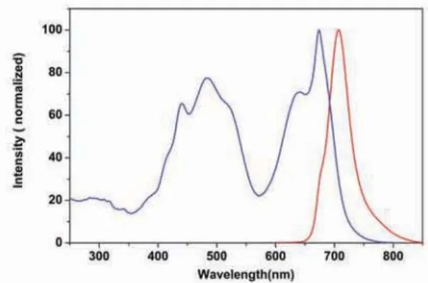
Figure 3.24 The excitation and emission spectra of iFluor™ 750 Goat Anti-Rabbit IgG conjugate (Cat# 16660) in PBS buffer (pH 7.2).
Figure 3.26 Detection of phosphatidylserine binding activity in Jurkat cells. Jurkat cells were stained with Annexin V-iFluor™ 750 conjugate (Cat# 20076) for 30 minutes. Blue: untreated live cells; Red: apoptotic cells treated with 1 μM staurosporine for 4 hours.
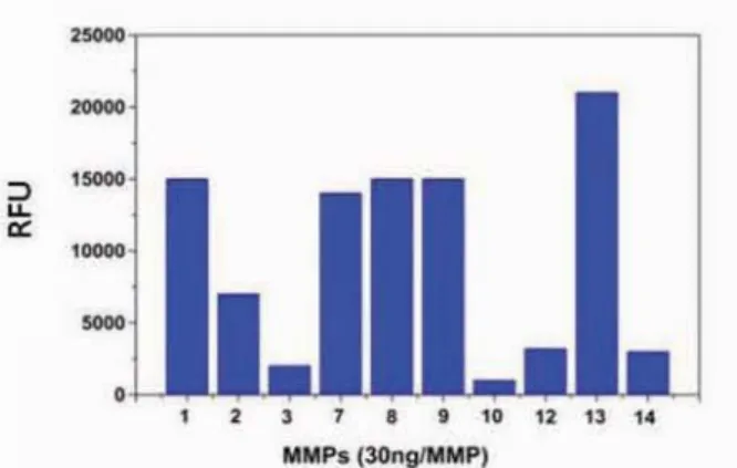
Figure 3.25 Detection of MMPs using MMP Infrared™ substrate, an infrared FRET substrate consisting of iFluor™ 750 (donor) and Tide Quencher™ 7 (acceptor). The APMA-activated MMPs, 30 ng each, were mixed with MMP Infrared™ substrate (10 μM). The fluorescence signal was monitored at 1 hour upon starting the enzyme reaction using a filter set of Ex/Em = 740/780 nm. The MMP Infrared™ substrate can detect the activity of sub-nanogram of all MMPs (n=3). This novel infrared FRET substrate can also be used for in vivo detection of MMP activities.
Ex (nm) 753
Em (nm) 779
EC (cm-1M-1) ~250,000
CF280 nm* 0.114
CF260 nm** 0.114
* CF280 nm = EC280 nm/ECdye max (Correction factor for peptides and proteins) ** CF260 nm = EC260 nm/ECdye max (Correction factor for oligos and nucleic acids)
Quick Summary
Product Ordering Information
www.aatbio.com
iFluor™ 790 Dyes
an Excellent Replacement for IRDye®800 and Alexa Fluor® 790 Dyes
Spectrally similar to IRDye®800 and Alexa Fluor® 790 dyes, iFluor™ 790 dyes are the longest-wavelength iFluor™ dyes that AAT Bioquest currently offers. Its fluorescence emission maximum around 810 nm is well separated from commonly used far-red fluorophores, including iFluor™ 647, iFluor™ 680 or allophycocyanin (APC), facilitating the rapidly growing NIR biological analysis. iFluor™ 790 is one of the brightest NIR fluorescent labeling dyes with fluorescence quantum yield close to 0.1. Its conjugates have been successfully used for NIR fluorescent probes-based in vivo imaging analysis.
Cat #
Size
Product Name
Alternative to
1360 5 mg iFluor™ 790 acid IRDye® 800, Alexa Fluor® 790
1362 1 mg iFluor™ 790 amine IRDye® 800, Alexa Fluor® 790
16507 200 μg iFluor™ 790 goat anti-mouse IgG (H+L) IRDye® 800, Alexa Fluor® 790
16587 200 μg iFluor™ 790 goat anti-mouse IgG (H+L) *Cross Adsorbed* IRDye® 800, Alexa Fluor® 790
16661 200 μg iFluor™ 790 goat anti-rabbit IgG (H+L) IRDye® 800, Alexa Fluor® 790
16721 200 μg iFluor™ 790 goat anti-rabbit IgG (H+L) *Cross Adsorbed* IRDye® 800, Alexa Fluor® 790
1366 1 mg iFluor™ 790 maleimide IRDye® 800, Alexa Fluor® 790
36801 1 mg iFluor™ 790 RGD conjugate IRDye® 800, Alexa Fluor® 790
1368 1 mg iFluor™ 790 succinimidyl ester IRDye® 800, Alexa Fluor® 790
23131 300 tests Phalloidin-iFluor™ 790 conjugate IRDye® 800, Alexa Fluor® 790
1265 1 kit ReadiLink™ iFluor™ 790 protein labeling kit IRDye® 800, Alexa Fluor® 790
iFluor™ 790 Dyes
3
iF
luor™ F
luor
esc
ent Labeling D
yes
Figure 3.27 The excitation and emission spectra of iFluor™ 790 Goat Anti-Rabbit IgG conjugate (Cat# 16661) in PBS buffer (pH 7.2).
Figure 3.28 Detection of caspase-3 activity in Jurkat cells with Z-KKK(iFluor™ 790)RKVGKDEVDKKC(TQ-8). Jurkat cells were treated with or without 20 PM camptothecin for 5 hours, and/or 5 PM of the caspase inhibitor AC-DEVD-CHO for 10 min. The caspase-3 assay solution was added and incubated at room temperature for 1 hour. The fluorescence intensity was measured at Ex/Em = 780/810 nm. This iFluor™ 790-based NIR fluorogenic FRET substrate was also used for in vivo apoptosis detection.
Figure 3.29 Mice with tumor were intravenously injected with 10 nmol of 2-Deoxyaminoglucose (DG)-iFluor™ 790 conjugate or control iFluor™ 790 acid and imaged at the 6th hour. Mouse injected with the DG-iFluor™ 790 showed specific accumulation of the iFluor™ 790 probe to the tumor site.
Ex (nm) 782
Em (nm) 811
EC (cm-1M-1) ~250,000
CF280 nm* 0.224
CF260 nm** 0.251
* CF280 nm = EC280 nm/ECdye max (Correction factor for peptides and proteins) ** CF260 nm = EC260 nm/ECdye max (Correction factor for oligos and nucleic acids)
Quick Summary
Product Ordering Information
ReadiLink
TMFluorescence Labeling Kits
www.aatbio.com
22
5FMt'BY
TBMFT!BBUCJPDPNtJOGP!BBUCJPDPN Unless otherwise specified, all products are for Research Use Only.Not for use in diagnostic or therapeutic procedures.
ReadiLink™ iFluor™ Protein Labeling Kits
ReadiLink™ iFluor™ Protein Labeling Kits provide a convenient way to label proteins using the stable reactive form of the iFluor™ dyes. The reactive iFluor™ dyes show good reactivity and selectivity with the aliphatic amines of proteins and forms a carboxamide bond, which is identical to, and is as stable as the natural peptide bond. iFluor™-protein conjugates may be used for immunofluorescent staining, fluorescent in situ hybridization, flow cytometry and other biological applications. Each kit comes with all the essential compo-nents for performing the conjugation reaction and for purifying the iFluor™-protein conjugates. ReadiLink™ Kits only require two simple mixing steps to produce the desired conjugates for flow cytometry and fluorescence imaging applications.
Product Ordering Information
Cat #
Size
Product Name
Alternative to
1220 1 kit ReadiLink™ iFluor™ 350 protein labeling kit Lightning-Link® dye labeling kits
1255 1 kit ReadiLink™ iFluor™ 488 protein labeling kit Lightning-Link® dye labeling kits
1227 1 kit ReadiLink™ iFluor™ 555 protein labeling kit Lightning-Link® dye labeling kits
1230 1 kit ReadiLink™ iFluor™ 594 protein labeling kit Lightning-Link® dye labeling kits
1260 1 kit ReadiLink™ iFluor™ 633 protein labeling kit Lightning-Link® dye labeling kits
1235 1 kit ReadiLink™ iFluor™ 647 protein labeling kit Lightning-Link® dye labeling kits
1240 1 kit ReadiLink™ iFluor™ 680 protein labeling kit Lightning-Link® dye labeling kits
1245 1 kit ReadiLink™ iFluor™ 700 protein labeling kit Lightning-Link® dye labeling kits
1250 1 kit ReadiLink™ iFluor™ 750 protein labeling kit Lightning-Link® dye labeling kits
1265 1 kit ReadiLink™ iFluor™ 790 protein labeling kit Lightning-Link® dye labeling kits
Key Features of ReadiLink™ Kits :
t Complete, all the components provided in the kits.
t Simple, only two mixing steps required.
t Rapid, less than 10 minutes hands-on time.
3
iF
luor™ F
luor
esc
ent Labeling D
yes
ReadiLink™ Kits Start Reaction 1st MixingStop Reaction and Quench Undesired Fluorescence
2nd Mixing
Desired Conjugate
t
Biotinylation of proteins and antibodies with ReadiViewTM biotin.t
Labeling proteins and antibodies with iFluorTM , mFluorTM, trFluorTM and other dyes.t
Labeling proteins and antibodies with APC, RPE and PerCP.t
Labeling proteins and antibodies with enzymes.t
Lableing small bioactive compounds.24
Hours Turnaround*
The Best Custom Bioconjugation Service
Together We ShineSM
* APC and RPE custom conjugation services require 3 working days.
mF
luor™ F
luor
esc
ent Labeling D
yes
mFluor™ Fluorescent Labeling Dyes
4
5FMt'BY
TBMFT!BBUCJPDPNtJOGP!BBUCJPDPNUnless otherwise specified, all products are for Research Use Only. Not for use in diagnostic or therapeutic procedures.
24
mFluor™ Fluorescent Labeling Dyes
www.aatbio.com
mFluor
TM
Labeling Dyes at-a-glance
Violet
(405 nm)
Blue
(488 nm)
Green
(532 nm)
Yellow
(561 nm)
Red
(633 nm)
Blue
mFluor™ 450
Green
mFluor™ 510
Orange
mFluor™ 540
mFluor™ Blue 570
Red
mFluor™ Green 620 mFluor™ Yellow 630
Far Red
mFluor™ Red 700
Infrared
mFluor™ Red 780
Excitation Laser Emission Color