Original Article
MiR-130a regulates the proliferation and
metastasis of HCC cells through targeting ZEB1/2
Ting Wang1, Jue Wei2, Ling Xu2, Rongrong Jia2, Chuanyong Guo1
1Medical College of Soochow University, Suzhou, Jiangsu, China; 2Department of Gastroenterology, Shanghai Tongren Hospital, Jiaotong University of Medicine, Shanghai, China
Received December 8, 2016; Accepted December 25, 2016; Epub March 1, 2017; Published March 15, 2017
Abstract: Accumulating evidence indicates that microRNA-130a (miR-130a) function as a tumor suppressor, in-volved in many biological processes including tumor initiation, development and metastasis. However, the function and underlying molecular mechanism of miR-130a in hepatocellular carcinoma (HCC) still remains to be further
elucidated. During to the resistant to chemo-/radio-therapy and high recurrence/metastatic rate after surgery, five-year survival rate of patients diagnosed with HCC is still unsatisfied. Thus, exploring the underlying mechanism of HCC and finding new treatment targets is essential for improving the survival rate of HCC patients. In the present
study, we measured the dysregulated level of miR-130a in clinical HCC tissues and HCC cells, and to investigate its function and underlying mechanisms in the initial and progression of HCC. The current study revealed that
miR-130a expression was significantly downregulated in HCC tissues and cell lines. Gain-of-function and loss-of-function
assays indicated that forced expression of miR-130a in HCC cells inhibited cell proliferation and migration/invasion.
Bioinformatics and luciferase reporter assays confirmed that ZEB1/2 was targets gene of miR-130a. The results of
the present study indicated that miR-130a could be a potential target for treating HCC.
Keywords: HCC, miR-130a, proliferation, metastasis, ZEB1/2
Introduction
Liver cancer is the second most common cause of cancer-associated death worldwide [1]. As a subtype of liver cancer, hepatocellular carcino-ma occupies 70-90% of pricarcino-mary liver cancer. During to the resistant to chemo-/radio-therapy and high recurrence/metastatic rate after sur-gery, five-year survival rate of patients diagnos- ed with HCC is still unsatisfied [2, 3]. Therefore, exploring the underlying mechanism of HCC and finding new treatment targets is essential for improving the survival rate of HCC patients. MicroRNAs (miRNAs), a group of small noncod-ing RNAs, have been reported as critical regu- lators involving in a variety of physiological and pathological processes through binding to the 3’-untranslated region (UTR) of the target Mrna [4]. MiR-130a has been shown to both as onco-gene and anti-oncoonco-gene in multiple cancers [5-8]. Li B et al. has reported that miR-130a down-regulated in hepatocellular carcinoma and associates with poor prognosis, however,
the underlying mechanism has not been com-pletely elucidated [9]. Thus, the function of miR-130a in HCC needs to be further illustrated. In the current study, we investigate the expre- ssion of miR-130a in HCC tissues and HCC cells. We also identified zinc finger E-box-bind- ing homeobox 1 and 2 (ZEB1 and ZEB2) as target gene of miR-130a involved in the func-tion of miR-130a exerted in the proliferafunc-tion and metastasis of HCC cells. We demonstrate that miR-130a acts as a tumor suppressor in HCC, and targeting miR-130a may be a novel pathway for HCC treatment.
Method and materials
Patients
Cell lines
Three HCC cell lines (SMMC-7721, HepG2, Hep3B), and a normal liver epithelium cell line (L02) were purchased from the Institute of Biochemistry and Cell Biology of the Chinese Academy of Sciences (Shanghai, China). All the cells were cultured in DMEM (GIBCO-BRL) medium supplemented with 10% fetal bovine serum (10% FBS), 100 U/mL penicillin, and 100 mg/mL streptomycin in humidified air at 37°C with 5% CO2.
Real-time quantitative reverse-transcription polymerase chain reaction (qRT-PCR)
Total RNA from tissues and cells was isolated with Trizol reagent (Invitrogen, CA, USA) under the manufacturer’s instructions. Reverse tran-scription was performed with PrimeScript RT reagent Kit (Takara, Japan) according to the manufacturer’s protocol. qRT-PCR was per-formed with SYBR Prime Script RT-PCR Kits (Takara, Japan) based on the manufacturer’s instructions. The miR-130a and EZH1/2 level was calculated with the 2-ΔΔCt method, which
was normalized to u6 and GAPDH, respective- ly. All assays were performed in triplicate. The expression levels were relative to the fold change of the corresponding controls, which were defined as 1.0.
Cell viability
Cell viability was assessed via 3-(4,5-dimethyl-thiazol-2-yl)-2, 5-diphenyl-trtrazolium bromide (MTT) assay. 5 × 10³ cells/well were seeded in
a 96-well flat-bottomed plate for 24 h, then transfected with indicated vectors and cultured in normal medium. At 0, 24, 48, 72 h and 96 h after transfection, the MTT solution (5 mg/ml, 20 μl) was added to each well. Following in- cubation for 4 h, the media was removed and 100 μl DMSO were added to each well. The rel-ative number of surviving cells was assessed by measuring the optical density (O.D.) of cell lysates at 560 nm. All assays were performed in triplicate.
Colony formation assay
Cells (500 cells/well) were plated in 6-well plates and incubated in normal medium at 37°C. After two weeks, the cells were fixed and stained with 0.1% crystal violet. The number of visible colonies was counted manually.
Wound healing assays
Cell migration capacity was calculated by wound healing assay. 2 × 10⁵ cells with or
without transfection were plated into 12- well plates and incubated in DMEM with 10% FBS at 37°C. After reaching 100% confluence, cells were wounded by scraping with a 200 μl tip, following washed 3 times in serum-free medium and incubated in regular medium. Wounds were observed at 0 and 48 h. The cell migration distance was calculated by sub-tracting the wound width at each time point from the wound width at the 0 h time point. Three independent assays were assayed. Cell migration and invasion assays
Cell migration/invasion assays were detected by transwell chamber (8 um pore size, Corn- ing). 48 h after transfection, cells in serum-free media were placed into the upper chamber, while medium containing 10% FBS was added into the lower chamber. After 48 h incubation, cells remaining in upper membrane were wiped off, and cells that migrated were fixed in metha-nol, and stained with 0.1% crystal violet. Cells were counted under a microscope. Three inde-pendent experiments were carried out.
Cell attachment and detachment assay
For attachment assay, cells were seeded in 24-well plates at 5 × 10⁴ cells per well. Unat-
tached cells were removed after 1 h incuba- tion, and the attached cells were counted after trypsinization. The data were presented as a percentage of the attached cells compared to total cells. For cell detachment assay, after 24 h incubation, the cells were incubated with 0.05% trypsin for 3 min to detach the cells. Then, the culture medium was added to inacti-vate the trypsin and the detached cells were collected. The remaining cells were incubated with 0.25% trypsin to detach and counted. The data were presented as a percentage of the detached cells to total cells. Three independent experiments were carried out.
Cell transfection
were two-sided. P<0.05 was considered statis-tically significant.
Results
Expression of miRNA-130a, ZEB1, and ZEB2 in HCC tissues and HCC cell lines
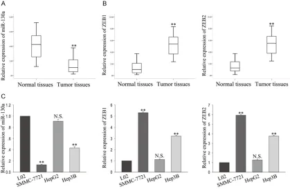
To investigate the role of miR-130a in HCC, we collected 36 pairs HCC tissues and adja-cent normal tissues, and performed qRT-PCR to measure the expression of miR-130a. As shown in Figure 1A, the average expression level of miR-130a was significantly downregu-lated in HCC tissue samples compared with adjacent controls. Additionally, we also ana-lyzed the putative targets of ZEB1, and ZEB2 in HCC tissues and corresponding normal tis-sues. The mRNA levels of ZEB1 and ZEB2 were significantly upregulated in HCC tissues com-pared with the adjacent tissues (Figure 1B). Moreover, the similar results were observed in the HCC cell lines. We examined the level of miR-130a and ZEB1/2 in three HCC cell lines (HepG2, Hep3B, SMMC-7721), and a normal liver epithelium cell line L02. As is shown in
Figure 1C, the lowest level of miR-130a (and ZEB1/2) was observed in SMMC-7721 (and HepG2) cells, while the highest level of miR-130a (and ZEB1/2) was obtained in HepG2 (and SMMC-7721) cells. These results indicat-ed that miR-130a and ZEB1/2 might be invol- ved in the progression of HCC.
ZEB1/2 was regulated by miRNA-130a both in transcription and translation level
Two putative targets of miR-130a screened by two bioinformatic analysis websites (http:// www.targetscan.org/cgi-bin/targetscan/vert_ 61/view_gene.cgi?taxid=9606&rs=NM_0010 83588&members=&showcnc=0&shownc=0& showncf=), and (http://mirdb.org/cgi-bin/tar-get_detail.cgi?targetID=1804872) and dual luciferase reporter assay was performed to make further confirm, as present in Figure 2A
(top), the 3’UTR region of both ZEB1 and ZE- B2 contains miR-130a binding site, and Figure 2A (bottom) showed that miR-130a mimics re- duced the luciferase activity of wild-type (WT) ZEB1 and ZEB2 reporter vector but not that of mutant reporter vector (MUT). To further con-firm the putative targets of miR-130a, we trans-fected miR-130a mimics or miR-130a inhibitor the Lipofectamine 2000 kit (Invitrogen)
accord-ing to the manufacturer’s instructions. Dual luciferase reporter assay
pmirGLO-ZEB1wt and pmirGLO-ZEB1-mut (miR-130a) (or pmirGLO-ZEB2wt and pmirGLO-ZEB2-mut (130a) was co-transfected with miR-130a mimics or miRNA NC into HEK 293 cells by Lipofectamine-mediated gene transfer. The relative luciferase activity was normalized to Renilla luciferase activity 48 h after transfec-tion. The data were relative to the fold change of the corresponding control groups defined as 1.0.
Western bolt analysis and antibodies
Total protein lysates were separated in 10% sodium dodecyl sulfate-polyacrylamide gel electrophoresis (SDS-PAGE), and were electro-phoretically transferred to polyvinylidene difluo-ride membranes (Roche). Protein loading was estimated using mouse anti-GAPDH monoclo-nal antibody. The membranes were blotted with 10% non-fat milk in TBST for 2 h at room tem-perature, washed and then probed with the rabbit anti-human ZEB1 (1:2000 dilution), ZE- B2 (1:2000 dilution), and GAPDH (1:3000 dilu-tion), overnight at 4°C, followed by treatment with secondary antibody conjugated to horse-radish peroxidase for 2 h at room temperature. The proteins were detected using an enhan- ced chemiluminescence system and exposed to X-ray film. All antibodies were purchased from Abcam (USA).
Statistical analysis
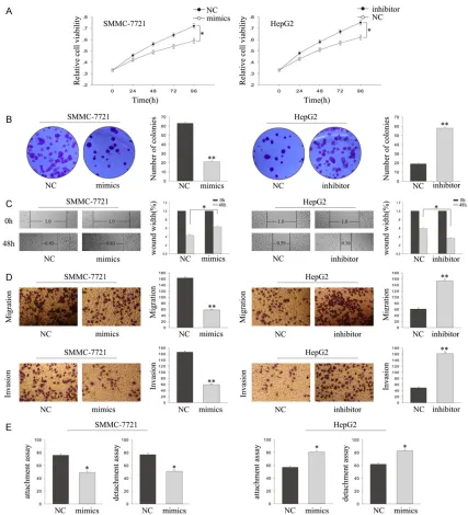
130a decrease cells viability, while downre- gulation of miR-130a increased cell viability. Consistent with MTT, colony formation show- ed that forced expression of miR-130a signifi-cantly reduced the proliferation ability of HCC cells, and vice vasa in cells knockdown miR-130a (Figure 3B). Then, results from wound healing assay, transwell and attachment/de- tachment assays showed that forced expres-sion of miR-130a significantly inhibited HCC cells metastasis, while deletion of miR-130a obviously facilitated cells metastasis (Figure 3C-E). These results together indicate that miR-130a could suppress the proliferation and metastasis of HCC cells.
The effect of miRNA-130a is dependent on ZEB1/2
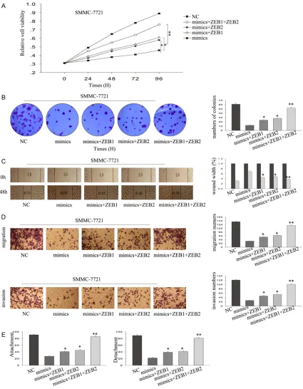
To further confirm that the role of miR-130a played in HCC is mediated by ZEB1/2, res- cue assays were performed. SMMC-7721 cells were transfected with empty vector, miR-130a into SMMC-7721 cells and HepG2 cells to
over-express or delete the over-expression of miR-130a (Figure 2B). After miR-130a mimics or inhibitor transfection, we analyzed the mRNA and pro-tein expression level of ZEB1 and ZEB2. As shown in Figure 2C, the mRNA and protein lev-els of ZEB1 and ZEB2 were markedly decreas- ed by miR-130a mimic transfection and obvi-ously increased by miR-130a inhibitor trans- fection compared with the negative control, re- spectively. These results determined that miR-130a directly targeted ZEB1 and ZEB2 and regulates their expression at transcriptional and translational levels.
The effects of miRNA-130a on cell prolifera-tion, metastasis of HCC cells
Then we explore the function of miR-130a on
proliferation and metastasis of HCC cells. Cell proliferation was measured by using MTT and colony formation assays. As shown in Figure 3A, MTT revealed that overexpression of
miR-Figure 1. Expression of miRNA-130a, ZEB1, and ZEB2 in HCC tissues and HCC cell lines. A, B. qRT-PCR was per
-formed to measure the miR-130a and ZEB1/2 expression in 36 pairs of HCC tissues and corresponding histologi -cally normal tissues. C. The level of miR-130a in four HCC cell lines (HepG2, Hep3B, SMMC-7721), and a normal liver epithelium cell line L02. Error bars represent the mean ± SEM of at least three independent experiments. N.S.:
[image:4.612.93.512.77.350.2]mimics and pcDNA3.1/ZEB1 or ZEB2. As sh- own in Figure 4A and 4B, MTT and colony for-mation assays revealed that single used of ZEB1 or ZEB2 can’t obviously reverse the anti-proliferation function of miR-130a, while co-transfected with ZEB1/2 and miR-130a could significantly abolished anti-proliferation func-tion of miR-130a. Addifunc-tionally, wound healing assays, transwell assays, and attachment/de-
[image:5.612.95.520.67.537.2]tachment assays described that single trans-fected with ZEB1 or ZEB2 can’t markedly re- verse the anti-metastasis effect of miR-130a, while co-transfected with ZEB1/2 and miR-130a could significantly reversed anti-metas- tasis function of miR-130a (Figure 4C-E). These findings suggest that miR-130a could signifi-cantly inhibit the proliferation and metastasis of HCC cells in a ZEB1/2-dependent way. Figure 2. ZEB1/2 was regulated by miRNA-130a both in transcription and translation level. A. The binding site between miR-130a and ZEB1/2 (top), luciferase reporter assays (bottom), luciferase reporter assays (bottom). B. Satisfactory transfection efficiency of miR-130a mimics and inhibitor. C. The level of ZEB1/2 both in mRNA and pro -tein level in response to the level of miR-130a. Error bars represent the mean ± SEM of at least three independent
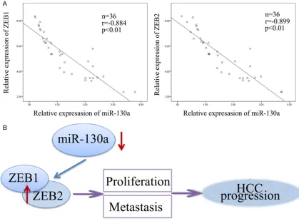
miR-130a expression level, respectively (2-tail- ed Spearman’s correlation, r=-0.884 and r= -0.899, P<0.01; Figure 5A). Collectively, these findings indicated a regulatory signal pathway in which miR-130a regulated ZEB1/2, eventu-ally causing decreased proliferation ability and metastasis capacity in HCC cells (Figure 5B). ZEB1/2 was negatively correlated with
miR-130a
[image:6.612.92.519.71.541.2]Finally, we analyzed the expression relation- ship between miR-130a and ZEB1/2. The re- sults showed that the level of ZEB1 and ZE- B2 was negative correlation with the level of
Figure 3. The effects of miRNA-130a on cell proliferation, metastasis of HCC cells. A, B. MTT and colony formation assays were performed to measure the effect of miR-130a on HCC cells proliferation. C-E. Wound healing assays, transwell assays and attachment/detachment assays were performed to detect the function of miR-130a on HCC cells migration capacity. Error bars represent the mean ± SEM of at least three independent experiments. N.S.: no
Figure 4. The effect of miRNA-130a is dependent on ZEB1/2. A, B. MTT and colony formation rescue assays. C-E.
Wound healing, transwell, and attachment/detachment rescue assays. Error bars represent the mean ± SEM of at
least three independent experiments. N.S.: no significant *P<0.05, **P<0.01 vs. control group.
Discussion
In the present study, we demonstrated that the decreased miR-130a was critical for the
Shun Meng et al. demonstrated that downregu-lation of miRNA-130a contributes to endotheli-al progenitor cell dysfunction in diabetic pa- tients via targeting Runx3 [18]. What’s more, miRNA-130a was identified by Ningwei Li et al. as novel regulators of cisplatin resistance in human ovarian cancer A2780 cells in 2015 [19]. In our study, we found that ectopic of miR-130a could inhibitor HCC cell proliferation and metastasis. Meanwhile, deletion of miR-130a could significantly promote the proliferation ability and metastatic potential of HCC cells. Additionally, we found that the 3’-UTR of ZEB1 and ZEB2 gene contained binding sites for miR-130a by utilizing TargetScan, and miRBase. ZEB1 and ZEB2, two members of the ZEB family of transcription factors, are characterized by the presence of two zinc finger clusters and a centrally located homeodomain [20]. The expression of ZEB1/2 is regulated by multiple signaling pathways, including WNT, transform-ing growth factor beta, and miRNAs [21]. We Hepatocellular carcinoma (HCC) is the third
[image:8.612.94.513.77.387.2]leading cause of cancer associated mortality. Although the improvement of mechanism study and clinical treatment has been made, the sur-vival of patients with HCC still remains unsatis-fied. Accumulating evidence has shown that microRNAs (miRNAs) function as critical factors for tumor recurrence and metastasis. MiR-130a has been identified down-regulated in hepatocellular carcinoma and associates with poor prognosis; however, the underlying mech-anism has not been completely elucidated. To investigate the function of miR-130 in HCC, we measure the level of miR-130 in HCC tissues and HCC cells. Results from qRT-PCR showed that miR-130a was significantly down-regulated in HCC tissues and HCC cells. MiR-130 has been reported in many cancers and function both as oncogene and tumor suppressor [5, 10-16]. In 2016, Quan Li et al. revealed that miRNA-130a regulates cell malignancy by tar-geting RECK in chronic myeloid leukemia [17].
Figure 5. ZEB1/2 was negatively correlated with miR-130a. A. The level of ZEB1/2 was negative correlation with the
level of miR-130a expression level (Spearman’s correlation analysis, r=-0.884 and r=-899, P<0.01). B. Schematic overview of miR-130 regulatory signaling. Error bars represent the mean ± SEM of at least three independent
and proliferation by targeting RUNX3. Oncol Rep 2015; 34: 1153-1161.
[8] Pan Y, Wang R, Zhang F, Chen Y, Lv Q, Long
G,Yang K. MicroRNA-130a inhibits cell prolif-eration, invasion and migration in human breast cancer by targeting the RAB5A. Int J Clin Exp Pathol 2015; 8: 384-393.
[9] Li B, Huang P, Qiu J, Liao Y, Hong J, Yuan Y. MicroRNA-130a is down-regulated in hepato-cellular carcinoma and associates with poor prognosis. Med Oncol 2014; 31: 230.
[10] Chen C, Hu Y, Li L. NRP1 is targeted by miR-130a and miR-130b, and is associated with multidrug resistance in epithelial ovarian can-cer based on integrated gene network analy-sis. Mol Med Rep 2016; 13: 188-196. [11] Chen C, Wang HJ, Yang LY, Jia XB, Xu P, Chen J,
Liu Y. [Expression of MiR-130a in serum sam-ples of patients with epithelial ovarian cancer and Its association with platinum resistance]. Sichuan Da Xue Xue Bao Yi Xue Ban 2016; 47: 60-63.
[12] Cui YL, Wang B, Gao HM, Xing YH, Li J, Li HJ, Lin
Z, Wang YQ. Interleukin-18 and miR-130a in
severe sepsis patients with thrombocytopenia. Patient Prefer Adherence 2016; 10: 313-319. [13] Han B, Lian L, Li X, Zhao C, Qu L, Liu C, Song J,
Yang N. Chicken gga-miR-130a targets HOXA3 and MDFIC and inhibits Marek’s disease lym-phoma cell proliferation and migration. Mol Biol Rep 2016; 43: 667-676.
[14] Signorelli SS, Li Volsi G, Pitruzzella A, Fiore V, Mangiafico M, Vanella L, Parenti R, Rizzo M, Li Volti G. Circulating miR-130a, miR-27b, and
miR-210 in patients with peripheral artery dis-ease and their potential relationship with oxi-dative stress: a pilot study. Angiology 2016; [Epub ahead of print].
[15] Song CL, Liu B, Shi YF, Liu N, Yan YY, Zhang JC, Xue X, Wang JP, Zhao Z, Liu JG, Li YX, Zhang
XH, Wu JD. MicroRNA-130a alleviates human coronary artery endothelial cell injury and
in-flammatory responses by targeting PTEN via
activating PI3K/Akt/eNOS signaling pathway. Oncotarget 2016; 7: 71922-71936.
[16] Tang G, Peng L, Qian G, Wang S, Hu H, Zhang X, Song G, Yao M, Zhai C. Resveratrol increases
microRNA-130a expression to promote angio-genesis and improve heart functions in mice after myocardial infarction. Exp Mol Pathol 2016; [Epub ahead of print].
[17] Li Q, Wu Y, Zhang J, Yi T, Li W. MicroRNA-130a
regulates cell malignancy by targeting RECK in chronic myeloid leukemia. Am J Transl Res 2016; 8: 955-967.
[18] Meng S, Cao J, Zhang X, Fan Y, Fang L, Wang C, Lv Z, Fu D, Li Y. Downregulation of
microRNA-130a contributes to endothelial progenitor cell
revealed that miR-130a can directly target to 3’-UTR of ZEB1 and ZEB2 and regulate their expression both at transcriptional and transla-tional levels. In HCC cells, miR-130a interacts with the oncogene ZEB1/ZEB2 at their 3’-UTR, and silenced the level of miR-130 results in high level of ZEB1/ZEB2 expression, while ecto-pic expression of miR-130a can inhibit the pro-gression of HCC cells.
In summary, our study was the first to investi-gate the role of miR-130a/ZEB1/ZEB2 in HCC cells. Our results showed that miR-130a was down-expression in HCC tissues and HCC cells and these results might provide a strong ratio-nale for its potential use as a therapeutic target for treating HCC.
Acknowledgements
The authors thank the laboratory members.
Disclosure of conflict of interest
None.
Address correspondence to: Chuanyong Guo, Me-
dical College of Soochow University, Suzhou
215-004, Jiangsu, China. E-mail: [email protected] References
[1] Torre LA, Bray F, Siegel RL, Ferlay J, Lortet-Tieulent J, Jemal A. Global cancer statistics, 2012. CA Cancer J Clin 2015; 65: 87-108. [2] Kuo MT. Redox regulation of multidrug
resis-tance in cancer chemotherapy: molecular mechanisms and therapeutic opportunities. Antioxid Redox Signal 2009; 11: 99-133. [3] Forner A, Llovet JM, Bruix J. Hepatocellular
car-cinoma. Lancet 2012; 379: 1245-1255. [4] Filipowicz W, Bhattacharyya SN, Sonenberg N.
Mechanisms of post-transcriptional regulation by microRNAs: are the answers in sight? Nat Rev Genet 2008; 9: 102-114.
[5] Chen J, Yan D, Wu W, Zhu J, Ye W, Shu Q.
MicroRNA-130a promotes the metastasis and epithelial-mesenchymal transition of osteosar-coma by targeting PTEN. Oncol Rep 2016; 35: 3285-3292.
[6] Liu Y, Li Y, Wang R, Qin S, Liu J, Su F,Yang Y,
Zhao F, Wang Z, Wu Q. MiR-130a-3p regulates
cell migration and invasion via inhibition of Smad4 in gemcitabine resistant hepatoma cells. J Exp Clin Cancer Res 2016; 35: 19. [7] Jiang H, Yu WW, Wang LL, Peng Y. miR-130a
[21] Pan Y, Zhang J, Fu H, Shen L. miR-144 func -tions as a tumor suppressor in breast cancer
through inhibiting ZEB1/2-mediated epithelial
mesenchymal transition process. Onco Targets Ther 2016; 9: 6247-6255.
dysfunction in diabetic patients via its target Runx3. PLoS One 2013; 8: e68611.
[19] Li N, Yang L, Wang H, Yi T, Jia X, Chen C, Xu P. MiR-130a and MiR-374a function as novel regulators of cisplatin resistance in human ovarian cancer A2780 cells. PLoS One 2015; 10: e0128886.
[20] Zhang P, Sun Y, Ma L. ZEB1: at the crossroads