Original Article
CAT104 silence behaves as a tumor
suppressor in human leukemia cells
by down regulating miR-182 expression
Chengfang Zhang1*, Guanli Song3*, Guanbo Song4*, Ruolei Li1, Min Gao1, Haiguo Zhang2
Departments of 1Clinical Laboratory, 2Hematology, Jining No. 1 People’s Hospital, Jining 272011, Shandong,
China; 3Department of Preventive and Health Care, Guang’anmen Hospital, China Academy of Chinese Medical
Sciences, Beijing 100053, China; 4Department of Clinical Laboratory, Jining Chinese Medicine Hospital, Jining
272000, Shandong, China. *Equal contributors.
Received November 15, 2017; Accepted November 28, 2017; Epub December 1, 2017; Published December 15, 2017
Abstract: Background: LncRNAs and miRNAs are found to play crucial roles in the tumorigenesis of acute myeloid leukemia (AML). We aimed to investigate the functions and mechanisms of lncRNA-CAT104 and miR-182 in AML. Methods: Expression of CAT104, miR-182, and ZEB1 in K562 and HL60 cell lines was respectively or synchronously altered by transfection. Expressions of CAT104, miR-182 and ZEB1 in cell were then analyzed by qRT-PCR. Cell
vi-ability, migration, invasion and apoptosis were evaluated by MTT, transwell assays and flow cytometry, respectively. Protein expressions of ZEB1 and factors related with apoptosis and two signal pathways (Wnt/β-catenin and JNK)
were detected by western blot. Results: CAT104 expressed highly in K562 and HL60 cells compared to embryonic kidney cell line HEK293 (P < 0.001). Knockdown of CAT104 inhibited cell viability, migration and invasion, but increased cell apoptosis of K562 and HL60 cells through inhibitionof miR-182 (P < 0.05). miR-182 promoted cell survival, migration and invasion through upregulatingthe expression of ZEB1 (P < 0.05). miR-182 silence
deacti-vated Wnt/β-catenin and JNK signal pathways by downregulating the expression of ZEB1 in K562 and HL60 cells.
Conclusion: LncRNA-CAT104 expressed highly in leukemia cells and its silence inhibited cell survival, migration and invasion by downregulating miR-182 expression. miR-182 functioned as an oncogene by upregulating ZEB1 via
which miR-182 silence deactivated Wnt/β-catenin and JNK signal pathways in leukemia cells.
Keywords: Acute myeloid leukemia, CAT104, miR-182, ZEB1, Wnt/β-catenin, JNK
Introduction
Acute myeloid leukemia (AML) is a cancer of the myeloid line of blood cells, characterized by that abnormal white blood cells in the bone marrow grow rapidly and interfere with the pro-duction of normal blood cells [1]. AML is the most common acute leukemia affecting adults, but is rare in children [2]. Chromosomal
abnor-malities, mutations in specific gene and non -coding RNAs such as microRNAs (miRNAs) have been considered to be possible causes for AML development [3-5]. Cure rates of AML in clinical trials ranged from 20-45% [6]. Thus, a compre-hensive understanding of the pathogenesis of AML is critical for developing novel therapeutic strategies.
Noncoding RNAs are found to show abnormal expression patterns in cancerous tissues which
sequester-The roles of CAT104 and miR-182 in AML
ing miRNAs [15, 16]. Therefore, lncRNA contrib-uted to a large range of functions such as mod-ulation of apoptosis and invasion [17], repro-gramming of induced pluripotent stem cells [18] and marker of cell fate [19]. A recent study found that a new lncRNA, CAT104, expressed highly in breast cancer cells and was related with the survival of breast cancer cells [20]. However, the effect of CAT104 on other cancers especially AML has not been revealed so far. In the present study, we aimed to investigate the role of CAT104 in leukemia cells. In addi-tion, we also explored the regulatory effect of miR-182 on leukemia cells. Our study indicates that CAT104 silence functions as a tumor sup-pressor in leukemia cells by downregulating miR-182 expression. miR-182 plays an onco-genic role in AML by upregulating ZEB1 expres-sionvia which miR-182 silence deactivates JNK
and Wnt/β-catenin signal pathways. Our study
may open new avenues in the research of AML pathogenesis for developing novel therapeutic strategies.
Materials and methods
Cell culture
Human leukemia cell lines K562 and HL60, human osteosarcoma cell line MG63, human osteoblast cell line hFOB1.19 and human em- bryonic kidney cell line HEK293 were purchas- ed from the American Type Culture Collection (ATCC, Rockville, MD, USA). MG63, hFOB1.19 and HEK293 cells were cultured in the Dulbe-
cco’s Modified Eagle’s Medium (DMEM, Gibco,
Grand Island, NY, USA), and K562 and HL60 cells were maintained in RPMI 1640 medium (Gibco). Both DMEM and RPMI 1640 media contained 10% of fetal bovineserum (FBS, Gibco), 100 U/ml of penicillin (Gibco) and 100
μg/ml of streptomycin (Gibco). All cells were cultured at 37°C in a humidified 5% CO2
incubator. Cell transfection
The short-hairpin RNA (shRNA) directly against human lncRNA-CAT104 was ligated into the U6/GFP/Neo plasmid (GenePharma, Shanghai, China) and was referred as sh-CAT104. The plasmid carrying a non-targeting sequence was used as a negative control (NC) of sh-CAT104 and was referred as sh-NC. The sequences of
full-length ZEB1 were constructed in the pEX plasmid (GenePharma) and were referred as pEX-ZEB1. The empty vector was used as a NC of pEX-ZEB1 and was referred as pEX. The short interfering RNAs (siRNA) targeting ZEB1 was referred as si-ZEB1. Non-targeting siRNA for negative control was referred as si-NC. si-ZEB1, si-NC, miR-182 mimic, mimic control, miR-182 inhibitor and inhibitor control were all synthe-sized by GenePharma Co. K562 and HL60 cells were transfected using Lipofectamine 3000 reagent (Invitrogen, Carlsbad, CA, USA) accord-ing to the manufacturer protocol. After 72 h of transfection, the stably transfected cells were selected by the culture medium containing 0.5 mg/ml G418 (Sigma-Aldrich, St Louis, MO, USA).
Real-time quantitative reverse-transcriptase PCR (qRT-PCR)
Total RNA was extracted from cells using Trizol reagent (Invitrogen) according to the manufac-turer’s instructions. For the test of miR-182 level, the Taqman MicroRNA Reverse Transcri- ption Kit and Taqman Universal Master Mix II (Applied Biosystems, Foster City, CA, USA) were used in turns for reverse transcription and qPCR with manufacturer’s protocols. U6 (Appli- ed Biosystems) was used as an internal stan-dard. To test the expression levels of CAT104 and mRNA of ZEB1, we used The ExiLERATE universal cDNA synthesis and SYBR® Green Master Mix Kits (Exiqon, Vedbaek, Denmark) in turns according to manufacturer’s protocols. The GAPDH (Exiqon) was used for normalizing. The changes in expression were calculated using the 2-ΔΔCt method.
Cell viability assay
For cell viability assay, a 3-(4, 5-dimethylthiazol- 2-yl)-2,5-diphenyl tetrazolium bromide (MTT) assay was used. Cells (5 × 10³) were seeded in
a 96-wells plate and 30 μl of MTT (5 mg/ml;
Klamar, Shanghai, China) solution was added in the cell culture for 4 h incubation at 37°C and 5% CO2 conditions. The media were discarded,
Cell apoptosis assay
Cell apoptosis analysis was performed us- ing Annexin V-FITC/PI apoptosis kit (Dojindo
Molecular Technologies, Kyushu, Japan). Briefly,
cells were washed in cold phosphatebuffered
saline (PBS) and resuspended in 100 μl of bind
-ing buffer contain-ing 5 μl of Annexin V-FITC and 5 μl of PI. Cells were then incubated for 10 min
at room temperature in the dark. Flow cytome-try analysis was done by using a FACS can (Beckman Coulter, Fullerton, CA, USA). The number of early apoptotic cells (Annexin-V posi-tive and PI negaposi-tive) were analyzed by using the FlowJo software (Flowjo LLC, Ashland, OR, USA). Cell migration and invasion assays
Cell migration was determined by using a
24-wells transwell culture chamber with 8 μm
PET membranes (Millipore, Bedford, MA, USA). Cells (1 × 10⁵) were suspended in 200 μl of serum-free DMEM medium on the upper com-partment, and 600 ml of complete medium containing 10% FBS (Gibco) was added to the lower compartment. After incubation at 37°C for 24 h according to the manufacturer’s proto-col, the chamber was carefully removed. Non-traversed cells on the upper surface of the membrane were wiped off carefully with a cot-ton swab. Traversed cells on the lower side
were fixed with methanol and then stained with
crystal violet. We counted the number of
tra-versed cells microscopically in five randomly chosen fields and got the average.
Cell invasion was determined also using a
24-wells transwell culture chamber with 8 μm
PET membranes (Millipore). Diluted Matrigel (BD, San Jose, CA, USA) was added vertically at the center of the membrane in the upper cham-ber and incubated at 37°C for 4 h to facilitate gel formation. Cells (5 × 10⁴) in 200 μl of serum-free DMEM medium were plated onto the upper compartment, while complete medium contain-ing 10% FBS was added to the lower chamber. After processing the invasion chambers for 24 hours (37°C, 5% CO2) in accordance with the manufacturer’s protocol, the non-invading cells were removed with a cotton swab; the invading
cells were fixed in methanol, then stained with
crystal violet solution and counted
microscopi-cally from five randomly chosen fields to get
the average. The data are presented as relative migration or invasion rates.
Western blot
Proteins used for western blot was extracted using RIPA lysis buffer (Beyotime, Shanghai,
China) and quantified using the BCA Protein
Assay Kit (Pierce, Rockford, IL, USA). Protein bands were separated by sodium dodecyl sul-fate-polyacrylamide gel electrophoresis
(SDS-PAGE) and transferred to a polyvinylidene diflu -oride membrane (PVDF, Millipore). After 2 h of blocking with 5% skim milk (Nestlé, Shuang- cheng, China) in Tris Buffered Saline with Tween (TBST), the membrane was incubated with pri-mary antibodies (all from Abcam, Cambridge, UK) at 4°C overnight. Primary antibodies in- cluded anti-Bcl-2 (ab59348, 1:1000), anti- Bax (ab182733, 1:2000), anti-pro-caspase-3 (ab44976, 1:500), anti-cleaved-caspase-3 (ab- 13847, 1:500), anti-caspase-9 (ab202068, 1:2000), ZEB1 (ab203829, 1:1000), anti-JNK (ab179461, 1:1000), anti-p-anti-JNK (ab1249- 56, 1:5000), anti-c-Jun (ab32137, 1:5000), anti- p-c-Jun (ab32385, 1:5000), anti-Wnt3a (ab28- 472, 1:1000), anti-Wnt5a (ab72583, 1:500),
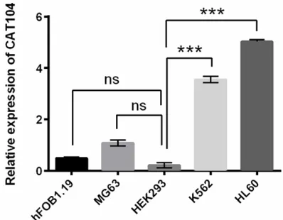
anti-β-catenin (ab6302, 1:4000) and anti-GAP -DH (ab8245, 1:1000). GAP-DH antibody was used as the endogenous protein for reference. After washing, the membrane was incubated with secondary antibody marked by horserad-ish peroxidase for 1 h at room temperature. Secondary antibodies included goat anti-mouse IgG (ab6789, 1:5000; Abcam) and goat anti-rabbit IgG (ab6721, 1:5000; Abcam). The sig-Figure 1. Different expression levels of CAT104 in
several cell lines. Expression levels of CAT104 were detected by qRT-PCR in human osteoblast cell line hFOB1.19, human osteosarcoma cell line MG63, hu-man embryonic kidney cell line HEK293 and huhu-man
leukemia cell line K562 and HL60. ns, no significant;
[image:3.612.89.288.73.228.2]The roles of CAT104 and miR-182 in AML
nals were developed by using enhanced
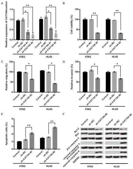
[image:4.612.89.522.69.618.2]chemi-luminescence (ECL) reagents (GE Healthcare, Little Chalfont, UK) according to the manufac-turer’s instructions. Figure 2. Knockdown of CAT104 inhibited cell viability, migration and invasion and promoted cell apoptosis in K562 and HL60 cells. A. Expression level of CAT104 was measured by qRT-PCR in K562 and HL60 cells with or without transfection with sh-CAT104/sh-NC. B. Cell viability was determined by a MTT assay in K562 and HL60 cells with or without transfection with sh-CAT104/sh-NC. C, D. Migration and invasion assays were used to determine the effect
of CAT104 on cell migration and invasion. E. Apoptosis rate was determined by flow cytometry in K562 and HL60
Statistical analysis
All experiments were repeated three times. Data are presented as the mean ± SD. Stati- stical analyses were performed by using SPSS 19.0 statistical software (SPSS, Chicago, IL, USA) and a one-way analysis of variance (ANOVA) to calculate the P-values. A P-value of < 0.05 was considered to be a statistically
sig-nificant result.
Results
Expression levels of CAT104 in several cell lines
We detected the expression levels of CAT104 in several cell lines which included human leuke-mia cell lines K562 and HL60, human osteo-sarcoma cell line MG63, human osteoblast cell line hFOB1.19 and human embryonic kid-ney cell line HEK293 by qRT-PCR. As shown in Figure 1, the expression of CAT104 in the cell line HEK293 was not statistically different from that expressed in cell lines hFOB1.19 and MG63 (P > 0.05). However, CAT104 expression was much higher in K562 and HL60 cell lines than HEK293 cell line (P < 0.001), indicating CAT104 was highly expressed in human leuke-mia cell lines K562 and HL60.
Knockdown of CAT104 inhibited cell viability, migration and invasion, but promoted cell apoptosis of leukemia cells
The sh-CAT104 was transfected into K562 and HL60 cell lines to knock down CAT104. After transfection, expression levels of CAT104 were
detected by qRT-PCR. As shown in Figure 2A,
transfection of sh-CAT104 significantly set off a
decrease in expression levels of CAT104 in both K562 and HL60 cells compared to trans-fectionof sh-NC (P < 0.05 or P < 0.01). The
high-er transfection efficiency was obshigh-erved in the
second group of sh-CAT104, which was select-ed for further experiments.
The effect of CAT104 on cell viability of K562 and HL60 cells was investigated by a MTT assay. As shown in Figure 2B, transfection of
sh-CAT104 significantly reduced cell viability
in both K562 and HL60 cells (P < 0.01). Cell migration and invasion of K562 and HL60 cells transfected with sh-CAT104 or sh-NC were de- tected by transwell assay with or without Matr- igel, respectively. Inhibition of CAT104 caused
significant decreases in therates of cell migra -tion and invasion of K562 and HL60 cells (P < 0.05, Figure 2C and 2D).
To reveal the effect of CAT104 on cell apop- tosis of K562 and HL60 cells, we performed an Annexin V FITC/PI apoptosis assay and detect-ed apoptosis relatdetect-ed proteins by western blot. Figure 2E demonstrated that transfection of
sh-CAT104 significantly caused increases in
the ratesof apoptosisin both K562 and HL60 cells (P < 0.01). Western blot showed that knockdown of CAT104 caused a decrease in the expression of the antiapoptotic protein Bcl-2 and increases in the expressions of pro-apoptotic protein Bax, executioner cleaved-cas-pase-3 and initiator cleaved-caspase-9 (Figure 2F). These results indicated that inhibition of CAT104 promoted cell apoptosis and reduced cell viability, migration and invasion of K562 and HL60 cells.
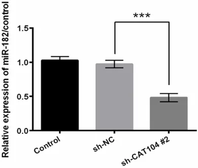
CAT104 positively regulated the expression of miR-182
miR-182 plays an important role in the process of many cancers [21, 22]. In the next step, we set out to identify the effect of CAT104 on miR-182. As shown in Figure 3, transfection of
sh-CAT104 significantly caused a decrease in the
expression of miR-182 (P < 0.001). This result supported that CAT104 positively regulated the expression of miR-182.
CAT104 silence behaved as a tumor suppres-sor in leukemia cells through downregulating the expression of miR-182
[image:5.612.91.286.73.239.2]We further investigated the association be- tween CAT104 and miR-182 in cell viability, Figure 3. CAT104 affected the expression of
The roles of CAT104 and miR-182 in AML
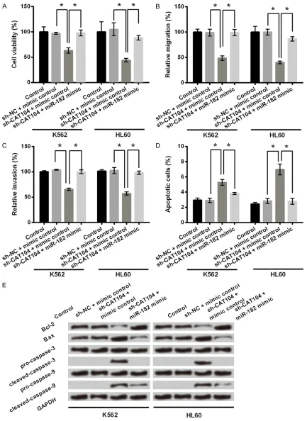
Figure 4. CAT104 silence inhibited cell viability, migration and invasion and promoted cell apoptosis in K562 and HL60 cells through regulating miR-182 expression. A. K562 and HL60 cells were co-transfected with sh-NC/sh-CAT104 and miR-182 mimic/mimic control. Cell viability was determined by a MTT assay. B, C. Migration and inva-sion assays were used to determine the effect of CAT104 and miR-182 on cell migration and invainva-sion. D. Apoptosis
migration, invasion and apoptosis of K562 and HL60 cells. Knockdown of CAT104 alone reduced cell viability of K562 and HL60 cells as described above (P < 0.05, Figure 4A). However, co-transfection of sh-CAT104 and miR-182 mi- mic reversed the effect of CAT104 inhibition on cell viability (P < 0.05). This reversal also occu- rred in cell migration, invasion and apoptosis of K562 and HL60 cells (Figure 4B-E). Knockdown of CAT104 alone reduced cell migrationand in- vasion of K562 and HL60 cells (P < 0.05), while
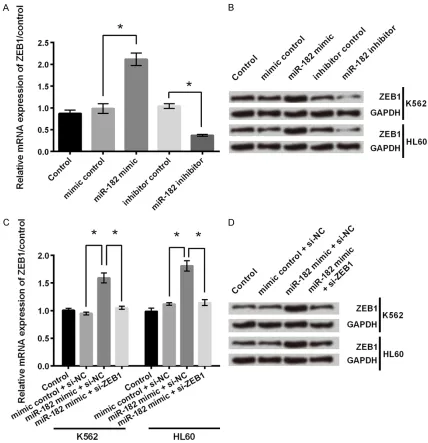
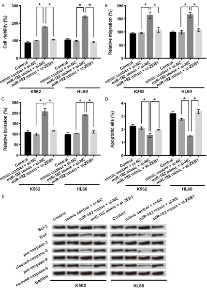
[image:7.612.92.520.71.514.2]The roles of CAT104 and miR-182 in AML
Figure 6. miR-182 affected cell viability, migration, invasion and apoptosis in K562 and HL60 cells through regu-lating ZEB1 expression. A-C. K562 and HL60 cells were co-transfected with si-NC/si-ZEB1 and miR-182 mimic/ mimic control. MTT, and transwell assays were used to determine cell viability, migration and invasion respectively
in co-transfected cells. D. Apoptosis rate was determined by flow cytometry in co-transfected cells. E. Western blot
and miR-182 mimic (P < 0.05). Considering CAT104 positively regulated the expression of
miR-182, these findings suggested that knock -down of CAT104 inhibited cell viability, migra-tion and invasion, and promoted cell apoptosis in leukemia cells maybe through downregulat-ing the expression of miR-182.
miR-182 upregulated the expression of ZEB1
ZEB1 is reported for its contributing role in cancer invasion and metastasis [23]. We next
identified the regulatory effect of miR-182 on
ZEB1 in both mRNA and protein levels. As shown in Figure 5A, overexpression of miR-182 caused an increase in the expression of ZEB1 mRNA, while inhibition of miR-182 had an inverse effect (P < 0.05). Western blot showed that the protein expression level of ZEB1 was increased by overexpression of miR-182, while was decreased by miR-182 suppression (Figure
cells, while miR-182 overexpression and inhibi-tion of ZEB1 expression simultaneously decr- eased cell viability (P < 0.05). The same trend was found in migration and invasion assays (Figure 6B and 6C). Overexpression of miR-182 alone increased cell migration and invasion rates in both K562 and HL60 cells (P < 0.05). In contrast, the migration and invasion rates of these two cell lines co-transfected with miR-182 mimic and si-ZEB1 were decreased (P < 0.05).
In apoptosis assay (Figure 6D), the apoptosis rates of K562 and HL60 cells transfected with
miR-182 mimic were significantly decreased
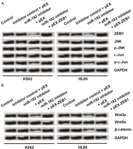
compared to that of cells transfected with mimic control (P < 0.05). On the contrary, co-transfection ofmiR-182 mimic and si-ZEB1 dra-matically resulted in increases of the apoptosis rates of these two cell lines (P < 0.05). The same trend was observed in analyzing expres-Figure 7. MiR-182 silence deactivated JNK and Wnt/β-catenin signaling
pathways through downgulating ZEB1 expression. A. K562 and HL60 cells were co-transfected with pEX/pEX-ZEB1 and miR-182 inhibitor/inhibitor control. Expressions of ZEB1 and proteins related to JNK signaling pathway were measured by western blot in transfected cells. B. Protein expression
levels of Wnt3a, Wnt5a and β-catenin in transfected cells were determined
by western blot.
5B). These results indicated that miR-182 upregulated the expression of ZEB1 in both mRNA and protein levels in K562 and HL60 cells.
miR-182 functioned as an oncogene in leukemia cells through upregulating the ex-pression of ZEB1
[image:9.612.90.365.70.378.2]The roles of CAT104 and miR-182 in AML
sion of apoptosis related proteins by western blot (Figure 6E). Overexpression of miR-182 alone increased the expression of the Bcl-2 and decreased expressions of Bax, cleaved-cas-pase-3 and cleaved-caspase-9. These effects were reversed by co-transfection of miR-182 mimic and si-ZEB1. These results suggested that overexpression of miR-182 promoted cell viability, migration and invasion and inhibited cell apoptosis in leukemia cells through upregu-lating ZEB1 expression.
MiR-182 silence deactivated JNK and
Wnt/β-catenin signal pathways through downregulat-ing ZEB1 expression
Finally, we sought to investigate the underling mechanisms in which miR-182 and ZEB1 in- volved. Firstly we measured the expression lev-els of JNK and c-Jun proteins in the case of miR-182 inhibition with or without ZEB1 overex-pression. As shown in Figure 7A, inhibition of miR-182 alone decreased expressions of ZEB1, p-JNK and p-c-Jun. However, co-transfection with miR-182 inhibitor and pEX-ZEB1 causedin-creases in expressions of ZEB1, p-JNK and p-c-Jun compared to that with miR-182 inhibitor and pEX. These results supported that knock-down of miR-182 deactivated JNK signal path-way possibly through downregulating ZEB1. Next, we measured the expression levels of
Wnt3a, Wnt5a and β-catenin proteins in the
case of miR-182 inhibition with or without over-expression of ZEB1. As shown in Figure 7B, inhibition of miR-182 alone decreased
expres-sions of Wnt3a and β-catenin proteins, while it
had no effect on Wnt5a. However, co-trans- fection with miR-182 inhibitor and pEX-ZEB1 caused increases in expressions of Wnt3a and
β-catenin proteins compared to that with
miR-182 inhibitor and pEX. These results supported that knockdown of miR-182 also could inhibit
Wnt/β-catenin signal pathway through down -regulating ZEB1.
Discussion
More and more lncRNAs were found to play important rolesin AML. For example, Wang et al
identified RUNXOR as a novel lncRNA involved
in a long range DNA interaction of RUNX1 in AML [24]. LncRNA-CCD26 has been shown to control the growth of myeloid leukemia cells through the regulation of KIT expression [25].
Another lncRNA (ZNF571-AS1) may be involved in AML via JAK/STAT signal pathways [26]. Com- pared to the huge number of lncRNA (about 27,919) [27], amount of lncRNAs which we have understood isonly the tip of the iceberg.
CAT104 was identified recently as a novel
lncRNA involved in predicting the survival of breast cancer [20]. However, the effect of CAT104 on AML is unknown. In the present
study, we for the first time found that CAT104
was upregulated in human leukemia cell lines (K562 and HL60). In addition, knockdown of
CAT104 significantly increased cell apoptosis
and inhibited cell viability, migration and
inva-sion of K562 and HL60 cells. These findings
supported that lncRNA-CAT104 might intervene in pathogenesis and function as an oncogene in AML.
Recent reports supported that lncRNAs could potentially interact with other classes of non-coding RNAs especially miRNAs and modulate their regulatory role [28]. For instance, lncRNA-CCAT1 negatively regulated miR-218-5p to pro-mote the development of gallbladder cancer [29]. However, how lncRNA-CAT104 exerts on- cogenic functions in AML is not clear. In the present study, we found CAT104 positively reg-ulated the expression of miR-182, which in turn affected cell survival, migration and invasion of leukemia cells.
In order to reveal the underling mechanism of miR-182 in AML, we tested the association be-
tween miR-182, ZEB1 and Wnt/β-catenin and
JNK signal pathways. Activated JNK regulates several important cellular functions including cell growth, differentiation, survival and apop-tosis by activating some molecules such as c-Jun [33]. However there is little research focused on the association between miR-182 and JNK signal pathway. We found miR-182 silence deactivated JNK signal pathways inleu-kemia cells. On the other hand, the canonical
Wnt-pathway can elevate the level of β-catenin to activate transcription of specific target genes and lead to tumor development [34].
Wnt/β-catenin signal pathway was reported to be acti-vated by miR-182-5p in human bladder cancer [35]. Similarly, we found miR-182 silence
deac-tivated Wnt/β-catenin signal pathway in leuke -mia cells. In addition, miR-182 silence
deacti-vated JNK and Wnt/β-catenin signal pathways
by downregulating ZEB1 in AML.
In conclusion, we demonstrated that lncRNA-CAT104 silence exerted anti-cancer functions in human leukemia cells by downregulating miR- 182 expression. miR-182 may play an oncogen-ic role in human leukemia cells through upregu-lating ZEB1 expression. Furthermore, miR-182
silence deactivates Wnt/β-catenin and JNK sig -nal pathways via downregulating ZEB1 expres-sion. The illustration of relationships among these molecules may promote understanding the pathogenesis of AML. In addition to trying to downregulate miR-182 levels, designing strategies to manipulate the expression of lncRNA-CAT104 may represent an alternative therapeutic approach for the treatment of AML. However, further research is still needed. Acknowledgements
This work was supported by the Science and Technology Development Projects of Jining City (Grant Number: 2015-57-102, 2015-57-134). Disclosure of conflict of interest
None.
Address correspondence to: Haiguo Zhang, Depart- ment of Hematology, Jining No. 1 People’s Hospital, No. 6, Jiankang Road, Jining 272011, Shandong, China. Tel: +86-0537-6056666; E-mail: [email protected]
References
[1] Tenen DG. Disruption of differentiation in hu-man cancer: AML shows the way. Nat Rev Cancer 2003; 3: 89.
[2] Jemal A, Thomas A, Murray T and Thun M. Cancer statistics, 2002. CA Cancer J Clin 2002; 52: 23.
[3] Heerema-Mckenney A and Arber DA. Acute my-eloid leukemia. Hematol Oncol Clin North Am 2009; 23: 633.
[4] Woods BA and Levine RL. The role of muta-tions in epigenetic regulators in myeloid malig-nancies. Immunol Rev 2012; 12: 599. [5] Emamdoost F, Khanahmad H,
Ganjalikhani-Hakemi M and Doosti A. The miR-125a-3p in-hibits TIM-3 expression in AML cell line HL-60 in vitro. Indian J Hematol Blood Transfus 2016; 1-6.
[6] Matthews JP, Bishop JF, Young GA, Juneja SK, Lowenthal RM, Garson OM, Cobcroft RG, Dodds AJ, Enno A and Gillett EA. Patterns of
failure with increasing intensification of induc -tion chemotherapy for acute myeloid leukae-mia. Br J Haematol 2015; 113: 727-736. [7] Spichtinger P and Cziczo DJ. MicroRNAs in
chronic lymphocytic leukemia: from causality to associations and back. Expert Rev Hematol 2012; 5: 579-581.
[8] Fu X, Ravindranath LN, Petrovics G and Sriva- stava S. Regulation of apoptosis by a
prostate-specific and prostate cancer-associated non -coding gene, PCGEM1. DNA Cell Biol 2006; 25: 135-141.
[9] Yang G, Lu X and Yuan L. LncRNA: a link be-tween RNA and cancer. Biochim Biophys Acta 2014; 1839: 1097.
[10] Bartel DP. MicroRNAs: genomics, biogenesis, mechanism, and function. Cell 2004; 116: 281-297.
[11] Lu J, Getz G, Miska EA, Alvarezsaavedra E, Lamb J, Peck D, Sweetcordero A, Ebert BL, Mak RH and Ferrando AA. MicroRNA
expres-sion profiles classify human cancers. Nature
2005; 435: 834-838.
[12] Calin GA and Croce CM. MicroRNA-cancer con-nection: the beginning of a new tale. Cancer Res 2006; 66: 7390.
[13] Bartels CL and Tsongalis GJ. MicroRNAs: novel biomarkers for human cancer. Clin Chem 2009; 55: 623.
[14] Hangauer MJ, Vaughn IW, McManus MT. Per- vasive transcription of the human genome
produces thousands of previously unidentified
long intergenic noncoding RNAs. PLoS Genet 2013; 9: e1003569.
interac-The roles of CAT104 and miR-182 in AML
tion with microRNA372 in liver cancer. Nucleic Acids Res 2010; 38: 5366-83.
[16] Wang K, Liu F, Luyu Z, Long B, Yuan SM, Wang Y, Liu CY, Teng S, Zhang XJ and Li PF. The long noncoding RNA CHRF regulates cardiac hyper-trophy by targeting miR-489. Circ Res 2014; 114: 1377-88.
[17] Mattick JS. The central role of RNA in human development and cognition. FEBS Lett 2011; 585: 1600.
[18] Li Z, Wang L, Wang Y, Liu L, Wang L, Li W, Zhou Q. Generation of an LncRNA Gtl2-GFP reporter for rapid assessment of pluripotency in mouse induced pluripotent stem cells. J Genet Genomics 2015; 42: 125-128.
[19] Flynn RA, Chang HY. Long noncoding RNAs in cell-fate programming and reprogramming. Cell Stem Cell 2014; 14: 752.
[20] Guo W, Wang Q, Zhan Y, Chen X, Yu Q, Zhang J, Wang Y, Xu XJ and Zhu L. Transcriptome se-quencing uncovers a three-long noncoding RNA signature in predicting breast cancer sur-vival. Sci Rep 2016; 6: 27931.
[21] Tang T, Wong HK, Gu W, Yu MY, To KF, Wang CC, Wong YF, Cheung TH, Chung TK and Choy KW. MicroRNA-182 plays an onco-miRNA role in cervical cancer. Gynecol Oncol 2013; 129: 199-208.
[22] Chiang CH, Hou MF and Hung WC. Up-regul-
ation of miR-182 by β-catenin in breast cancer
increases tumorigenicity and invasiveness by targeting the matrix metalloproteinase inhibi-tor RECK. Biochim Biophys Acta 2013; 1830: 3067-3076.
[23] Siebzehnrubl FA, Silver DJ, Tugertimur B, Deleyrolle LP, Siebzehnrubl D, Sarkisian MR, Devers KG, Yachnis AT, Kupper MD, Neal D, Nabilsi NH, Kladde MP, Suslov O, Brabletz S, Brabletz T, Reynolds BA and Steindler DA. The ZEB1 pathway links glioblastoma initiation, in-vasion and chemoresistance. EMBO Mol Med 2013; 5: 1196-1212.
[24] Wang H, Guo R, Hoffman AR, Hu J and Li W. Effect of long noncoding RNA RUNXOR on the epigenetic regulation of RUNX1 in acute myelo-cytic leukemia. J Clin Oncol 2015; 33: 7018-7018.
[25] Hirano T, Yoshikawa R, Harada H, Harada Y, Ishida A and Yamazaki T. Long noncoding RNA, CCDC26, controls myeloid leukemia cell growth through regulation of KIT expression. Mol Cancer 2015; 14: 90.
[26] Pan JQ, Zhang YQ, Wang JH, Xu P and Wang W. lncRNA co-expression network model for the prognostic analysis of acute myeloid leukemia. Int J Mol Med 2017; 39: 663-671.
[27] Hon CC, Ramilowski JA, Harshbarger J, Bertin N, Rackham OJ, Gough J, Denisenko E, Schm- eier S, Poulsen TM and Severin J. An atlas of human long non-coding RNAs with accurate 5’ ends. Nature 2017; 543: 199.
[28] Paraskevopoulou MD and Hatzigeorgiou AG. Analyzing MiRNA-LncRNA interactions. Metho- ds Mol Biol 2016; 1402: 271-286.
[29] Ma MZ, Chu BF, Zhang Y, Weng MZ, Qin YY, Gong W and Quan ZW. Long non-coding RNA CCAT1 promotes gallbladder cancer develop-ment via negative modulation of miRNA-218-5p. Cell Death Dis 2015; 6: e1583. [30] Hanke M, Hoefig K, Merz H, Feller AC, Kausch
I, Jocham D, Warnecke JM, Sczakiel G. A robust methodology to study urine microRNA as tum- or marker: microRNA-126 and microRNA-182 are related to urinary bladder cancer. Urol Oncol 2010; 28: 655-61.
[31] Brabletz S and Brabletz T. The ZEB/miR-200 feedback loop--a motor of cellular plasticity in development and cancer? EMBO Rep 2010; 11: 670.
[32] Wellner U, Schubert J, Burk UC, Schmalhofer O, Zhu F, Sonntag A, Waldvogel B, Vannier C, Darling D and Zur HA. The EMT-activator ZEB1 promotes tumorigenicity by repressing stem-ness-inhibiting microRNAs. Nat Cell Biol 2009; 11: 1487-1495.
[33] Dong C, Davis RJ and Flavell RA. Signaling by the JNK group of MAP kinases. J Clin Immunol 2001; 21: 253.
[34] Dihlmann S and Von KD. Wnt/beta-catenin-pathway as a molecular target for future anti-cancer therapeutics. Int J Cancer 2004; 113: 515-524.