Original Article
Clinical relevance of tag single
nucleotide polymorphisms within the CAT gene in
patients with PTSD in the Chongqing Han population
Zhao-Xia Duan1, Wei Li2, Jian-Yi Kang1, Jie-Yuan Zhang1, Kui-Jun Chen1, Bing-Cang Li1, Mei He3, Jian-Min Wang1
1State Key Laboratory of Trauma, Burns and Combined Injury, Department 6 of Institute of Surgery Research, Daping Hospital, Third Military Medical University, Chongqing 400042, China; 2Department of Neurology, Daping Hospital, Third Military Medical University, Chongqing 400042, China; 3Department of Clinical Psychology, South-west Hospital, Third Military Medical University, Chongqing 400038, China
Received January 20, 2014; Accepted February 4, 2014; Epub March 15, 2014; Published April 1, 2014
Abstract: Background: Free radical-induced oxidative damage of the brain has been implicated in a number of psychiatric disorders, including post-traumatic stress disorder (PTSD). Catalase (CAT) is a major antioxidant enzyme and a number of polymorphisms in CAT have been shown to be associated with several diseases, including hyper-tension, diabetes mellitus, Alzheimer’s disease, and vitiligo. The aim of this study was to evaluate the association of CAT gene polymorphisms with PTSD in a case-control study. Materials and methods: A total of 460 unrelated adult Chinese Han adults, including 287 healthy volunteers and 173 patients with PTSD. Six tag single-nucleotide polymorphisms (tSNPs) were selected from the entire CAT gene through construction of haplotype bins, and they were genotyped using an improved multiplex ligation detection reaction (iMLDR) technique. Allelic frequencies and clinical characteristics were compared in two independent Chinese Han populations. Results: Six tag SNPs were
identified in the Chinese Han population and all were common SNPs. However, we could detect no evidence of
genetic association between six tag SNPs in the CAT gene and PTSD in the Chinese Han population. Conclusions: This result suggests that six tag SNPs of the CAT gene may not be associated with PTSD, and that CAT gene might
not influence the development of PTSD in patients following exposure to a traumatic event, also may be the sample
sizes too small to allow a meaningful test.
Keywords: Catalase, tag single nucleotide polymorphisms, post-traumatic stress disorder, case-control study
Introduction
Posttraumatic stress disorder (PTSD) is
classi-fied as an anxiety disorder within the DSM-IV. It is defined as the development of symptoms fol -lowing exposure to an extreme traumatic event (criterion A). These symptoms are character-ized by the following three separate but inter-related symptom clusters: re-experiencing, avoidance and numbing, and hyper arousal (DSM 1994). Exposure to a traumatic event is the essential element in the development of PTSD. Approximately 50-70% of the U.S. popu-lation is exposed to a traumatic event some-time during their lifesome-time. However, only 5-12% develops PTSD [1, 2]. The reasons for these dif-ferences in prevalence are not completely cle- ar, but likely relate to the heterogeneity in the
Oxidative stress is a biological condition that is characterized by production of excessive amounts of oxidants, decreased levels of anti-oxidants or both. Excessive amount of free radi-cals may chemically react with cell membrane lipids, proteins and nucleic acids that conse-quently have the potential to damage the struc-ture of neuronal cells [5, 6]. In contrast, oxida-tive stress results from impaired oxidaoxida-tive defense mechanisms, such as depletion of enzymatic (e.g., superoxide dismutase, cata-lase and glutathione peroxidase, GPX) and non-enzymatic (e.g., glutathione, GSH, vitamins A, C, and E, and selenium) antioxidants. Either way, the consequence of oxidative stress is increased damage to all major groups of cellu-lar macromolecules including proteins, lipids, carbohydrates, and nucleic acids, which may result in apoptosis or necrosis [7, 8]. The theory of oxidative stress in mental disorders is based on vulnerability of the brain to oxidative dam-age [9]. Recently, several studies have suggest-ed an association between oxidative stress and psychopathology in anxiety disorders including panic disorder, obsessive-compulsive disorder and PTSD [5]. However, to the best of our knowl-edge there has been no study published yet investigating the oxidative stress markers in PTSD.
Catalase is a ubiquitous enzyme found in all known organisms. It is most abundant in the liver, kidneys, and erythrocytes. Together with superoxide dismutase (SOD) and GPX, catalase constitutes a primary defense against oxidative stress. Catalase has a very high turnover
num-hypertension, angle closure glaucoma, Alzhe- imer’s disease and vitiligo has been investigat-ed [12-14]. However, to our knowlinvestigat-edge, its genetic effects on PTSD have not been studied yet, despite the presumptively important role of CAT in anxiety disorders including PTSD. Therefore, we investigated the genetic effects of the entire CAT gene on the risk of PTSD. In this study, we hypothesized that the genetic variations in the CAT gene might affect the mor-bidity of PTSD in patients following exposure to a traumatic event. To assess the association of common genetic variants within the entire CAT gene with PTSD susceptibility comprehensively, we selected a set of tag SNPs (tSNPs) within the entire CAT gene and investigated their clini-cal relevance in relation to the development of PTSD in patients with exposure to traumatic events.
Materials and methods
Study design and data collection
A total of 460 unrelated Han Chinese adults, including 287 healthy volunteers and 173 patients with PTSD, were recruited from the Chongqing district for this study. The protocol of this study was approved by the Ethical and Protocol Review Committee of the Third Military
Medical University. The benefits and risks of
[image:2.612.90.347.96.280.2]the study participation were fully explained to each subject and written informed consent was obtained from the participants or their next of kin.
Table 1. The primer sequence and concentration in PCR mixture1
Primer Name Primer Sequence TemperatureAnnealing rs10836233 F TGCTATGGCCATCACCAGACAC 54.6°C rs10836233 R CCACAGCTCCATGACTCCTGTTC 56.5°C rs208679 F GCCCACTTTGTCACAGGCAGAA 54.6°C rs208679 R GGCTCTACACTGAGGGATTTTCCAATA 44.4°C rs2300182 F GTTGAAGCTTTCCTGCCCCACT 54.6°C rs2300182 R CCTCCTGCCCAATGAAGGAATC 54.6°C rs7104301 F TCAGCACTGATTTCACAACAGATCA 48.0°C rs7104301 R TGGAGTCCTCGAGATACTGGCATTT 49.0°C rs769217 F CCTTTTTGCCTATCCTGACACTCAC 48.0°C rs769217 R AGGGGGAGCCCAACGTCTTTAG 59.0°C rs7949972 F GCTGGTCTTTGGTTACCCTGGTATT 48.0°C rs7949972 R CATTCCCCAGGGATCACTCTGA 54.6°C
Healthy control group
The healthy volunteers control group consisted of 175 men and 112 women with a median age of 35 (18-55) years. All volunteers were nursing staff, medical staff, and university staff and students in the Daping Hospital. Formal screen-ing for psychological disorders was not under-taken in the control population. Therefore, the control subjects represented an unselected control group. Controls were recruited in the Chongqing region and all belonged to the Han population.
PTSD patients
A total of 173 unrelated Chongqing Han patients (103 men and 70 women) meeting the Diagnostic and Statistical Manual of Mental
Disorders-IV (DSM-IV) criteria for PTSD were
recruited for the study. Patients were assessed for PTSD by a consultant psychiatrist or a senior
psychiatric registrar using DSM-IV criteria.
Furthermore, every patient exceeded the clini-cal cutoff score of 94 on the Mississippi Sclini-cale for combat-related PTSD [15]. The patients’ clinical history and demographic data including the ethnic background were also obtained. None of the patients had received treatment
for a psychiatric disorder or had been treated with psychotropic medication. Following PTSD assessment with a psychiatrist, treatment was then commenced. Patients were only excluded if other major Axis I diagnoses (excepting major depression) were present, such as bipolar dis-order, schizophrenia or other psychotic disor-ders, and alcohol or substance abuse during the past 6 months. Patients were also excluded if there were any medical or psychiatric condi-tions, including pregnancy and lactation that could pose health risks during the course of the study. The patients had a mean age of 36 years
(± 6.9). All patients had sufficient comprehen -sion of Chinese and could understand the administered questionnaires. Patients were assessed for PTSD by a consultant psychiatrist
or a senior psychiatric registrar using DSM-IV
[image:3.612.92.527.89.345.2](American Psychiatric Association, 1994) crite-ria for a primary diagnosis of PTSD during the previous 6 months. All these patients were adm- itted to the Clinical Psychology Department of Southwest Hospital of Chongqing, Department of Psychiatry of the Mental Health Center of Chongqing, and outpatient in the Department of Psychiatry of the Chongqing Medical Unive- rsity, between the 1st of January 2011 and the 1st of June 2013.
Table 2. The probe sequence and concentration in probe mixture
Probe Name Target Allele Concen-Probe tration
PROBE SEQUENCE (5’ PHOSPHORATED)
Anneal-ing Tem-perature
rs10836233 FG G 1 μM TCTCTCGGGTCAATTCGTCCTTGCCAAATAGGCAGAGCAGAAGGTATCTG 69.3°C
rs10836233 FA A 1 μM TGTTCGTGGGCCGGATTAGTGCCAAATAGGCAGAGCAGAAGGTATCTA 67.2°C
rs10836233 FP 2 μM CTGCTACTGCATATAACACCACTGCTACTTTTTT 63.4°C
rs208679 FG G 1 μM TTCCGCGTTCGGACTGATATTTAGTGCCCTCAGATGTAAAGTGAAGGTAGGAG 66.4°C
rs208679 FA A 1 μM TACGGTTATTCGGGCTCCTGTTTAGTGCCCTCAGATGTAAAGTGAAGGTAGGAA 66.6°C
rs208679 FP 2 μM SAGTCCATTCCCYCTTAYAGGATTGTTATAATTTTTTTTTTTTTTTT 63.0°C
rs2300182 RA A 1 μM TCTCTCGGGTCAATTCGTCCTTTCCATTTCCCCACTCTAACAGTTAGATGAA 67.4°C
rs2300182 RT T 1 μM TGTTCGTGGGCCGGATTAGTTCCATTTCCCCACTCTAACAGTTAGATGAT 66.6°C
rs2300182 RP 2 μM TTGTCCTCATTTATATAACAGCTTTCCCCTTTTTT 65.2°C
rs7104301 RG G 1 μM TCTCTCGGGTCAATTCGTCCTTTCAAATTAAGAGTCTGGTAGYAGTTTACAGTTAGAC 66.2°C rs7104301 RA A 1 μM TGTTCGTGGGCCGGATTAGTTCAAATTAAGAGTCTGGTAGYAGTTTACAGTTAGAT 65.8°C
rs7104301 RP 2 μM RTGGAGTCGAAAGAAAGAAAATACCAYCTTTTTTTTTTTTTTTT 63.9°C
rs769217 FC C 1 μM TTCCGCGTTCGGACTGATATGAGTGGCCAACTACCAGCGTCAC 67.4°C
rs769217 FT T 1 μM TACGGTTATTCGGGCTCCTGTGAGTGGCCAACTACCAGCGTCAT 66.8°C
rs769217 FP 2 μM GRCCCGATRTGCATGCAGGATTTTTTTTTTTTTTTTTTTTTTTTTTTT 72.1°C
rs7949972 RC C 1 μM TTCCGCGTTCGGACTGATATCACGTTTTGAGCTGAGAATGGTCAG 66.3°C
rs7949972 RT T 1 μM TACGGTTATTCGGGCTCCTGTCACGTTTTGAGCTGAGAATGGTCAA 66.6°C
rs7949972 RP 2 μM GTTCAGTCTTCAAGATGTCTGGGAATTTTTTTTT 63.0°C
Table 3. Frequencies of CAT gene polymorphisms in healthy volunteers and patients with PTSD
NCBI rs# Location Position Variant MAF (Hapmap-HCB) healthy volunteers patients with PTSD
Genotype, N (287) MAF HWE Genotype, N (173) MAF HWE rs208679 5’ flanking -6411 A/G 0.22 AA 170 AG 100 GG 17 0.23 0.65 AA 93 AG 65 GG 15 0.27 0.45 rs10836233 5’ flanking -2900 G/A 0.30 GG 137 GA 118 AA 32 0.32 0.39 GG 94 GA 60 AA 19 0.28 0.06 rs2300182 Intron1 7376 T/A 0.23 TT 201 TA 81 AA 5 0.16 0.33 TT 121 TA 46 AA 6 0.17 0.53 rs769217 exon 9 (synon) 22436 T/C 0.49 TT 50 TC 157 CC 80 0.55 0.07 TT 37 TC 79 CC 57 0.56 0.33 rs7104301 3’ flanking 33166 A/G 0.23 AA 153 AG 113 GG 21 0.27 0.98 AA 101 AG 60 GG 12 0.24 0.46 rs7949972 3’ flanking 41570 C/T 0.45 CC 96 CT 138 TT 53 0.43 0.78 CC 61 CT 79 TT 32 0.42 0.47
Note: MAF indicates minor allele frequency; HWE indicates Hardy-Weinberg Equilibrium.
Table 4. Analyses of association of CAT gene polymorphisms with the risk of PTSD between healthy volunteers and patients with PTSD
NCBI rs# Location Variant Genotype
Frequency Codominant Dominant* Recessive
healthy
volunteers patients with PTSD (+) P OR (95% CI) P OR (95% CI) P OR (95% CI) rs208679 5’ flanking A/G AA 170 (59.20) 93 (53.76) 0.38 0.80 (0.59-1.09) 0.29 0.80 (0.55~1.17) 0.26 0.66 (0.32~1.37)
AG 100 (34.80) 65 (37.57) GG 17 (6.00) 15 (8.67)
rs10836233 5’ flanking G/A GG 137 (47.70) 94 (54.34) 0.35 0.85 (0.64-1.14) 0.18 1.30 (0.89~1.90) 1.00 1.02 (0.56~1.86) GA 118 (41.20) 60 (34.68)
AA 32 (11.10) 19 (10.98)
rs2300182 Intron1 T/A TT 201 (70.00) 121 (69.94) 0.49 1.07 (0.75-1.53) 1.00 1.00 (0.66~1.50) 0.35 0.49 (0.15~1.64) TA 81 (28.20) 46 (26.59)
AA 5 (1.80) 6 (3.47)
rs769217 exon 9 (synon) T/C TT 50 (17.40) 37 (21.39) 0.17 1.02 (0.78-1.34) 0.25 1.27 (0.85~1.91) 0.33 0.78 (0.48~1.25) TC 157 (54.70) 79 (45.66)
CC 80 (27.90) 57 (32.95)
rs7104301 3’ flanking A/G AA 153 (53.30) 101 (58.38) 0.56 1.15 (0.85-1.57) 0.33 1.23 (0.84~1.80) 1.00 1.06 (0.51~2.21) AG 113 (39.40) 60 (34.68)
GG 21 (7.30) 12 (6.94)
rs7949972 3’ flanking C/T CC 96 (33.40) 61 (35.47) 0.88 1.02 (0.78-1.34) 0.69 1.09 (0.74~1.63) 0.90 0.95 (0.59~1.54) CT 138 (48.10) 79 (45.93)
TT 53 (18.50) 32 (18.60)
[image:4.792.91.706.236.511.2]Selections of SNPs
The full sequence of the human CAT gene (Accession Number: NC-000011.9 Reference GRCh37.p5 Primary Assembly) observed in the current study included 10 kb upstream of the transcription start site all exons and introns and 10 kb downstream of the stop codon (33.235 kb total), which was pinpointed to chromosome 11, position 34460472-3449 3607 (data retrieved from Genbank in the Web site of NCBI). Genetic variation data for the entire CAT gene was obtained from the HapMap project for 45 healthy Chinese Han Beijing (CHB) adults (www.hapmap.org). From this
database, a total of 105 SNPs were identified,
of which, 44 SNPs with minor allele frequency (MAF) of more than or equal to 0.10 were selected for the analysis of htSNPs. Haplotype blocks were constructed throughout the entire CAT gene, using Haploview, version 4.0 (Broad Institute of MIT and Harvard, Cambridge, MA), a software package that provides computation of LD statistics and population haplotype pat-terns from genotype data [16]. Haplotype blocks represent regions inherited without sub-stantial recombination in the ancestors of the current population. The history of recombina-tion between a pair of SNPs can be estimated with the use of the normalized measure of allel-ic association D’ (value of D prime between the two loci) [17, 18]. The criteria for the selected SNPs to construct a haplotype block is that all SNPs in one region must be in strong LD with D’
>0.98 for the upper 95% confidence bound,
and >0.7 for the lower bound. A maximally infor-mative htSNP was then selected from each block using the software Tagger program (http://www.broad.mit.edu/mpg/haploview). This algorithm selects a subset of variants that capture all known common genetic variations in a gene, based on an LD threshold of r2≥0.8. The inverse of r2 represents the ratio of sample size needed to detect an indirect association with an un-analyzed SNP to direct association at the same power.
SNPs genotyping
The genomic DNA was extracted from periph-eral blood leukocytes using a Wizard Genomic
DNA Purification Kit (Promega, Madison,
Wis-consin, USA) following the manufacturer’s instructions. A multiplex polymerase chain rea- ction (PCR) -ligation detection reaction method
was used for genotyping the selected 6 tag SNPs. For each SNP, the alleles were
distin-guished by different fluorescent labels of allele specific oligonucleotide probe pairs. Different
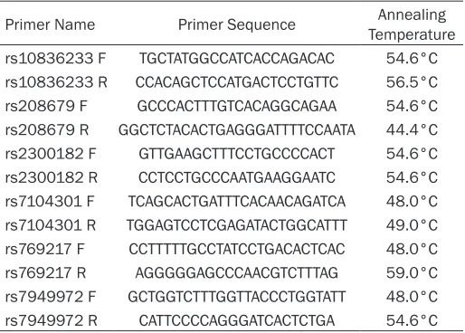
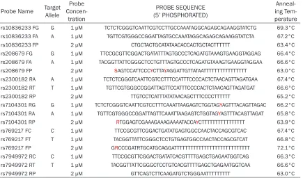
SNPs were further distinguished by different extended lengths at the 3’ end. The primer and probe information in two mixtures are described in Tables 1 and 2, respectively. Genotyping was performed in a blinded fashion without knowl-edge of the patients’ clinical data, and approxi-mately 10% of the samples were genotyp- ed in duplicate to monitor genotyping qua- lity.
Statistical analyses
Allele frequencies for each tSNP were deter-mined by gene counting. The genotype distribu-tion of each tSNP was analyzed for deviadistribu-tions
from the Hardy-Weinberg equilibrium using χ2 analyses. The extent of pairwise LD (r2-value) between polymorphisms was determined using the Haploview software version 4.2. Three genetic models were used (allele-dose, domi-nant, and recessive). A Fisher’s exact test was performed to identify statistical associations between the genotype and disease status. A
Pearson’s χ2 test was performed to identify sta-tistical associations between the allele and
dis-ease status. ORs with 95% confidence intervals
were calculated by using multivariate logistic regression models to estimate the relative risk of PTSD. Haplotypes were generated and ana-lyzed for linkage disequilibrium (LD) measures (D’ and r2) using JLIN [19]. All P-values were two-sided, and P<0.05 after the Bonferroni
cor-rection for multiple testing was defined as sta
-tistically significant. All statistical analyses
were carried out using the SPSS version 17.0 statistical software package (SPSS, Inc, Chicago, IL, USA).
Results
Construction of haplotype bins and selection of tag SNPs
SNPs with a minor allele frequency (MAF) ≥10%
within the CAT gene and the 10-kb up- and downstream regions. The selected six tag SNPs (tSNPs) are indicated by trigones. A LD plot of the 44 SNPs in the 33.235-kb region is dis-played by using an r2 black-and-white color scheme. Black represents very high LD correla-tion between SNPs (r2 = 0.8 to 1), and white indicates the absence of correlation between SNPs (r2 = 0 to 0.2).
Allele frequencies and genotype distribution of the CAT gene polymorphisms among partici-pants with potentially traumatic events
The genotyping success rates of the six tSNPs by an improved multiplex ligation detection reaction (iMLDR) technique ranged from 99.6% to 100% in our study cohort. The MAFs among the 287 healthy volunteers were 31.70% (rs10836233), 23.30% (rs208679), 15.90% (rs2300182), 27.00% (rs7104301), 44.80% (rs769217) and 42.50% (rs7949972). Among the 173 patients with PTSD they were 28.30% (rs10836233), 27.50% (rs208679), 16.80% (rs2300182), 24.30% (rs7104301), 44.20% (rs769217), and 41.90% (rs7949972), respec-tively, which were quite similar to those observed in the 45 unrelated CHB cohorts in the HapMap database. The genotype distribu-tion of all six tSNPs was in agreement with the Hardy-Weinberg equilibrium (P>0.05) (Table 3), indicating that the allele and genotype frequen-cies of these tSNPs in the population remain constant, i.e., they are maintained from genera-tion to generagenera-tion.
Clinical association of CAT gene polymor-phisms with development of PTSD in patients with potentially traumatic events
A total of 460 unrelated Han Chinese adul- ts, including 287 healthy volunteers and 173
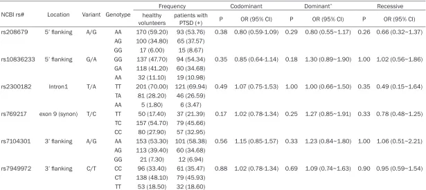
volunteers’ controls by logistic analysis. We found that none of six tag SNPs of the CAT gene
were significantly associated with the risk of
PTSD in all alternative analysis models (P>0.05) (Table 4).
Combination effects of the six tag SNPs
Because LD is highly structured as conserved blocks of sequences separated by hotspots of
recombination, the final function of a conserved
haplotype may be the result of interaction among polymorphisms within the block. The LD
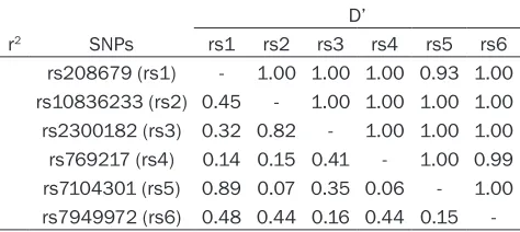
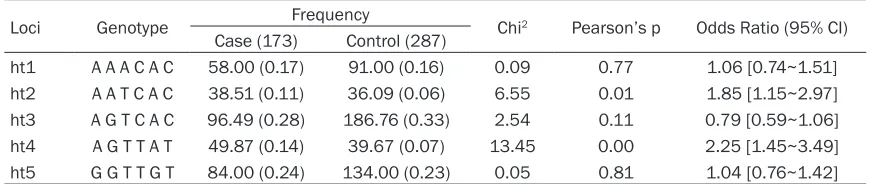
coefficient between polymorphisms was calcu -lated, which showed that all genotyped poly-morphisms are in strong LD (Tables 5, 6). Six tagging SNPs in the LD block were selected, haplotypes were reconstructed and the haplo-type frequencies were compared between the healthy volunteers and PTSD patients. Hapl- otype analysis showed that the haplotype ht2
(A-A-T-C-A-C) and ht4 (A-G-T-T-A-T) was signifi -cantly higher in patients than healthy volun-teers, suggesting that haplotype ht2 and ht4 had a risk effect on PTSD development (Table 6, P=0.01, odds ratio (95% CI) is 1.85 (1.15-2.97) for ht2, and P=0.000, odds ratio (95% CI) is 2.25 (1.45-3.49) for ht4). However, haplo-type association analysis did not provide a
higher OR value or a more significant result
than the OR and P values of single locus SNPs. These results suggest that gene polymor-phisms of the CAT gene, which is a major anti-oxidant enzyme, might not be associated with an increased susceptibility to PTSD in the Chongqing Han population.
Discussion
[image:6.612.90.327.109.215.2]PTSD is associated with exposure to a traumat-ic event outside the range of usual human experience, e.g., severe accidents, intense
Table 5. Pairwise LD tests among six tag SNPs in CAT gene between healthy volunteers and patients with PTSD
D’
r2 SNPs rs1 rs2 rs3 rs4 rs5 rs6
rs208679 (rs1) - 1.00 1.00 1.00 0.93 1.00 rs10836233 (rs2) 0.45 - 1.00 1.00 1.00 1.00 rs2300182 (rs3) 0.32 0.82 - 1.00 1.00 1.00 rs769217 (rs4) 0.14 0.15 0.41 - 1.00 0.99 rs7104301 (rs5) 0.89 0.07 0.35 0.06 - 1.00 rs7949972 (rs6) 0.48 0.44 0.16 0.44 0.15
-patients with PTSD were recruited from the Chongqing district for this study. The patient cohort met the Diagnostic and Statistical Manual of Mental
Disorders-IV (DSM-Disorders-IV) criteria for PTSD consisted of
173 consecutive Han Chinese patients, 103 male and 70 female, with a mean ± SD age of 35.6 ± 11.4 years. There were
no significant differences in age and sex ratio among patients stratified according
physical or sexual abuse, life-threatening medi-cal conditions, combats, and natural disasters [20]. Common life events such as bereave-ment, chronic illness, and divorce rarely pro-duce PTSD. Twin studies have shown that
genetic factors can influence the risk of devel -oping PTSD. As in combat veterans, the devel-opment of PTSD symptoms after non-combat trauma also seems to be moderately heritable [21]. Moreover, many of the same genes that
influence exposure to assault trauma appear to influence susceptibility to PTSD symptoms in
their wake.
In recent years, more and more evidence has suggested that oxidative stress is involved in the pathogenesis of various neuropsychiatry diseases, including anxiety disorders, major
depression, and PTSD [5]. Depletion of specific
antioxidants and decreased activity of antioxi-dant enzymes, as observed in the blood, leads to an impaired defense against oxidative stress. Consequences of oxidative stress include dam-age to macromolecules, of which lipid peroxida-tion has been extensively studied. Several mechanisms are likely involved in the increased generation of reactive species and impairment of oxidative defenses, both of which contribute to increased oxidative stress and result in dam-age to cellular macromolecules. Eventually the consequences will be increased apoptosis, neuronal degeneration, and brain damage, which contribute to the manifestation of neuro-psychiatry illness in susceptible individuals. However, the detailed mechanisms remain largely unknown.
CAT is an enzyme in the first line of antioxidant
defense and might be contribute to the etiology and development of PTSD. To the best of our
knowledge, we are the first investigators to
identify the potential clinical relevance of the
rs10836233, rs208679, rs2300182, rs7692 17, rs7104301 and rs7949972 polymorphisms,
which are identified as tSNPs of the entire CAT
gene in the Han Chinese population. tSNPs are a subset of all variants in a chromosome region within a disease study. The genetic effects of SNPs that are not genotyped in the study can be detected through LD with a tSNP [16]. In this case, the six tSNPs selected in the current
study theoretically reflect the biological signifi -cance of all the genetic variations across the entire CAT gene because of their strong LD with other unassayed variants.
A case-control study is a common and
conve-nient association study design for finding the
genetic basis of a disease. However, a major limitation of this approach is the potential for
population stratification when inappropriate
patient-control matching occurs, such as using healthy blood donors as the control group. To avoid confounding associations, we only select-ed patients with exposures to traumatic events and followed them prospectively to determine whether those who had genetic variants had a lesser or higher risk of PTSD. In addition, we limited the patients recruited into this study cohort to Han Chinese to avoid an artifact due to population admixture. In the context of a bio-logically relevant phenotype and a racially uni-form population, this might maximize the
likeli-hood of finding a meaningful genetic
asso-ciation.
As shown by our clinical association study (Tables 3-6) none of the six tSNPs selected in our study cohort had clinical relevance in rela-tion to the development of PTSD in patients with exposure to traumatic events, only two haplotype, ht2 (A-A-T-C-A-C) and ht4
(A-G-T-T-A-T) were significantly higher in PTSD patients
than healthy volunteers. This conclusion
essen-Table 6. Analyses of association of CAT gene haplotypes with the risk of PTSD between healthy volun-teers and patients with PTSD (+)
Loci Genotype Frequency Chi2 Pearson’s p Odds Ratio (95% CI)
Case (173) Control (287)
ht1 A A A C A C 58.00 (0.17) 91.00 (0.16) 0.09 0.77 1.06 [0.74~1.51] ht2 A A T C A C 38.51 (0.11) 36.09 (0.06) 6.55 0.01 1.85 [1.15~2.97] ht3 A G T C A C 96.49 (0.28) 186.76 (0.33) 2.54 0.11 0.79 [0.59~1.06] ht4 A G T T A T 49.87 (0.14) 39.67 (0.07) 13.45 0.00 2.25 [1.45~3.49] ht5 G G T T G T 84.00 (0.24) 134.00 (0.23) 0.05 0.81 1.04 [0.76~1.42]
[image:7.612.88.523.97.189.2]tially applies to the late onset type of the dis-ease, given the small number of early onset types in our PTSD population. However, as
already mentioned, age stratification between
or within populations did not affect our results.
Our failure to assign significance to the six tag
SNPs and haplotypes of CAT gene should be interpreted with caution for a number of rea-sons. One important reason might be due to polygenetic and multifactorial involvement in the pathogenesis of PTSD. In fact, any one gene might not determine the clinical phenotype of PTSD although the gene is a pivotal one. Therefore, a gene polymorphism should have much less power on the PTSD outcome. The susceptibility to PTSD might be the result of a combination of numerous genetic polymor-phisms. Another possible reason might be that we do not know whether these particular poly-morphisms have an upregulating effect in the brain and to what extent, and whether it increases the overall antioxidant capacity of the regions most affected by PTSD, especially given the relatively low levels of CAT in brain tis-sue. Even in the event that antioxidant capacity
is affected significantly, the result could mani -fest itself only as an effect in disease progres-sion, something that the present study was not designed to test. There are other antioxidant enzymes whose activity could be important in the human brain. The glutathione peroxidases constitute a family of enzymes invol GPX4 for its ability to reduce peroxidized phospholipids and cholesterol [22], are also promising candi-dates, provided that cved in hydrogen and lipid peroxide reduction, but the only common poly-morphism characterized so far, the GPX1 593C/T, does not result in an alteration in the enzymatic activity. Other known peroxidases, and especiallyommon polymorphisms altering
their activity or expression are identified in their
genes. Therefore, additional studies of other antioxidant enzyme genes, with a variety of eth-nic populations, will be needed before the
sig-nificance of the various components of the anti -oxidant machinery of the brain is elucidated in the context of PTSD onset and/or progression.
Conclusions
Here, we investigated the clinical relevance of the genetic variations within the entire CAT gene by means of constructing haplotype bins in patients with PTSD after potentially
traumat-ic events. We have demonstrated that none of the six tSNPs selected in our study cohort had clinical relevance in the development of PTSD in patients with exposure to potentially trau-matic events. It suggests that the CAT gene might not affect the development of PTSD, also may be the sample size may also be too small to allow a meaningful test.
Acknowledgements
We thank Dr. Han Luo, Qian-Ying Tand, Jun-Wei Guo and Shan-Shan Gu for their clinical help and Prof. Yi Ren, Rutgers, from The State University of New Jersey in the United States of America for reviewing the article. And we also thank Shanghai Genesky Bio-Tech Genetic Core Lab for assistance in genotyping tech-niques. This work was supported by the National Natural Science Foundation of China (81101422).
Disclosure of conflict of interest
The authors do not have a conflict of interest to
declare.
Address correspondence to: Dr. Jian-Min Wang, Research Institute of Surgery, Daping Hospital, Third Military Medical University, Daping, Chongqing 400042, China. Tel: 23-68757461; Fax: +86-23-68757461; E-mail: [email protected]; Mei He, Department of Clinical Psychology, Southwest Hos- pital, Third Military Medical University, Chongqing 400038, China.
References
[1] Ursano RJ, Zhang L, Li H, Johnson L, Carlton J, Fullerton CS, Benedek DM. PTSD and traumat-ic stress From gene to community and bench to bedside. Brain Res 2009; 1293: 2-12. [2] Sartor CE, McCutcheon VV, Pommer NE, Nel
-son EC, Grant JD, Duncan AE, Waldron M, Bu-cholz KK, Madden PA, Heath AC. Common ge-netic and environmental contributions to post-traumatic stress disorder and alcohol de-pendence in young women. Psychol Med 2011; 41: 1497-1505.
[3] Comings DE, Comings BG, Muhleman D, Dietz G, Shabbahrami B, Tast D. The dopamine D2 receptor locus as a modifying gene in neuro-psychiatric disorder. JAMA 1991; 266: 1793-1800.
[4] Sartor CE, Grant JD, Lynskey MT, McCutcheon
high-risk trauma, posttraumatic stress disor-der, and major depression. Arch Gen Psychia-try 2012; 69: 293-299.
[5] Hovatta I, Juhila J, Donner J. Oxidative stress in anxiety and comorbid disorders. Neuroscience Res 2010; 68: 261-275.
[6] Bouayed J, Rammal H, Soulimani R. Oxidative stress and anxiety: relationship and cellular pathways. Oxid Med Cell Longev 2009; 2: 63-67.
[7] Selek S, Bulut M, Ocak AR, Kalenderoglu A, Sa-vas HA. Evaluation of total oxidative status in
adult attention deficit hyperactivity disorder
and its diagnostic implications. J Psychiatr Res 2012; 46: 451-455.
[8] Berk M, Ng F, Dean O, Dodd S, Bush AI. Gluta-thione: a novel treatment target in psychiatry. Trends Pharmacol Sci 2008; 29: 346-351. [9] Wang X, Michaelis EK. Selective neuronal
vul-nerability to oxidative stress in the brain. Front Aging Neurosci 2010; 2: 12-16.
[10] Labiós M, Martínez M, Gabriel F, Guiral V, Dasi
F, Beltrán B, Muñoz A. Superoxide dismutase and catalase anti-oxidant activity in leucocyte lysates from hypertensive patients: effects of eprosartan treatment. J Renin Angiotensin Al-dosterone Syst 2009; 10: 24-30.
[11] Komina AV, Korostileva KA, Gyrylova SN, Belo -nogov RN, Ruksha TG. Interaction Between Single Nucleotide Polymorphism in Catalase Gene and Catalase Activity Under the Condi-tions of Oxidative Stress. Physiol Res 2012; 61: 655-658.
[12] Schallreuter K, Salem M, Gibbons N, Martinez A, Slominski R, Ludemann J, Rokos H. Blunted epidermal L-tryptophan metabolism in vitiligo affects immune response and ROS scavenging by Fenton chemistry, part 1: epidermal H2O2/ ONOO-mediated stress abrogates tryptophan hydroxylase and dopa decarboxylase activities, leading to low serotonin and melatonin levels. FASEB J 2012; 26: 247-2470.
[13] Abu-Amero KK, Azad TA, Mousa A, Osman EA, Sultan T, Al-Obeidan SA. A catalase promoter variant rs1001179 is associated with visual acuity but not with primary angle closure glau-coma in Saudi patients. BMC Med Genet 2013; 14: 84-90.
[14] Bulut H, Pehlivan M, Alper S, Tomatir AG, Onay H, Yüksel SE, Özkinay F, Pehlivan S. Lack of as-sociation between catalase gene polymor-phism (T/C exon 9) and susceptibility to vitiligo in a Turkish population. Genet Mol Res 2011; 10: 4126-4132.
[15] Keane TM, Caddell JM, Taylor KL. Mississippi Scale for Combat-Related Posttraumatic Stre- ss Disorder: three studies in reliability and va-lidity. J Consult Clin Psychol 1988; 56: 85-90. [16] Barrett JC, Fry B, Maller J, Daly MJ. Haploview:
analysis and visualization of LD and haplotype maps. Bioinformatics 2005; 21: 263-265. [17] Gabriel SB, Schaffner SF, Nguyen H. The
struc-ture of haplotype blocks in the human genome. Science 2002; 296: 2225-2229.
[18] Daly MJ, Rioux JD, Schaffner SF. High-resolu-tion haplotype structure in the human genome. Nat Genet 2001; 29: 229-232.
[19] Carter KW, McCaskie PA, Palmer LJ. JLIN: a Java based linkage disequilibrium plotter. BMC Bioinformatics 2006; 7: 60-68.
[20] Asmundson GJ, Taylor S. PTSD diagnostic crite-ria: understanding etiology and treatment. Am J Psychiatry 2009; 166 Suppl 6: 726-731. [21] Forsberg L, de Faire U and Morgenstern R.
Oxi-dative stress, human genetic variation, and disease. Arch Biochem Biophys 2001; 389: 84-93.
[22] Voisey J, Swagell CD, Hughes IP, Morris CP, van