z
RESEARCH ARTICLE
STUDY ON THE MOVEMENT OF THE EYE WITH APPLICATIONS IN ELECTROMECHANICAL
MICRODRIVES
*Andra-Maria Ciutac and Carmen-Gabriela Popa
ICPE-CA (The National Institute of Research who supported our work)
ARTICLE INFO ABSTRACT
This paper presents general aspects of the eye, along with a multitude of experiments performed in order to obtain the most suitable micro-drive. There are described theoretical aspects about the movement of the eye, as well as the results of each experiment and conclusions. Final models are also included in this paper. Research thesis -Given the types of eye movements and the range of values of this bio-drive, the purpose of this research theme involves: research, theoretical studies and technical experiments regarding new unconventional systems of electromechanical drive, based on micro motors and micro actuators with different operating principles: piezo, electrostrictive, electro thermal, electromagnetic, electrochemical as well as other operating principles that can have a great contribution to the drive of a spherical object, in different directions and with different mechanical parameters. Great emphasis will be put on the control of the direction, speed, acceleration, as these unconventional systems can also be applied in robotics, medicine, and bioengineering.
Domains of value of the micro drive:
●specific geometry and the structure of the micro muscular system
●angular displacement areas
●linear displacement areas
●micro forces of the drive
●mechanical micro couples
●micro forces
●micro couples of the micro biomechanical resistance Characteristics and mechanical parameters:
●Spherical diameters: 0-200 mm
●Linear areas: 0-5 mm
●Angular areas: 0-60
●Meridian plans
●Micro forces areas: 0-100 cN
●Micro biomechanical couples areas: 0-100 cNcm
●Power: 0-100 W
●Temperature areas: 0–60
●Speed area: 0-30 rpm
Copyright©2017, Andra-Maria Ciutac and Carmen-Gabriela Popa. This is an open access article distributed under the Creative Commons Attribution License, which permits unrestricted use, distribution, and reproduction in any medium, provided the original work is properly cited.
INTRODUCTION
Almost all the information that comes from the outside world into the brain depends on vision. The eye is one of the most complex organs in the entire body. Among its functions, one could mention the perception of size and texture of an object even before entering in contact with it or the perception of the distance at which it is situated. Its functions (in this case, the movement of the eyeball) are subjects that people take interest in due to their complexity and their paramount importance in our lives. These being stated, we express the intention to create a bionic system that resembles the eyeball, in order to obtain an electro-mechanical unconventional drive upon a spherical object, with different technical appliances.
*Corresponding author: Andra-Maria Ciutac,
ICPE-CA (The National Institute of Research who supported our work)
Eye movements (Dan Cristescu et al., 2014)
The eye globe is operated upon by six extraocular muscles, four of them called rectus and the others oblique. The rectus ones are responsible for moving the eye globe into the four cardinal directions: up, down, left and right, whereas the oblique muscles control the adjustment necessary for counteracting with head movements. In go/no-go tasks, humans initiate manual responses with average and minimum reaction times about 450ms and 250ms. Otherwise, the fastest eye movements can be initiated at a speed about 80-100ms. However, the fastest reliable eye movements are produced after 120ms.
There are four types of eye movements: (Guy Croton and Neil Adams, 2012)
● Saccades
● Smooth pursuit movements
ISSN: 0975-833X
International Journal of Current Research Vol. 9, Issue, 10, pp.58530-58536, October, 2017
INTERNATIONAL JOURNAL OF CURRENT RESEARCH
Article History: Received 07thJuly, 2017
Received in revised form 10thAugust, 2017
Accepted 27thSeptember, 2017
Published online 17thOctober, 2017
Citation: Andra-Maria Ciutac and Carmen-Gabriela Popa, 2017. “Study on the movement of the eye with applications in electromechanical Microdrives”, International Journal of Current Research, 9, (10), 58530-58536.
Available online at http://www.journalcra.com
Key words:
Bionic external muscle, Eye movement, Saccadic movement, Electrostrictive actuator, Eletrothermic actuator, Piezoelectric actuator, Electromagnetic actuator. z
RESEARCH ARTICLE
STUDY ON THE MOVEMENT OF THE EYE WITH APPLICATIONS IN ELECTROMECHANICAL
MICRODRIVES
*Andra-Maria Ciutac and Carmen-Gabriela Popa
ICPE-CA (The National Institute of Research who supported our work)
ARTICLE INFO ABSTRACT
This paper presents general aspects of the eye, along with a multitude of experiments performed in order to obtain the most suitable micro-drive. There are described theoretical aspects about the movement of the eye, as well as the results of each experiment and conclusions. Final models are also included in this paper. Research thesis -Given the types of eye movements and the range of values of this bio-drive, the purpose of this research theme involves: research, theoretical studies and technical experiments regarding new unconventional systems of electromechanical drive, based on micro motors and micro actuators with different operating principles: piezo, electrostrictive, electro thermal, electromagnetic, electrochemical as well as other operating principles that can have a great contribution to the drive of a spherical object, in different directions and with different mechanical parameters. Great emphasis will be put on the control of the direction, speed, acceleration, as these unconventional systems can also be applied in robotics, medicine, and bioengineering.
Domains of value of the micro drive:
●specific geometry and the structure of the micro muscular system
●angular displacement areas
●linear displacement areas
●micro forces of the drive
●mechanical micro couples
●micro forces
●micro couples of the micro biomechanical resistance Characteristics and mechanical parameters:
●Spherical diameters: 0-200 mm
●Linear areas: 0-5 mm
●Angular areas: 0-60
●Meridian plans
●Micro forces areas: 0-100 cN
●Micro biomechanical couples areas: 0-100 cNcm
●Power: 0-100 W
●Temperature areas: 0–60
●Speed area: 0-30 rpm
Copyright©2017, Andra-Maria Ciutac and Carmen-Gabriela Popa. This is an open access article distributed under the Creative Commons Attribution License, which permits unrestricted use, distribution, and reproduction in any medium, provided the original work is properly cited.
INTRODUCTION
Almost all the information that comes from the outside world into the brain depends on vision. The eye is one of the most complex organs in the entire body. Among its functions, one could mention the perception of size and texture of an object even before entering in contact with it or the perception of the distance at which it is situated. Its functions (in this case, the movement of the eyeball) are subjects that people take interest in due to their complexity and their paramount importance in our lives. These being stated, we express the intention to create a bionic system that resembles the eyeball, in order to obtain an electro-mechanical unconventional drive upon a spherical object, with different technical appliances.
*Corresponding author: Andra-Maria Ciutac,
ICPE-CA (The National Institute of Research who supported our work)
Eye movements (Dan Cristescu et al., 2014)
The eye globe is operated upon by six extraocular muscles, four of them called rectus and the others oblique. The rectus ones are responsible for moving the eye globe into the four cardinal directions: up, down, left and right, whereas the oblique muscles control the adjustment necessary for counteracting with head movements. In go/no-go tasks, humans initiate manual responses with average and minimum reaction times about 450ms and 250ms. Otherwise, the fastest eye movements can be initiated at a speed about 80-100ms. However, the fastest reliable eye movements are produced after 120ms.
There are four types of eye movements: (Guy Croton and Neil Adams, 2012)
● Saccades
● Smooth pursuit movements
ISSN: 0975-833X
International Journal of Current Research Vol. 9, Issue, 10, pp.58530-58536, October, 2017
INTERNATIONAL JOURNAL OF CURRENT RESEARCH
Article History: Received 07thJuly, 2017
Received in revised form 10thAugust, 2017
Accepted 27thSeptember, 2017
Published online 17thOctober, 2017
Citation: Andra-Maria Ciutac and Carmen-Gabriela Popa, 2017. “Study on the movement of the eye with applications in electromechanical Microdrives”, International Journal of Current Research, 9, (10), 58530-58536.
Available online at http://www.journalcra.com
Key words:
● Vergence movements
● Vestibule-ocular movements.
Fig.1. Extraocular muscles (Gyorgy Weber et al., 2003)
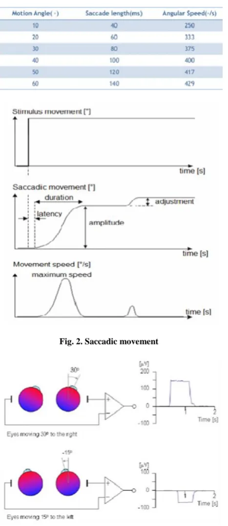
Saccades movements are rapid and ballistic and they abruptly change the point of fixation. They can be elicited voluntarily (following a target, for instance) or involuntarily (while sleeping and when opening the eyes.) Smooth pursuit movements are slow and help the eye keep a moving stimulus on the fovea. Such movements are under voluntary control. (Holle Kirchner and Simon J. Thorpe, 2006) (Fig. 2) Although the saccadic movement is saltatory, it seems that the images are followed continuously, because the brain compresses them. The average angular speed is between 375◦/s -400◦/s, with a
duration exceeding slightly 80ms. Smooth pursuit movements are slow and help the eye keep a moving stimulus on the fovea. Such movements are under voluntary control. Vergence movements align the fovea of each eye with the targets located at different distances from the observer. This type of movement is disjunctive; they involve either a convergence or divergence of the lines of sight of each eye to see an object that is nearer or farther away. Vestibule-ocular movements stabilize the eyes relative to the external world. Therefore, it compensates with head movements and prevents the loss of an image. (Dale Purves et al., 2001) The movement of the eye is generated by a change in potential that varies from 0.5-1 mV. The eye actually represents a dipole with the positive pole towards the cornea and the negative pole in the back of the eye. The maximum angular movement is 70˚ and the amplitude fluctuates in
between 5-20μV, while the resting potential ranges from
0.4mV to 1mV. As we mentioned above the eye represents a sphere that is slightly positively charged at one pole and slightly negatively charged at the other. When such an action potential occurs, the eye moves either to the right (when this change in voltage is positive) or to the left (when the change in voltage is negative).
Control methods
A micromechanical equivalent scheme of linear muscle is presented next. The scheme also includes elastic elements. Interpretation:
k–spring scale of the linear muscle c–damping factor of the linear muscle
[image:2.595.321.555.72.610.2]Since the elastic system in in series,the equivalent elastic constant is: kech= (k1+k2)/k1*k2
Table 1. Saccadic movement
Fig. 2. Saccadic movement
Fig. 3. The movement of the eye in accordance with a change in potential
And it can be defined by the following equation (Voinea et al., 1975):
F = x * kech,where x is the movement of the muscle.
The forces exerted on the linear muscles of a real eye measure between 0.01mN and 5cN.
The main equation of an electromechanical drive (conventional or unconventional) is: (Ignat and Ardelean, 2006)
Fa- Fr= Fj= dv/dt, where
[image:2.595.65.267.87.279.2]Fr - resistance force (appears on the sclera) Fj - inertial force
m - the mass of the eye globe
v–the linear speed of the actuating device
Eye movement represents an angular field of and an angular
speed of ± α and an angular speed of Ω.
Fig. 4. Scheme of linear muscle
Fig. 5. Eye movement explained
Table 2. Elastomer deformation
Length(cm) Force(cN)
10.5 0
11.0 50
11.5 100
12.0 150
11.5 100
11.0 50
10.6 0
Fig. 6. Elastomer deformation
Experiments
In order to decide which the best material to replace the eye muscles is, we have tested several materials, such as elastomer, carbon fibers and copper fibers, for thermal and mechanical endurance.
Fig. 7. Elastomer experiment setup
Table 3. Copper experiment data
Fig. 8 . Carbon fibers experiment setup
Table 4. Carbon fibers experiment data
58532 International Journal of Current Research, Vol. 9, Issue, 10, pp.58530-58536, October, 2017
Fr - resistance force (appears on the sclera) Fj - inertial force
m - the mass of the eye globe
v–the linear speed of the actuating device
Eye movement represents an angular field of and an angular
speed of ± α and an angular speed of Ω.
Fig. 4. Scheme of linear muscle
Fig. 5. Eye movement explained
Table 2. Elastomer deformation
Length(cm) Force(cN)
10.5 0
11.0 50
11.5 100
12.0 150
11.5 100
11.0 50
10.6 0
Fig. 6. Elastomer deformation
Experiments
In order to decide which the best material to replace the eye muscles is, we have tested several materials, such as elastomer, carbon fibers and copper fibers, for thermal and mechanical endurance.
Fig. 7. Elastomer experiment setup
Table 3. Copper experiment data
Fig. 8 . Carbon fibers experiment setup
Table 4. Carbon fibers experiment data
Fig. 9. Elastic fiber experiment setup
Table 5. Result at high voltage
Table 6. Result at lower current
Fig. 10. One of the two coils
Table 7. Experiments on coil 1
Table 8. Experiments on coil 2
Fig. 11. Coil experiment setup
Fig. 12. Model with elastic fibers and nickeline Fig. 9. Elastic fiber experiment setup
Table 5. Result at high voltage
Table 6. Result at lower current
Fig. 10. One of the two coils
Table 7. Experiments on coil 1
Table 8. Experiments on coil 2
Fig. 11. Coil experiment setup
Fig. 12. Model with elastic fibers and nickeline Fig. 9. Elastic fiber experiment setup
Table 5. Result at high voltage
Table 6. Result at lower current
Fig. 10. One of the two coils
Table 7. Experiments on coil 1
Table 8. Experiments on coil 2
Fig. 11. Coil experiment setup
Fig. 13. Elastomer model
Fig. 14. Model with magnets and elastic fiber
Fig. 15. Coil model
Fig. 16. Magnet model
Fig. 17. Magnet model without turning on the power source
Elastomers
● have shown a good contractility
● have almost recovered at their initial stage
● Poisson’s ratio is 0.5; elastomers behave partly like
liquids
● E = 3G (E–Young modulus, G–level of harshness)
In the graphic it can be seen that while increasing the force, the length of the elastomer ascends as well. A similar process is seen when diminishing the force. Therefore we can state that the force is directly proportional with the length of the elastomer.
58534 International Journal of Current Research, Vol. 9, Issue, 10, pp.58530-58536, October, 2017
Fig. 13. Elastomer model
Fig. 14. Model with magnets and elastic fiber
Fig. 15. Coil model
Fig. 16. Magnet model
Fig. 17. Magnet model without turning on the power source
Elastomers
● have shown a good contractility
● have almost recovered at their initial stage
● Poisson’s ratio is 0.5; elastomers behave partly like
liquids
● E = 3G (E–Young modulus, G–level of harshness)
In the graphic it can be seen that while increasing the force, the length of the elastomer ascends as well. A similar process is seen when diminishing the force. Therefore we can state that the force is directly proportional with the length of the elastomer.
Copper fibers
● The diameter of the copper fibers is 0,2mm
● It has responded to electrical tests by heating up to 50ºc and dilating up to 2mm at a voltage of 0,60v and intensity of 5a
Carbon fibers
● There were 45 carbon fibers, having a diameter of 0,15 cm all together, sharing a resistivity of 60Ὠ.
● When applying current at 1V and 0,2A the force decreased with 10 cn and then remained unchanged
Elastic fibers with nickeline
● One of the elastic fibers had been wrapped in nickeline for this experiment
● The results from the tables belong to two different tests, with different voltage currents
● In both cases the force (cn) decreased, even if with very little
● The initial dimensions of the material were: height=11.5mm,width=1.34mm, thickness=1.27mm and rnickeline=13.3ω
● While experimenting with elastic and nickeline fibers we have tried to form thermistors corresponding to the four rectus muscles. The active part of the thermistor is the nickeline fiber that is connected to a power source. As an effect the elastic heats up, dilating, as can be seen in Tables 5 and 6
Coils
We performed experiments on two coils, with different characteristics. One had 2100 spires with diameter of 2 mm (coil 1) and the other one had 4650 spires with a diameter of 3 mm (coil 2). We measured, with a dynamometer, the force needed to detach a thick needle attracted by the coil, connected to a power sources. We have the concluded the following experimental data:
At a voltage lower than 16V, coil 1 could not attract the needle, and coil 2 could not attract the needle at a voltage lower than 20V.
Experimental models
The model in Fig. 12 contains a sponge ball as the spherical object, which has attached to it four sets of elastic fibers, each set being composed of three elastic fibers carefully wrapped in nickeline, which facilitates the passing of current and therefore dilates the elastic fiber. These represent the four rectus muscles of the eye and together form an electro-thermal actuator. At the
bottom of each ‘muscle’ there is an entry port at which the
power source is connected. The model also has an adjustable stand a weight in order to keep the ‘muscles’ in tension all the
time. Experiments have been performed on this model and we have concluded that it is functional and a movement of 5-10° can be noticed when it is connected to power source of approximately 20 volts. Similar to the model with elastic fibers and nickeline, however functioning on another principle -electro-strictive- the next model uses four elastomers as muscles. Elastomers were suitable in terms of elasticity and hardness, as shown in the experiments. As seen in Fig. 28 &
29, each elastomer has a curved striped of copper attached to it, in order to facilitate the passing of the current through it. This results in their expansion and in the movement of the spherical object, represented by an orange plastic ball. The model is functional and it presents a movement of 5-10°. Apart from the previous two models – with elastic fibers with neckline and elastomers with copper - the model with magnets also has the oblique muscles represented and functional with the aid of magnets. Another model is made up of two coils, a spherical object and a thick needle, as shown in Fig. 32. When one of the coils is connected to a power source, it attracts the needle and therefore causes the movement of the spherical object with approximately 15 degrees. (Fig. 15) The coils were used in another model, in which we also simulated the ocular cavity. We used once again a spherical object on which we attached five round magnets, with a space of approx. 72 degrees between one another. For the ocular cavity we used a plastic ball cut in half as positioned with an opening of 90 degrees. At the back of the cavity, on the outside, we attached one of the coils and connected it to a power source. Afterwards, we positioned the spherical objects with the magnets inside the cavity. When turning the power source on, the coil attracts the magnets and determines the movement of the spherical object inside the cavity. We have measure a movement of approx. 20 degrees. Also, this model was designed in such a way to allow us to recreate the irrigation system of the eye. We plan on making a system that dips tiny drops into the cavity. For the model, we used NaCl to grease the inside of the cavity.
Future plans
We plan on working on the previously detailed models in order to improve their functionality, as well as to try and come up with new ideas for our future models. We will continue to perform experiments on different materials and, if found suitable, create an eye model using that material as muscles. One of our future models could be inspired by the movement of viruses, which we will begin to study. Additionally, we will participate in as many research presentations and contests as possible, in order to gain access to a larger public, be given feedback and suggestions, all for the improvement of the project.
Conclusion
The eye movements are extremely complex and précised. A model showing how a sphere can be moved by using conventional or unconventional drives would have a didactic purpose, as the ones presented in this project. There are a multitude of materials which can be used as the eye globe or the external muscles, the materials of the latter having the property of modifying their size and reducing or increasing the force at which they are subjected to, according to difference in voltage. Apart from the didactic purpose, a model showing how a sphere can be moved is yet to continue being studied in order to develop and bring innovations in bionics. The final result can be used for retinal prosthesis, as Argus II, the first retinal prosthesis accepted by both Europe and USA, lacks the mobility of a real eye.
Acknowledgements
Scientific Research “Alexandru Proca” for the materials
offered and for the places in which to conduct experiments. In particular, we are extremely grateful to our mentor, PhD Mircea Ignat for the moral support and guidance offered. Also,
we are grateful for the help offered by our sponsor, Mr. Cătălin
Chivu, who ensured our participation at Intel ISEF 2017. Additionally, we thank Neurosurgeon Dop Radu for the interest and the guidance offered for this project. Last, but not least, we want to mention our class teacher, Claudia Preda, as she was very responsive and appreciative of our research
theme, along with our high school “George Cosbuc”.
REFERENCES
Dale Purves, George J Augustine, David Fitzpatrick, Lawrence C Katz, Anthony-Samuel LaMantia, James O McNamara,
and S Mark Williams, ’Neuroscience-second edition’,
Sinauer Associates, 2001
Dan Cristescu, Carmen Salavastru, Bogadan Voiculescu, Cezar Niculescu, Radu Carmaciu, Anatomy Student Book, Corint Publishing House, Bucharest 2014
Guy Croton, Neil Adams, ‘The Human Body- A Family
Reference Guide’, Parraton Publishing House, 2012 Holle Kirchner, Simon J. Thorpe, ‘Vision Research’, Volume
46, Issue 11, Pages 1762-1772-‘Visual processing speed’,
2006
http://www.tedmontgomery.com/the_eye/
Ignat, M. and I. Ardelean, S.A, ’Microactionari neconventionale’, Ed. Electra, Bucuresti, 2006
PhD, Andrea Ferencz MD, PhD, Gabor Jancso MD, PhD, Sandor Ferencz MD, Szabolcs Horvath MD, Hossein Haddadzadeh Bahri MD, Ildiko Takacs MD, Borbala Balatonyi MD
Voinea, R., D.Voiculescu,V.Ceausu, ‘Mecanica’, Ed.
Didactica si pedagogica, Bucuresti, 1975