RESEARCH ARTICLE
SURFACE CHARACTERIZATION OF DECIDUOUS TEETH SUBJECTED TO HIGH
TEMPERATURE: AN AID TO FORENSICS
*Dr. Nimish Salunkhe, Dr. Shweta Chaudhary, Dr. Alok Patel and Dr. Mayur Chaudhary
Department of Paedodontics and Preventive Dentistry, Bharti Vidyapeeth Dental College and Hospital, Pune
ARTICLE INFO ABSTRACT
Background: Teeth exposed to thermal stress have the potential of not only aiding in identification
but also understanding circumstances surrounding the fire. Paucity of research on the effect of deciduous teeth exposed to high temperatures necessitates research in this area.
Aim: The present study therefore aims to investigate the type and extent of microscopic and
macroscopic changes that can occur in deciduous teeth when exposed to high temperatures.
Procedure: 66 deciduous teeth, extracted as a part of routine clinical treatment, were exposed to
temperatures ranging from 100̊C to 1100̊C for 30 minutes. After exposure to high temperatures, teeth were analyzed for microscopic and macroscopic colorimetric changes.
Result: Macroscopic colorimetric changes ranged from white and pale yellow crown and root
respectively at 100 ̊C to neutral white with pink tinge and neutral white for crown and root respectively at 1100̊C whereas the microscopic changes ranged from crazing of surface enamel with no changes in dentin and cementum at 100̊C to decreased visibility of enamel crystals and dentinal tubules and molten cementum at 1100̊C.
Conclusion: We conclude that these results would further add to the knowledge of forensic
investigators as a part of identification of teeth.
Copyright©2016, Dr. Nimish Salunkhe et al. This is an open access article distributed under the Creative Commons Attribution License, which permits unrestricted use, distribution, and reproduction in any medium, provided the original work is properly cited.
INTRODUCTION
The value of dental characteristics to identify a deceased individual has been well recognized since ancient times. Forensic odontology is now considered to be a specialized and reliable science for identification of deceased. The modern era of forensic odontology is said to have commenced with the identification of the victims ‘Bazar de la Charite’ fire which occurred in Paris. (Taylor, 2009) However, Dr. Oscar Amoedo, deserves the credit as the Father of Forensic Odontology for his pathbreaking work in the field. Fire remains one of the major causes of morbidity and mortality throughout the world and identification of a body from the fatal fire remains a daunting task. Forensic odontology in particular has been seen to be useful when damage has been caused by heat. Teeth have often been considered to be an indestructible part of human remains and have the highest resistance to most environmental effects like fire, desiccation and decomposition because of their particular resistant composition and protection by soft tissues. (Chetan A Pol and Suchitra R Gosavi, 2014) In a report by the National Fire Protection Association (USA), it was suggested that children are twice as likely as adults to become victims of
*Corresponding author: Dr. Nimish Salunkhe,
Department of Paedodontics and Preventive Dentistry, Bharti Vidyapeeth Dental College and Hospital, Pune
a house fire as they don’t have the ability to safely evacuate from the place of fire. Literature review reveals that most of the research related to burnt remains is done on permanent teeth and very few of the published data on incinerated deciduous teeth is available. This necessitates research in this area. (Karkhanis et al., 2009) This study therefore aims to find out the macroscopic and microscopic changes that could be seen on deciduous teeth subjected to high temperatures. It also correlates the colour change with the condition of tooth.
MATERIALS AND METHODS
A total of 66 deciduous teeth were obtained by extraction as a part of routine clinical treatment in the department of pediatric and preventive dentistry, Bharati Vidyapeeth Dental College, Pune. The extracted teeth covered with blood deposits and salivary coating were rinsed with normal saline. After rinsing, teeth were stored in 10% formalin till further procedure was carried out. (Dominici et al., 2001; Kumar et al., 2005) Later these teeth were subjected to sudden thermal shock for 30 minutes in a furnace in Praj Metallurgy Lab, Pune. Temperatures used ranged from 100°C to 1100°C. A group of 6 teeth were selected randomly and employed for heating at each temperature setting. Teeth were loaded on a ceramic brick (Fig. 1) then placed into the furnace (Fig. 2). Ceramic brick is a
ISSN: 0975-833X
International Journal of Current Research
Vol. 8, Issue, 11, pp.42423-42428, November, 2016
INTERNATIONAL JOURNAL OF CURRENT RESEARCH
Article History:
Received 13thAugust, 2016 Received in revised form 30thSeptember, 2016
Accepted 18thOctober, 2016
Published online 30thNovember, 2016
Key words:
Surface characterization of deciduous teeth, Incinerated teeth,
Colorimetric changes,
Microscopic and macroscopic changes.
Citation: Dr. Nimish Salunkhe, Dr. Shweta Chaudhary, Dr. Alok Patel and Dr. Mayur Chaudhary, 2016.“Surface characterization of deciduous teeth
subjected to high temperature: An aid to forensics”,International Journal of Current Research, 8, (11), 42423-42428.
RESEARCH ARTICLE
SURFACE CHARACTERIZATION OF DECIDUOUS TEETH SUBJECTED TO HIGH
TEMPERATURE: AN AID TO FORENSICS
*Dr. Nimish Salunkhe, Dr. Shweta Chaudhary, Dr. Alok Patel and Dr. Mayur Chaudhary
Department of Paedodontics and Preventive Dentistry, Bharti Vidyapeeth Dental College and Hospital, Pune
ARTICLE INFO ABSTRACT
Background: Teeth exposed to thermal stress have the potential of not only aiding in identification
but also understanding circumstances surrounding the fire. Paucity of research on the effect of deciduous teeth exposed to high temperatures necessitates research in this area.
Aim: The present study therefore aims to investigate the type and extent of microscopic and
macroscopic changes that can occur in deciduous teeth when exposed to high temperatures.
Procedure: 66 deciduous teeth, extracted as a part of routine clinical treatment, were exposed to
temperatures ranging from 100̊C to 1100̊C for 30 minutes. After exposure to high temperatures, teeth were analyzed for microscopic and macroscopic colorimetric changes.
Result: Macroscopic colorimetric changes ranged from white and pale yellow crown and root
respectively at 100 ̊C to neutral white with pink tinge and neutral white for crown and root respectively at 1100̊C whereas the microscopic changes ranged from crazing of surface enamel with no changes in dentin and cementum at 100̊C to decreased visibility of enamel crystals and dentinal tubules and molten cementum at 1100̊C.
Conclusion: We conclude that these results would further add to the knowledge of forensic
investigators as a part of identification of teeth.
Copyright©2016, Dr. Nimish Salunkhe et al. This is an open access article distributed under the Creative Commons Attribution License, which permits unrestricted use, distribution, and reproduction in any medium, provided the original work is properly cited.
INTRODUCTION
The value of dental characteristics to identify a deceased individual has been well recognized since ancient times. Forensic odontology is now considered to be a specialized and reliable science for identification of deceased. The modern era of forensic odontology is said to have commenced with the identification of the victims ‘Bazar de la Charite’ fire which occurred in Paris. (Taylor, 2009) However, Dr. Oscar Amoedo, deserves the credit as the Father of Forensic Odontology for his pathbreaking work in the field. Fire remains one of the major causes of morbidity and mortality throughout the world and identification of a body from the fatal fire remains a daunting task. Forensic odontology in particular has been seen to be useful when damage has been caused by heat. Teeth have often been considered to be an indestructible part of human remains and have the highest resistance to most environmental effects like fire, desiccation and decomposition because of their particular resistant composition and protection by soft tissues. (Chetan A Pol and Suchitra R Gosavi, 2014) In a report by the National Fire Protection Association (USA), it was suggested that children are twice as likely as adults to become victims of
*Corresponding author: Dr. Nimish Salunkhe,
Department of Paedodontics and Preventive Dentistry, Bharti Vidyapeeth Dental College and Hospital, Pune
a house fire as they don’t have the ability to safely evacuate from the place of fire. Literature review reveals that most of the research related to burnt remains is done on permanent teeth and very few of the published data on incinerated deciduous teeth is available. This necessitates research in this area. (Karkhanis et al., 2009) This study therefore aims to find out the macroscopic and microscopic changes that could be seen on deciduous teeth subjected to high temperatures. It also correlates the colour change with the condition of tooth.
MATERIALS AND METHODS
A total of 66 deciduous teeth were obtained by extraction as a part of routine clinical treatment in the department of pediatric and preventive dentistry, Bharati Vidyapeeth Dental College, Pune. The extracted teeth covered with blood deposits and salivary coating were rinsed with normal saline. After rinsing, teeth were stored in 10% formalin till further procedure was carried out. (Dominici et al., 2001; Kumar et al., 2005) Later these teeth were subjected to sudden thermal shock for 30 minutes in a furnace in Praj Metallurgy Lab, Pune. Temperatures used ranged from 100°C to 1100°C. A group of 6 teeth were selected randomly and employed for heating at each temperature setting. Teeth were loaded on a ceramic brick (Fig. 1) then placed into the furnace (Fig. 2). Ceramic brick is a
ISSN: 0975-833X
International Journal of Current Research
Vol. 8, Issue, 11, pp.42423-42428, November, 2016
INTERNATIONAL JOURNAL OF CURRENT RESEARCH
Article History:
Received 13thAugust, 2016 Received in revised form 30thSeptember, 2016
Accepted 18thOctober, 2016
Published online 30thNovember, 2016
Key words:
Surface characterization of deciduous teeth, Incinerated teeth,
Colorimetric changes,
Microscopic and macroscopic changes.
Citation: Dr. Nimish Salunkhe, Dr. Shweta Chaudhary, Dr. Alok Patel and Dr. Mayur Chaudhary, 2016.“Surface characterization of deciduous teeth
rectangular block which could withstand high temperatures and provide ease for placement and removal of teeth from the furnace. After 30 minutes, teeth were removed from the furnace and were allowed to cool at room temperature. Each incinerated tooth was examined under a stereomicroscope to assess the extent of heat induced alterations in the crown and root of the teeth. This was followed by colorimetric analysis of the incinerated teeth based on Munsell soil colour chart. Scanning electron microscopic analysis (FEI QUANTA 200) was carried out under low vacuum mode. Incinerated tooth may develop cracks in SEM so low vacuum mode was used. A double sided carbon tape was used to secure the teeth on a glass slide, which was then placed on the microscope stage. During the imaging procedure, standardized values for the different working parameters such as spot size, working distance and kV were not used, as each image had to be optimised individually depending upon the magnification.
RESULTS
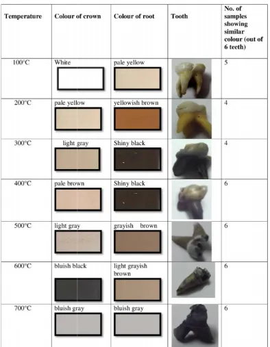
[image:2.595.102.497.289.797.2]All the teeth subjected to various temperatures showed colour changes as well as surface changes based on stereomicroscope and SEM findings. Table 1 (a, b) shows colour changes based on Munsell soil colour chart. At 100°C, crazing on enamel surface was seen while there were no cemental and dentinal changes. At 200°C, surface bubbling on root surface of teeth was seen. Crazing was still evident on enamel surface. At 300°C, fissures could be observed on enamel surface. The predentinal surface had developed globule like structures. (Fig. 3) At 400°C, enamel and dentin started separating which progressed from cervical margin. Enamel and cementum showed deep fissures. (Fig. 4, 5) At 500°C, there was complete separation of enamel and dentin. One more notable feature was that dentinal tubular diameter started reducing. (Fig. 6) At 600°C, gradual reduction in dentinal tubular diameter was still seen.
Table 1. Colour changes based on the Munsell soil colour chart, following colour changes were seen
rectangular block which could withstand high temperatures and provide ease for placement and removal of teeth from the furnace. After 30 minutes, teeth were removed from the furnace and were allowed to cool at room temperature. Each incinerated tooth was examined under a stereomicroscope to assess the extent of heat induced alterations in the crown and root of the teeth. This was followed by colorimetric analysis of the incinerated teeth based on Munsell soil colour chart. Scanning electron microscopic analysis (FEI QUANTA 200) was carried out under low vacuum mode. Incinerated tooth may develop cracks in SEM so low vacuum mode was used. A double sided carbon tape was used to secure the teeth on a glass slide, which was then placed on the microscope stage. During the imaging procedure, standardized values for the different working parameters such as spot size, working distance and kV were not used, as each image had to be optimised individually depending upon the magnification.
RESULTS
All the teeth subjected to various temperatures showed colour changes as well as surface changes based on stereomicroscope and SEM findings. Table 1 (a, b) shows colour changes based on Munsell soil colour chart. At 100°C, crazing on enamel surface was seen while there were no cemental and dentinal changes. At 200°C, surface bubbling on root surface of teeth was seen. Crazing was still evident on enamel surface. At 300°C, fissures could be observed on enamel surface. The predentinal surface had developed globule like structures. (Fig. 3) At 400°C, enamel and dentin started separating which progressed from cervical margin. Enamel and cementum showed deep fissures. (Fig. 4, 5) At 500°C, there was complete separation of enamel and dentin. One more notable feature was that dentinal tubular diameter started reducing. (Fig. 6) At 600°C, gradual reduction in dentinal tubular diameter was still seen.
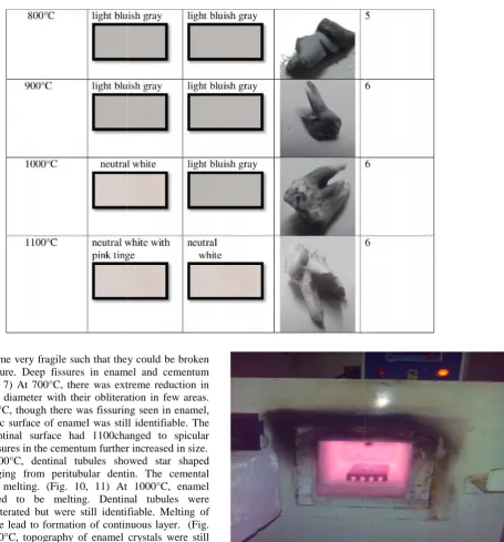
Teeth had become very fragile such that they could be broken by finger pressure. Deep fissures in enamel and cementum were seen. (Fig 7) At 700°C, there was extreme reduction in dentinal tubular diameter with their obliteration in few areas. (Fig. 8) At 800°C, though there was fissuring seen in enamel, the characteristic surface of enamel was still identifiable. The globular predentinal surface had 1100changed to spicular appearance. Fissures in the cementum further increased in size. (Fig. 9) At 900°C, dentinal tubules showed star shaped structure emerging from peritubular dentin. The cemental surface started melting. (Fig. 10, 11) At 1000°C, enamel crystal appeared to be melting. Dentinal tubules were completely obliterated but were still identifiable. Melting of cemental surface lead to formation of continuous layer. (Fig. 12, 13) At 1100°C, topography of enamel crystals were still seen in some areas. Dentinal tubules were also still identifiable. Root surface developed vesicles. Cementum was unidentifiable and had melted completely. (Fig. 14, 15)
[image:3.595.53.277.592.778.2]Fig. 1. Ceramic brick
Fig. 2. Ceramic brick containing teeth placed in the furnace
Fig. 3. SEM image at 300°C showing globule like formation on predentinal surface
Teeth had become very fragile such that they could be broken by finger pressure. Deep fissures in enamel and cementum were seen. (Fig 7) At 700°C, there was extreme reduction in dentinal tubular diameter with their obliteration in few areas. (Fig. 8) At 800°C, though there was fissuring seen in enamel, the characteristic surface of enamel was still identifiable. The globular predentinal surface had 1100changed to spicular appearance. Fissures in the cementum further increased in size. (Fig. 9) At 900°C, dentinal tubules showed star shaped structure emerging from peritubular dentin. The cemental surface started melting. (Fig. 10, 11) At 1000°C, enamel crystal appeared to be melting. Dentinal tubules were completely obliterated but were still identifiable. Melting of cemental surface lead to formation of continuous layer. (Fig. 12, 13) At 1100°C, topography of enamel crystals were still seen in some areas. Dentinal tubules were also still identifiable. Root surface developed vesicles. Cementum was unidentifiable and had melted completely. (Fig. 14, 15)
[image:3.595.311.559.602.764.2]Fig. 1. Ceramic brick
Fig. 2. Ceramic brick containing teeth placed in the furnace
Fig. 3. SEM image at 300°C showing globule like formation on predentinal surface
Teeth had become very fragile such that they could be broken by finger pressure. Deep fissures in enamel and cementum were seen. (Fig 7) At 700°C, there was extreme reduction in dentinal tubular diameter with their obliteration in few areas. (Fig. 8) At 800°C, though there was fissuring seen in enamel, the characteristic surface of enamel was still identifiable. The globular predentinal surface had 1100changed to spicular appearance. Fissures in the cementum further increased in size. (Fig. 9) At 900°C, dentinal tubules showed star shaped structure emerging from peritubular dentin. The cemental surface started melting. (Fig. 10, 11) At 1000°C, enamel crystal appeared to be melting. Dentinal tubules were completely obliterated but were still identifiable. Melting of cemental surface lead to formation of continuous layer. (Fig. 12, 13) At 1100°C, topography of enamel crystals were still seen in some areas. Dentinal tubules were also still identifiable. Root surface developed vesicles. Cementum was unidentifiable and had melted completely. (Fig. 14, 15)
Fig. 1. Ceramic brick
Fig. 2. Ceramic brick containing teeth placed in the furnace
Fig. 4. SEM image at 400°C showing fissures in cementum
Fig. 5. SEM image at 400°C showing fissures in enamel
Fig. 6. SEM imgae at 500°C showing reduction in dentinal tubular diameter
Fig.7. SEM image at 600°C showing deep fissures in enamel
Fig. 8. SEM image at 700°C showing extreme reduction in dentinal tubular diameter
Fig. 9. SEM image at 800°C showing spicular appearance of predentinal surface
Fig. 10. SEM image at 900°C showing star shaped structures from peritubular dentin
Fig. 11. SEM image at 900°C showing melting of cemental surface Fig. 4. SEM image at 400°C showing fissures in cementum
Fig. 5. SEM image at 400°C showing fissures in enamel
Fig. 6. SEM imgae at 500°C showing reduction in dentinal tubular diameter
Fig.7. SEM image at 600°C showing deep fissures in enamel
Fig. 8. SEM image at 700°C showing extreme reduction in dentinal tubular diameter
Fig. 9. SEM image at 800°C showing spicular appearance of predentinal surface
Fig. 10. SEM image at 900°C showing star shaped structures from peritubular dentin
Fig. 12. SEM image at 1000°C showing obliterated dentinal tubules
Fig. 13. SEM image at 1000°C showing melting of cemental surface
Fig. 14. SEM image at 1100°C showing complete melting of cemental surface and is unidentifiable
Fig. 15. SEM image at 1100°C showing identifiable dentinal structure
DISCUSSION
The results obtained in this study are in accordance with the study carried out by Karkhanis et al. in 2009. The teeth although resistant to most physical trauma can become brittle and fragile when subjected to increased temperature. Knowledge of effect of high temperatures on teeth can aid forensic dentists to handle such fragile teeth and help in gathering the evidence. It is seen that the anterior teeth are more thermally damaged as compared to posteriors as posteriors are more protected from perioral musculature that are tongue and cheeks. Teeth had become brittle and fragile. Teeth exposed to higher temperatures show tendency to fragment into smaller pieces as compared to those exposed to lower temperatures which fragmented into larger pieces. At lower temperatures the teeth fragmented primarily due to fissures parallel to the long axis of the teeth. (Karkhanis et al., 2009) It was seen that colour changes could give the idea of fragility of teeth. Lighter the colour of teeth, more fragile the teeth were. This confirms previous research which suggests that blackened teeth are less fragile in comparison with remains that are grey or white in colour. (Delattre V. Burned, 2000) Teeth subjected to lower temperatures were darker in colour as compared to those subjected to higher temperatures. The overall differences in colour and microstructure observed in the deciduous teeth, as compared to the permanent teeth may be related to the fact that the deciduous enamel is more porous and has a larger area of organic/inorganic surface interface compared to the permanent teeth. (Wilson and Beynon, 1989) At 200-300 °C, surface bubbling was observed on the root surface. These similar changes were seen at 300-400°C by Muller et al in permanent teeth. The reason for early changes in deciduous teeth can be due to the fact that deciduous teeth are hypomineralised as compared to permanent teeth. (Masatoshi et al., 2001) At 400-500°C, enamel and dentin separation was seen. This reason could be attributed to the dehydration taking place in enamel and dentin which causes them to shrink leading to separation. In addition to this separation, Karkhanis et al also observed disintegration of separated enamel into smaller fragments which was not seen in the present study. At 500-700°C, there was gradual reduction in dentinal tubular diameter. These changes are in conjunction with the study by Chetan et al where he noticed similar changes at 600°C. Tubules were obliterated in some areas especially at incisal edges and cuspal tips. The reason could be that dentinal tubules are more closely arranged in these areas. At 900-1000°C, cemental surface was unidentifiable due to melting leading to formation of continuous layer. At 1000-1100°C, enamel and cementum had melted. Dentinal tubules were completely obliterated but their tubular morphology was still identifiable due to the higher mineral content of peritubular dentin. Root surface had developed vesicles. This feature was also observed by Chetan et al at 1000°C. Based on these changes, the temperature at which the teeth and body were subjected to could be identified which could help in understanding circumstances surrounding the fire.
Conclusion
This study throws light on preliminary analysis of changes in deciduous teeth subjected to high temperatures. A further insight on heat induced changes in deciduous teeth is still required. Further research could help forensic investigators develop concrete methods in handling fragile teeth subjected to high temperatures and ways to solve the crimes involving fire.
Fig. 12. SEM image at 1000°C showing obliterated dentinal tubules
Fig. 13. SEM image at 1000°C showing melting of cemental surface
Fig. 14. SEM image at 1100°C showing complete melting of cemental surface and is unidentifiable
Fig. 15. SEM image at 1100°C showing identifiable dentinal structure
DISCUSSION
The results obtained in this study are in accordance with the study carried out by Karkhanis et al. in 2009. The teeth although resistant to most physical trauma can become brittle and fragile when subjected to increased temperature. Knowledge of effect of high temperatures on teeth can aid forensic dentists to handle such fragile teeth and help in gathering the evidence. It is seen that the anterior teeth are more thermally damaged as compared to posteriors as posteriors are more protected from perioral musculature that are tongue and cheeks. Teeth had become brittle and fragile. Teeth exposed to higher temperatures show tendency to fragment into smaller pieces as compared to those exposed to lower temperatures which fragmented into larger pieces. At lower temperatures the teeth fragmented primarily due to fissures parallel to the long axis of the teeth. (Karkhanis et al., 2009) It was seen that colour changes could give the idea of fragility of teeth. Lighter the colour of teeth, more fragile the teeth were. This confirms previous research which suggests that blackened teeth are less fragile in comparison with remains that are grey or white in colour. (Delattre V. Burned, 2000) Teeth subjected to lower temperatures were darker in colour as compared to those subjected to higher temperatures. The overall differences in colour and microstructure observed in the deciduous teeth, as compared to the permanent teeth may be related to the fact that the deciduous enamel is more porous and has a larger area of organic/inorganic surface interface compared to the permanent teeth. (Wilson and Beynon, 1989) At 200-300 °C, surface bubbling was observed on the root surface. These similar changes were seen at 300-400°C by Muller et al in permanent teeth. The reason for early changes in deciduous teeth can be due to the fact that deciduous teeth are hypomineralised as compared to permanent teeth. (Masatoshi et al., 2001) At 400-500°C, enamel and dentin separation was seen. This reason could be attributed to the dehydration taking place in enamel and dentin which causes them to shrink leading to separation. In addition to this separation, Karkhanis et al also observed disintegration of separated enamel into smaller fragments which was not seen in the present study. At 500-700°C, there was gradual reduction in dentinal tubular diameter. These changes are in conjunction with the study by Chetan et al where he noticed similar changes at 600°C. Tubules were obliterated in some areas especially at incisal edges and cuspal tips. The reason could be that dentinal tubules are more closely arranged in these areas. At 900-1000°C, cemental surface was unidentifiable due to melting leading to formation of continuous layer. At 1000-1100°C, enamel and cementum had melted. Dentinal tubules were completely obliterated but their tubular morphology was still identifiable due to the higher mineral content of peritubular dentin. Root surface had developed vesicles. This feature was also observed by Chetan et al at 1000°C. Based on these changes, the temperature at which the teeth and body were subjected to could be identified which could help in understanding circumstances surrounding the fire.
Conclusion
As teeth are one of the last structures to be destroyed by fire, pediatric dentists and forensic dentists are the most preferred ones who can detect the changes caused by fire and help in detecting the temperature and circumstances surrounding the fire.
REFERENCES
Chetan A Pol, Suchitra R Gosavi. 2014. Scanning electron microscopic analysis of incinerated teeth: An aid to forensic identification. Journal of Oral and Maxillofacial Pathology, Jan–Apr., 18(1): 32-35.
Delattre V. 2000. Burned beyond recognition: systematic approach to the dental identification of charred human remains. J Forensic Sci., 45: 589-596.
Dominici JT, Eleazer PD, Clark SJ, Staat RH, Scheetz JP. 2001. Disinfection/ sterilization of extracted teeth for dental student use. J Dent Educ., 65:1278-1280.
Karkhanis, S., J Ball, D Franklin. 2009. Macroscopic and microscopic changes in incinerated deciduous teeth. J Forensic Odontostomatol., 27(2):9-19.
Kumar M, Sequeira PS, Peter S, Bhat GK. 2005. Sterilisation of extracted human teeth for educational use. Indian J Med Microbiol., 23: 256-258.
Masatoshi A, Monique H, Bruce S, George S. 2001. Comparative study to quantify demineralized enamel in deciduous and permanent teeth using laser and light induced fluorescence techniques. Caries Research, 35: 464-470.
Taylor. J. 2009. A brief history of forensic odontology and disaster victim identification practices in Austrailia. J Odontostomato, 27(2):64-74.
Wilson P, Beynon A. 1989. Mineralisation differences between human deciduous and permanent enamel measured by quantitative microradiography. Arch Oral Biol., 34:85-88.