O R I G I N A L A R T I C L E
Expression of a SOX1 overlapping transcript in neural
differentiation and cancer models
Azaz Ahmad1•Stephanie Strohbuecker1• Cristina Tufarelli2• Virginie Sottile1
Received: 28 January 2017 / Revised: 4 June 2017 / Accepted: 26 June 2017
ÓThe Author(s) 2017. This article is an open access publication
ABSTRACT SOX1 is a member of the SOXB1 subgroup of transcription factors involved in early embryogenesis, CNS development and maintenance of neural stem cells. The structure and regulation of the humanSOX1locus has been less studied than that ofSOX2, another member of the SOXB1 subgroup for which an overlapping transcript has been reported. Here we report that theSOX1locus harbours a SOX1 overlapping transcript (SOX1-OT), and describe expression, splicing variants and detection ofSOX1-OTin different stem and cancer cells. RT-PCR and RACE experiments were performed to detect and characterise the structure of SOX1-OT in neuroprogenitor cultures and across different cancer cell lines. SOX1-OTwas found to present a complex structure including several unannotated exons, different transcript variants and at least two poten-tial transcription start sites. SOX1-OT was found to be highly expressed in differentiated neural stem cells across different time points of differentiation, and its expression correlated with SOX1 gene expression. Concomitant
expression ofSOX1andSOX1-OTwas further observed in several cancer cell models. While the function of this transcript is unknown, the regulatory role reported for other lncRNAs strongly suggests a possible role forSOX1-OTin regulating SOX1 expression, as previously observed for SOX2. The elucidation of the genetic and regulatory con-text governing SOX1 expression will contribute to clarifying its role in stem cell differentiation and tumorigenesis.
Keywords Stem cellLncRNASOX1-OT Expression pattern Cancer modelSOX1
Introduction
SOX1 and SOX2 are two closely related transcription factors belonging to the SOXB1 subgroup of the high mobility group box (HGM-box) family greatly involved in the regulation of pluripotent stem cells and neural stem cells [1]. In human, the SOX2 gene maps to Chr3q26.3, within an intron of a long non-coding RNA (LncRNA) called SOX2 overlapping transcript (SOX2-OT; Fig.1a) [2]. LncRNAs, defined as non-coding RNAs (ncRNAs) that are more than 200 nucleotides long, have been suggested to play a role in several biological processes including nuclear organisation, epigenetic regulations and post-translational modifications [3,4]. This structure is conserved between mouse and human, and in both species theSOX2 overlap-ping transcripts are reported to have multiple transcription start sites (TSS) and to be transcribed into several alter-native transcript variants [5]. Recently, concomitant gene expression of SOX2 and SOX2-OT has been reported in breast, lung and oesophageal carcinoma [6–8]. Current studies suggest a positive role for SOX2-OTin regulating Electronic supplementary material The online version of this
article (doi:10.1007/s00018-017-2580-3) contains supplementary material, which is available to authorized users.
Azaz Ahmad and Stephanie Strohbuecker contributed equally to the work.
& Cristina Tufarelli
[email protected] & Virginie Sottile
1 Wolfson STEM Centre, Division of Cancer & Stem Cells,
School of Medicine, University of Nottingham, Nottingham, UK
2 Division of Graduate Entry Medicine & Health, School of
Medicine, University of Nottingham, Derby, UK
SOX2, and concordant gene expression has been reported in cellular differentiation, pluripotency and carcinogenesis [5–7, 9]. SOX2-OT is differentially spliced into multiple transcript variants in stem and cancer cells, and has been proposed to play a role in regulating expression ofSOX2 [9,10].
SOX1, another SOXB1 member closely related toSOX2, is involved in early embryogenesis, CNS development and maintenance of neural stem cells [11]. SOX1 and SOX2 originated from a common ancestor by gene duplication
during the course of evolution and exhibit similar sequences, expression patterns and overexpression pheno-types [12]. The structure and regulation of the humanSOX1 locus has been studied far less than that ofSOX2, and there has been no report of any overlapping transcript for this gene. Here we address this question and describe the complex structure of the SOX1locus which was found to harbour an overlapping transcript, and describe expression, splicing variants and detection in different stem cell and cancer cell models.
Fig. 1 Structural similarity between SOX2 and SOX1 loci.a Snap-shot images of the humanSOX2locus on human chromosome 3 taken from the UCSC genome browser showing theSOX2gene itself (top) and the zoomed out region (below) to emphasise the length and alternative isoforms of theSOX2-OTnon-coding gene within which SOX2 lies. b Snapshots of the human SOX1 locus on human
[image:2.595.59.543.70.261.2]Materials and methods
Reagents were purchased from ThermoFisher (UK) unless otherwise stated.
Cell sample preparation
Cells lines used in this study are described in Table1. Ntera2, hMSCs, HeLa, SH-SY5Y and HOS cell lines were grown in Dulbeco’s Modified Eagle Medium (DMEM) supplemented with 10% foetal calf serum (FCS), 1% L-glutamine, 1% non-essential amino acids and 0.5% Peni-cillin/streptomycin, and incubated in a humidified incubator in an atmosphere of 5% CO2 at 37°C. Cell
culture conditions for MDA-MB-361, MDA-MB-231 and T47D are defined in [13], CaCo2, HCT116 and MCF7 in [14], Hs578T in [15]. For RNA extraction, cell monolayers were washed with PBS, detached with 0.05% trypsin/ EDTA and pelleted for 5 min. Cell pellets were stored in TRI reagent (Sigma-Aldrich) at-80°C.
For neural differentiation samples, human immortalised neuroprogenitor cells (ReNcell Merck Millipore, referred to as ‘ReN’) were cultured according to manufacturer’s instructions. Cells were seeded on laminin (Trevigen) in ReNcell NSC Maintenance Medium (Merck Millipore) supplemented with 20 ng/mL FGF2 and 20 ng/mL EGF. After 24 h incubation (day 0, D0), cells were treated with medium deprived of FGF and EGF to induce differentia-tion for up to day 6 (D6). At stated time points, RNA was harvested and processed as described below.
Immunostaining
Cells were fixed with 4% paraformaldehyde (PFA), washed with PBS and incubated in PBS?0.1% Triton X-100 for
10 min before blocking with a 10% serum dilution. After PBS wash, samples were incubated with a dilution of pri-mary antibody against NESTIN (1:100, cat. # IC1259F, R&D Systems, Abingdon, UK) or MAP2 (1:100, MT-01, ExBio, Vestec, Czech Republic) overnight at 4°C, before extensive washing, incubation in a FITC-labelled sec-ondary antibody solution (1:500, FI-2000, Vector laboratories, Peterborough, UK) for 1 h at room tempera-ture, further washing and finally mounting with DAPI-containing Vectashield (H-1200, Vector laboratories).
RNA extraction
Total RNA was extracted using 0.5 mL of TRI-Reagent (Sigma-Aldrich) per 1–59106 cells according to the manufacturer’s protocol, followed by RNA purification from the aqueous phase using the RNA Clean & Concen-trator-25 kit (Zymo Research). RNA concentration was determined spectrophotometrically and the samples were stored at-80°C.
cDNA synthesis and RT-PCR
[image:3.595.52.550.525.716.2]RNA samples were subjected to DNAse-I treatment using the DNase-I, Amplification grade kit according to the manufacturer’s protocol, using 1 U/lL of DNase-I for each 1 lg of RNA at 25°C for 20 min. After DNase-I treat-ment, 2lg RNA was used to synthesise cDNA by reverse transcription using 200 units/lL of SuperScript III Reverse Transcriptase in 30lL of total reaction volume, including 100 pmol/lL of random 15mer primers (MWG Biotech), 0.5 mM dNTP and 0.1 mM DTT. Tubes containing the reaction mix without reverse transcriptase (‘-RT’) were used as negative control. cDNA samples were cleaned up
Table 1 Cell lines used in the experimental study
No. Cell line Tissue Cell type References
1 Ntera2 Testis Pluripotent embryonal carcinoma [16]
2 hMSCs Bone marrow Mesenchymal progenitors [17]
3 ReN cells Brain Neural progenitors [18]
4 HeLa Cervix Adenocarcinoma [19]
5 SH-SY5Y Bone marrow Neuroblastoma [20]
6 HOS Bone Osteosarcoma [21]
7 CaCo2 Colon Colorectal Adenocarcinoma [22]
8 HCT116 Colon Colorectal Carcinoma [23]
9 MCF7 Mammary gland Adenocarcinoma [24]
10 MDA-MB-361 Mammary gland Adenocarcinoma [25]
11 MDA-MB-231 Mammary gland Adenocarcinoma [25]
12 Hs578T Mammary gland Carcinoma [26]
using MinElute PCR purification kit (Qiagen) and stored at
-20°C.
PCR amplification of cDNA was performed in a volume of 20lL using Platinum Taq DNA polymerase. Thermal cycler conditions used after heating at 95°C for 10 min involved 40 cycles of denaturation at 95°C for 30 s, annealing at 55°C for 60 s and extension at 72°C for 60 s, followed by a final 7-min extension step at 72°C. PCR reactions set using either water instead of cDNA (‘H2O’) or
-RT samples as template were used as controls to rule out any contamination issues.
PCR products were analysed by electrophoresis on 2% agarose gels. Primers used for theSOX1-OTamplification are shown in Supplementary Table 1. All fragments detected by RT-PCR were sequenced (Source BioScience, Nottingham, UK) to confirm specificity and map their position.
Quantitative polymerase chain reaction (qRT-PCR)
For gene quantification by real-time PCR, Taqman qPCR assays were performed in 20lL reaction volumes con-taining 10lL Taqman Gene Expression Master Mix (Applied Biosystems), 1lL Taqman gene expression assay and 5lL distilled water. Taqman assays used were Ref. Hs01057642_s1 for SOX1 and three reference genes
YWHAZ (Ref: Hs03044281_g), GAPDH (Ref:
Hs02758991_g1) and HPRT1 (Ref: Hs02800695_m1). qPCR was performed on an Applied Biosystem Fast 7500, with 50 cycles including a hold stage at 94°C for 5 min followed by denaturation step at 94 C for 30 s and then annealing at specific primer temperatures for 45 s, fol-lowed by extension at 72 C for 1 min.
Statistical analysis
For relative gene quantification ofSOX1mRNA at different time points of neural differentiation, qPCR Ct values were normalised to the geometric mean of those of three reference genes (GAPDH, HPRT1 andYWHAZ) according to MIQE guidelines [28]. Fold changes in gene expression were nor-malised to ReN cells at day 0 (2-DDCt). One-way ANOVA with post hoc Tukey test was carried out for multiple comparison. Three technical replicates were used (n=3), p value obtained\0.0001, 95% confidence interval, error bars represent±RQ, Statistical software Graphpad prism 6 was used: ***p\0.001; ****p\0.0001.
Bioinformatics analysis
The UCSC Blat [29] was used for the alignment of RT-PCR/50RACE product sequences and the Blat results were then visualised using UCSC genome browser (https://
genome.ucsc.edu) [30], Human Feb. 2009 (GRCh37/hg19) assembly [31]. FANTOM5 project tracks (http://fantom. gsc.riken.jp/5/) provided the TSS activities in individual biological states and the regions identified by CAGE (Cap Analysis Gene Expression) [32]. The ECR browser (http:// ecrbrowser.dcode.org/) [33] was used to visualise and analyse Evolutionary Conserved Regions (ECRs) ofSOX1 -OTsequence across different species.
For RNA sequencing analysis, publicly available data-sets were downloaded from the European nucleotide archive (ENA, http://www.ebi.ac.uk/ena). Paired-end RNA-seq [non-stranded, Poly(A)-enriched] was obtained in biological triplicates for a neural differentiation from H1 human neural progenitors on day 0, 1, 2, 4, 5, 11 and 18 (Array Express: E-GEOD-56785) [34]. After trimming the data using Trimmomatic [35] (first 10 bp, quality trim-ming), the reads were mapped to the human reference (Ensembl GRCh38) using HISAT2 [36]. For each sample, the transcriptome was assembled using StringTie [37] imposing a 2 read minimum for each splice site. The obtained transcriptomes were merged for the biological replicates and visualised using the IGV browser [38].
50RACE
50RACE experiments were carried out using the 50RACE System for Rapid Amplification of cDNA Ends, version 2.0. All the steps were carried out according to manufac-turer’s protocol using 2.5lg DNAase I digested RNA from ReN cells differentiated at day 6, and SOX1-OT -specific primer pairs were GSP1, GSP2 and GSP3 with an annealing temperature of 60°C (sequences available upon request). 50RACE products were gel-purified with a gel extraction kit (Qiagen) and cloned using the TA cloning kit (Promega) according to the manufacturer’s instructions. Positive clones were analysed by Sanger sequencing (Source Biosciences); sequences obtained were aligned to the UCSC Genome Browser on Human Feb 2009 (GRCh37/hg19) Assembly.
Results
Comparative analysis of human and mouseSox1-OT
structure
transcript v1 showed overlap with theSOX1gene (Fig.1b). LINC00403 is annotated as a 135.706 genomic region found on human chromosome 13: 112626624-112762329, giving rise to a 704 bp long RNA [30]. The LINC00403 structure has a validated status in RefSeq, and the reference sequences were derived from three different tissues, amygdala (GenBank: DA195709.1), foetal eye (GenBank: BQ184460.1.1) and Lung-carcinoid (GenBank: AI693652.1).
Given the evolutionary conservation of the human SOX2-OT [5], multiple sequence alignment of the anno-tated human SOX1-OT genomic locus against different vertebrate species was carried out to evaluate the level of evolutionary conservation of the transcript (Fig.2a) [33]. The comparative sequence alignment revealed some evo-lutionary conserved regions (ECR) across different vertebrate species, including an ECR towards the 30end of SOX1-OTcorresponding to an exon of the annotated mouse Sox1overlapping transcript GM5607 that was not found in the human annotation (Fig.2b). Human–mouse alignment of this region demonstrated high level of sequence con-servation ([99%; Supplementary Fig. 1A), which allowed the design of primers compatible with both human and mouse templates for experimental validation. RT-PCR performed using these primers confirmed that this region was expressed in mouse embryonic and neural stem cells (mESC and mNSC, respectively), and also revealed its expression in human cells with neural differentiation potential (NTera, ReN, SH-SY5Y) (Supplementary Fig. 1B, C). The detection of this yet unannotated exon, together with the presence of the several ECR highlighted by the cross-species comparison, suggested a possible conserved role for this transcript.
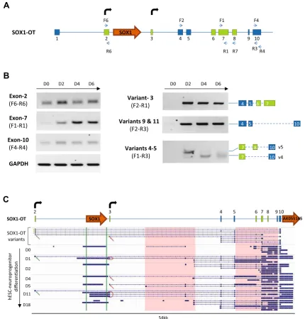
Structural architecture ofSOX1-OTin ReN cells
Based on the initial strong signal for SOX1-OT in ReN cells, these cells were further used to characterise the structure ofSOX1-OTusing two parallel and complemen-tary approaches: RT-PCR using primers in annotated exons ofSOX1-OT, and 50RACE to identify the transcription start site (TSS) of SOX1-OT (Fig.3). RT-PCR revealed the presence of three new exons (in green in variants 3–6, Fig.3b). 50RACE primed in the last annotated exon uncovered two additional exons at the 50 end of the tran-script (in green in variants 8–11, Fig.3b), the furthest 50of which was further validated by RT-PCR (variant 7, Fig.3b).
The 50RACE analysis revealed two main TSS forSOX1 -OTlocated in close genomic proximity to theSOX1gene (Fig.3c, bent arrows). To confirm the regulatory tran-scriptional potential of these two TSS, an online bioinformatics analysis was performed by aligning the
SOX1-OT sequence to the FANTOM5 project tracks through the UCSC genome browser (Fig.3c) [39]. The FANTOM5 project provides genome-wide mammalian gene expression data by mapping TSS, promoter regions and enhancers in human and mouse primary cells, cell lines and tissues [32]. The alignment highlighted two potential transcriptional start sites with high peaks of Cap analysis for gene expression (CAGE) reads that matched with the TSS experimentally identified by 50RACE, providing fur-ther support for the identity of theSOX1-OTTSS found in ReN cells (Fig.3c, red tracks).
SOX1-OTandSOX1are co-expressed during neural differentiation
To determine whether the differentSOX1-OTvariants were expressed at different levels during neuronal differentia-tion, ReN neuroprogenitor cells were differentiated over a 6-day time course, and tested as undifferentiated (D0) or after 2, 4 and 6 days of neural differentiation. ReN cell differentiation was confirmed by immunofluorescence at D0 and D6 showing loss of the undifferentiated marker Nestin and increase in MAP2 expression (a neuronal marker) (Fig.4a). Relative quantification of SOX1 expression in ReN cells over the 6-day differentiation showed thatSOX1mRNA significantly increased at D2, D4 and D6 of differentiation compared to D0 (Fig.4b).
Fish
Frog
Chicken
Opossum
Mouse
Dog
Chimpanzee
SOX1
SOX1-OT
Intergenic
Coding
Repeats
ECRs
UTRs
Intronic
A
B
Fig. 2 Cross-species comparative analysis of SOX1 overlapping transcript loci. a Evolutionary conserved regions revealed in the cross-species alignment of human assembly hg19 region chr13:112626600–112765500 generated by the ECR browser (http:// ecrbrowser.dcode.org). ECR evolutionary conserved region, UTR untranslated region. b Snapshot images of the SOX1 overlapping transcript loci on human (hg19 chr13:112626600–112765500, top panel) and mouse (mm9, chr8: 12,300,135–12,439,035) taken from
[image:6.595.64.538.59.592.2]AK055145 annotated just 30 to the last exon of LINC00403 may be part of some SOX1-OT transcript variants in this cell type (Fig.5c). This observation appeared to better mirror the data obtained for the
annotated mouse Sox1-ot transcript Gm5607 (see human and mouse locus comparison in Fig.2b). The analysis was extended to human neural tissue, through transcriptome analysis of RNA-seq data available for developing human
A
B
R3 Variant 1
Variant 2
F2 F1
R1
GSP1 SOX1
C
Variant 4
Variant 6
Variant 7
Variant 3
Variant 5 RT-PCR
R7
5’RACE
F6
Variant 8
Variant 10 Variant 9
Variant 11
Variant 2
BLAT TSS
Fig. 3 SOX1-OT structure in ReN human neuroprogenitor cells.
a Schematic diagram showing SOX1-OT Variants 1 and 2 as annotated in the UCSC human genome browser, showing the evolutionary conserved region identified by cross-species comparison (shaded green box), the positions (blue arrows) of the RT-PCR (F1, R1, F2 and R3) and 50RACE (GSP1) primers, and the location of SOX1(orange arrow). b Transcript variants identified by RT-PCR and 50RACE. Variants 3–5 were identified by RT-PCR using primers within the conserved sequence (primers F1 and R1) in combination with primers in annotated exons; variant 6 was amplified using primer R7 in one of the newly identified exons from variant 6 and primer F2,
while variant 7 was amplified using primer F6 in the first exon upstream of SOX1 identified by 50RACE (variants 8, 9 and 10). 50RACE products were amplified by anchoring the 50RACE in the most 30 exons of the annotated LINC0403 (primer GSP1 in a).
[image:7.595.88.511.58.524.2]cortex [40]. This analysis identified a greater variety of SOX1-OT transcript variants over increasing gestational time points in developing cortex tissue than in the cell samples used in this study (Supplementary Fig. 2). Nev-ertheless, many were found to initiate at a transcription start site (TSS) very close to, if not identical to, the TSS detected by 50RACE. Evidence from this transcriptome analysis also suggested thatSOX1-OTmay extend further than its currently annotated 30 end.
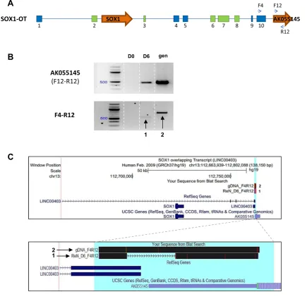
Protein-coding gene AK55145 is part ofSOX1-OT
To further investigate the 30extent of the humanSOX1-OT transcript, primers within AK055145 (F12 and R12, Fig.6a) were used alone or in combination with primer F4
in the last annotated exon of LINC00403 to test expression in D0 and D6 ReN cells (Fig. 6a).
RT-PCR detection of AK55145 gene expression (primer pair F12–R12) showed that AK55145 was only detected in differentiated ReN cells (Fig.6b, top panel). Using primer pair F4–R12, a product was amplified in D6 ReN cells suggesting that the last exon from SOX1-OT and the AK55145 gene may be part of the same transcript. To confirm our findings, the PCR fragments amplified from D6 cDNA and from gDNA were sequenced and aligned to the genome using BLAT [29], confirming that theSOX1 -OT transcript extended to include the AK55145 gene. Therefore, these results indicated that the locus ofSOX1 -OT extends further downstream than the currently anno-tated SOX1-OT transcript as shown in the UCSC genome browser-generated images (Fig.6c).
A
B
Day
0
Day
6
MAP2
NESTIN
D
a
p
i
i
p
a
D
*** ***
**** SOX1mRNA
Fig. 4 Differentiation of the ReN human neuroprogenitor cell line.
aImaging of day 0 (control) and day 6 (differentiated) ReN cultures in brightfield (left panels) and following immunostaining for NESTIN and MAP2 (green) with corresponding dapi counterstain (blue).Bar
50lm. bRelative quantification (RQ) ofSOX1mRNA expression analysed by quantitative RT-PCR across the different time points of ReN cells differentiation (day 0, 2, 4 and 6). Error barsrepresent
[image:8.595.89.516.44.451.2] [image:8.595.91.510.67.233.2]SOX1-OTexpression correlates withSOX1gene expression in different cancer cell lines
Recently, several reports have suggested SOX1 involve-ment in cancer developinvolve-ment [41–43], and the present study has investigated whether SOX1 gene expression may correlate with expression ofSOX1-OT in cancer. To
achieve this, SOX1 and SOX1-OT expression was anal-ysed in a variety of cancer cell lines by RT-PCR using different combinations of primer pairs across the locus (Fig.7a). Primer pair F4-R4 was used to detect the last annotated exon of LINC00403 that is shared by several SOX1-OT variants (see Fig.3b). Expression of the SOX1 amplicon (primers F13–R13) was co-detected with that of
A
B
C
F2
3 7
2 SOX1 6 8 9 10
F6
R3 F1
R1 4 5
R7 R6
F4
R4 1
SOX1-OT
Exon-10 (F4-R4)
D0 D2 D4 D6
Exon-2 (F6-R6)
GAPDH Exon-7 (F1-R1)
Variant- 3
(F2-R1) 4 5 6 7
Variants 4-5
(F1-R3) 7 10 v4
v5 7 8 10 Variants 9 & 11
(F2-R3)
10 4 5
D0 D2 D4 D6
D0
D2
D11
D18 SOX1-OT
variants
hE
SC-neuro
pro
g
enito
r
differenao
n
54kb 3
2 4 5 6 7 8 910
SOX1-OT SOX1 AK055145
D1
D4 D5
Fig. 5 Expression profile ofSOX1-OT during human neuroprogen-itor differentiation.aComposite structure of theSOX1-OTgenomic locus including the exons (green boxes) and additional TSS (bent arrows) newly identified in ReN cells; primers used in the expression profiling are shown asblue arrows.bRT-PCR detection ofSOX1-OT exons 2, 7, 10 and transcript variants 3, 4, 5, 9, 11 in undifferentiated ReN cells (D0) and at day 2 (D2), 4 (D4) and 6 (D6) of neural differentiation. GAPDH was used as positive control for RT reaction.
cSchematic diagram of theSOX1-OTlocus in ReN cells (top) with indicated exons (numbered boxes) and TSS (bent arrows), and IGV
[image:9.595.82.513.58.508.2]the SOX1-OT F4–R4 region in most of the cell lines analysed (Fig.7B).SOX1andSOX1-OTwere co-detected in teratocarcinoma (NTera) and some breast cancer (MCF7, T47D) cell lines, but not in colon (HCT116, CaCo-2), some breast (MDA-MB-231/361, Hs578T) and cervical (HeLa) cancer cells (Fig.7b). The exception to this pattern was the osteoblast HOS cell line, which expressed the SOX1 gene but not SOX1-OT, and the neuroblastoma SH-SY5Y cell line which presented the opposite pattern. Using primer pair F6–R3, we detected SOX1-OT variants 8–10 that span theSOX1 gene, but no
SOX1-OT variant spanning the SOX1 gene was detected in the cancer cell lines tested (Fig.7c). Transcriptome tracks for HeLa and MCF7 cells available through the ENCODE project annotations were analysed and indi-cated patterns consistent with our RT-PCR results, showing HeLa cells negative throughout this region, while some transcription could be seen in MCF7 cells across the locus (Supplementary Fig. 3). These findings suggested that SOX1-OT variants spanning the SOX1 gene are expressed in MCF7 cells, but these appeared to have a different structure to those found in ReN cells.
A
B
C
2 1
1 2
3 7
2 4 5 6 8 9 10
F4
1
F12
R12
SOX1-OT SOX1 AK055145
D0 D6 gen
F4-R12
1 2
AK055145 (F12-R12)
Fig. 6 LINC00403 and AK055145 are part of SOX1-OT.
aSchematic diagram showing the positions of primers used in exon 10 of SOX1OT and within the annotated gene AK055145.bRT-PCR product obtained using cDNA from ReN cells at day 0 (D0) and 6 (D6) of differentiation; genomic DNA (gen) was used as a positive control for PCR. PCR F12-R12 was co-linear to the genomic DNA and gave rise to a 462-bp product only detectable in D6 cells; F4–R12 amplicons spanned the region from exon 10 of LINC00403 and
[image:10.595.84.520.61.485.2]A
F2
3 7
2 6 8
SOX1
10 9 F6
R3 F1
R1 4 5
1 SOX1-OT
F4
R4 F13
R13
B
C
Variants 8-10
(F6-R3)
Variants 9 & 11
(F2-R3)
10 4 5
GAPDH
10 4 5
2
10 5
2
2 10
300
300
500
500
300
300
+RT
-RT
+RT
-RT
+RT
-RT SOX1
SOX1-OT Exon 10
(F4-R4) GAPDH
+RT
-RT
+RT
-RT
+RT
-RT 300
300 500 500 300
300
Fig. 7 SOX1andSOX1-OTexpression in a panel of human cancer cell lines.aSchematic diagram of theSOX1-OTlocus showing the positions of the primer pairs used to detectSOX1and different regions ofSOX1-OTin cancer cell lines.bRT-PCR detection ofSOX1and SOX1-OT(exon 10) in the stated human cancer cell lines and in day 6 differentiated ReN cells as a positive control.cRT-PCR detection of
SOX1-OTvariants 8–11 and exon 7 detected with the indicated primer combinations in the stated human cell lines. PCRs performed on total RNA after reverse transcription with (?RT) or without (-RT) reverse transcriptase;MDNA size standard;H2Ono template PCR negative
[image:11.595.87.509.55.598.2]Discussion
Characterisation of the structure ofSOX1-OT
Genome-wide studies have reported large numbers of non-coding RNAs whose function and significance are not clear. To understand the complex transcriptome architecture, expression and regulation of genetic information, it has become necessary to distinguish between mRNA and ncRNA transcripts [44]. Here we show that theSOX1-OTtranscript, annotated as a long intergenic non-coding mRNA-like tran-script with no inferred coding potential, could be detected in human cells. SOX1-OT has a complex structure including several unannotated exons, different transcript variants, and at least two potential TSS. Our data identified a total of 10 exons for humanSOX1-OT, 5 of which (exon2, 3, 6–8) are novel and previously unknown. In addition to the two annotated tran-script variants (V1–V2), we report 9 newSOX1-OTtranscript variants (V3–V11) not previously reported in the literature. Therefore,SOX1-OT presents complex transcriptional fea-tures, whose potential functions and biological significance remain to be explored. The TSS identified for humanSOX1 -OTis located in close genomic proximity to and upstream of theSOX1gene (Fig.1a), suggesting a possible role in regu-latingSOX1 gene expression. The likelihood of SOX1-OT acting as a regulator ofSOX1is supported by similarities with theSOX2locus. The multi-exon, non-codingSOX2-OT tran-script overlappingSOX2has recently been shown to give rise to multiple splice variants from different TSS, and is attributed a positive regulatory role inSOX2transcription [9].
Our results show that the first exon of the RefSeq-an-notated transcript LINC00403 is either absent or expressed at levels below the present detection limits in ReN cells. However, it is important to note that the current annotated structure of SOX1-OT has been obtained by combining information collected from three different tissues types (amygdala, eye, carcinoid); this might explain the differ-ences with the present study, which focused on characterising the transcript in a well-defined neural cell type. Interestingly, the newly experimentally characterised structure of humanSOX1-OT resembles that of the anno-tated mouseSox1-ot. Both have TSS upstream of and near to theSOX1coding gene; moreover, although the 30end of the mouse overlapping transcript extends further down-stream compared to the current annotation of the human SOX1-OT, our findings extend humanSOX1-OTto include the downstream AK55145 gene, in line with the mouse transcript 30 end. Our results suggest this 30 end might be used in differentiated ReN cells and not in undifferentiated cells; further work will be required to determine whether Transcription Termination End (TTE) usage is regulated in a cell type/tissue/differentiation stage specific manner.
Potential role ofSOX1-OTin neural differentiation as a regulator ofSOX1
SOX1-OT was found to be highly expressed in differenti-ated neural stem cells, and its expression appeared to correlate withSOX1 gene expression. DifferentSOX1-OT transcript variants were differentially detected during the course of neural differentiation. Our observed correlation between SOX1 and SOX1-OT expression during neural differentiation is similar to that reported forSox2andSox2 -ot during mouse neurosphere differentiation in vitro; however, in this case both Sox2 andSox2-otwere upreg-ulated after day 2 and then slightly downregupreg-ulated at day 7 of neural differentiation [5], while here SOX1andSOX1 -OT were upregulated at day 2 and expression remained upregulated towards day 6 of neural differentiation in vitro. It is therefore possible that co-expression ofSOX1-OTand SOX1 during neural differentiation might indicate a co-regulatory role in pathways regulating neural differentia-tion. Furthermore, we observed a switch between transcript variants 4 and 5 from day 2 to 4, further supporting a possible regulatory role during neural differentiation. Fur-ther experiments testing the effect of forced expression or downregulation of the new transcript will be required to determine if SOX1-OT plays a functional role in neural differentiation and a possible link toSOX1expression.
SOX1-OTandSOX1are concomitantly expressed in different cancerous cell lines
SOX1 expression has been already reported in several cancer types [45–48]. We detected co-expression ofSOX1 -OT and SOX1 RNAs in NTera, T47D and MCF7 cancer cell lines. Concomitant expression of SOX2 and its LncRNASOX2-OThas been described in different cancer types, and it was shown that SOX2 gene expression is regulated bySOX2-OTin this context. For example,SOX2 -OT is upregulated together with SOX2 and OCT4 in oesophageal squamous cell carcinoma [6]. Moreover, co-expression of SOX2-OT and SOX2 has been previously reported in the NTera cell line, and SOX2-OT has been functionally associated with theSOX2gene in pluripotency and tumorigenesis [9]. Also, concordant expression of SOX2andSOX2-OThas been reported in breast cancer and both are upregulated in cell suspension culture conditions that favour stem cell expansion [7].
Therefore, SOX1-OT might have a potential role in cancer by promoting SOX1 expression; its expression in different cancer types in which SOX1 has already been reported will need further investigation. Based on our data and in silico analysis, it is possible that cells from different tissues and/or different cell types from the same tissue may express different repertoires of transcript variants, so fur-ther larger scale expression analyses will be required to identify all the isoforms, their structures and polyA sites. Indeed, analysis of Poly-A seq data from both mouse and human brain samples confirmed the presence of a vari-ability of poly-A signals (Suppl. Figure 4). In the context of cancer, the structure of alternative SOX1-OT variants expressed in cancer types that express SOX1 will also require consideration, in order to identify the repertoire of transcript variants expressed through large-scale gene expression and RACE analyses. Interestingly, the osteosarcoma cell line HOS has been shown to express SOX1but not SOX1-OT, while in contrast the neuroblas-toma cell line SH-SY5Y showed signal forSOX1-OTbut notSOX1. This observation indicates thatSOX1andSOX1 -OTexpression might be independent of each other in these cancer types, or there might be another regulatory mech-anism for these two transcripts, which requires further exploration.
Our results also indicate that theSOX1-OTlocus extends further downstream than the currently annotatedSOX1-OT transcript, suggesting that the gene AK055145 annotated just 30to the last exon of LINC00403 may be part of some SOX1-OT transcript variants. Therefore, further experi-ments such as 30RACE will be necessary to confirm this initial observation.
Conclusion
In conclusion, we report the expression of an overlapping transcript at theSOX1 locus, and have demonstrated that SOX1-OThas a complex structure with two potential TSS and multiple transcript variants. These transcript variants are highly expressed in differentiating neuroprogenitors, where their expression coincides with that of SOX1. Fur-thermore, we have shown co-expression ofSOX1-OTand SOX1 RNA in neural and cancer cell lines, suggesting a possible role forSOX1-OTin stem cell differentiation and cancer. Further work is now needed to determine the function of SOX1-OT and its potential regulatory link to SOX1expression.
Acknowledgements We gratefully acknowledge support from the Alzheimer’s Society (S. Strohbuecker) and the University of Not-tingham Vice Chancellor’s scholarship for research excellence (A. Ahmad). We are grateful to Dr P. Collier and Dr A. Grabowska for the kind gift of RNA samples.
Compliance with ethical standards
Conflict of interest The authors declare that they have no conflicting interests.
Open Access This article is distributed under the terms of the Creative Commons Attribution 4.0 International License (http:// creativecommons.org/licenses/by/4.0/), which permits unrestricted use, distribution, and reproduction in any medium, provided you give appropriate credit to the original author(s) and the source, provide a link to the Creative Commons license, and indicate if changes were made.
References
1. Zhang S, Cui W (2014) Sox2, a key factor in the regulation of pluripotency and neural differentiation. World J Stem Cells 6:305–311
2. Fantes J, Ragge NK, Lynch SA, McGill NI, Collin JR, Howard-Peebles PN et al (2003) Mutations in SOX2 cause anophthalmia. Nat Genet 33:461–463
3. Quinodoz S, Guttman M (2014) Long noncoding RNAs: an emerging link between gene regulation and nuclear organization. Trends Cell Biol 24:651–663
4. Cao J (2014) The functional role of long non-coding RNAs and epigenetics. Biol Proced Online 16:11
5. Amaral PP, Neyt C, Wilkins SJ, Askarian-Amiri ME, Sunkin SM, Perkins AC, Mattick JS (2009) Complex architecture and regu-lated expression of the Sox2ot locus during vertebrate development. RNA 15:2013–2027
6. Shahryari A, Rafiee MR, Fouani Y, Oliae NA, Samaei NM, Shafiee M et al (2014) Two novel splice variants of SOX2OT, SOX2OT-S1, and SOX2OT-S2 are coupregulated with SOX2 and OCT4 in esophageal squamous cell carcinoma. Stem Cells 32:126–134
7. Askarian-Amiri ME, Seyfoddin V, Smart CE, Wang J, Kim JE, Hansji H et al (2014) Emerging role of long non-coding RNA SOX2OT in SOX2 regulation in breast cancer. PLoS One 9:e102140
8. Gu¨re AO, Stockert E, Scanlan MJ, Keresztes RS, Ja¨ger D, Altorki NK et al (2000) Serological identification of embryonic neural proteins as highly immunogenic tumor antigens in small cell lung cancer. Proc Natl Acad Sci USA 97:4198–4203
9. Shahryari A, Jazi MS, Samaei NM, Mowla SJ (2015) Long non-coding RNA SOX2OT: expression signature, splicing patterns, and emerging roles in pluripotency and tumorigenesis. Front Genet 6:196
10. Saghaeian Jazi M, Samaei NM, Ghanei M, Shadmehr MB, Mowla SJ (2016) Identification of new SOX2OT transcript variants highly expressed in human cancer cell lines and down regulated in stem cell differentiation. Mol Biol Rep 43:65–72 11. Kan L, Israsena N, Zhang Z, Hu M, Zhao LR, Jalali A et al (2004)
Sox1 acts through multiple independent pathways to promote neurogenesis. Dev Biol 269:580–594
12. Archer TC, Jin J, Casey ES (2011) Interaction of Sox1, Sox2, Sox3 and Oct4 during primary neurogenesis. Dev Biol 350:429–440
13. Huguet EL, McMahon JA, McMahon AP, Bicknell R, Harris AL (1994) Differential expression of human Wnt genes 2, 3, 4, and 7B in human breast cell lines and normal and disease states of human breast tissue. Cancer Res 54:2615–2621
metastasis suppressor gene TFPI-2 in cancer. Nucleic Acids Res 41:6857–6869
15. Colston KW, Perks CM, Xie SP, Holly JM (1998) Growth inhi-bition of both MCF-7 and Hs578T human breast cancer cell lines by vitamin D analogues is associated with increased expression of insulin-like growth factor binding protein-3. J Mol Endocrinol 20:157–162
16. Andrews PW, Damjanov I, Simon D, Banting GS, Carlin C, Dracopoli NC, Føgh J (1984) Pluripotent embryonal carcinoma clones derived from the human teratocarcinoma cell line Tera-2. Differentiation in vivo and in vitro. Lab Invest 50:147–162 17. France LA, Scotchford CA, Grant DM, Rashidi H, Popov AA,
Sottile V (2014) Transient serum exposure regimes to support dual differentiation of human mesenchymal stem cells. Tissue Eng Regen Med 8:652–663
18. Hoffrogge R, Mikkat S, Scharf C, Beyer S, Christoph H, Pahnke J et al (2006) 2-DE proteome analysis of a proliferating and dif-ferentiating human neuronal stem cell line (ReNcell VM). Proteomics 6:1833–1847
19. Scherer WF, Syverton JT, Gey GO (1953) Studies on the prop-agation in vitro of poliomyelitis viruses. IV. Viral multiplication in a stable strain of human malignant epithelial cells (strain HeLa) derived from an epidermoid carcinoma of the cervix. J Exp Med 97:695–710
20. Biedler JL, Roffler-Tarlov S, Schachner M, Freedman LS (1978) Multiple neurotransmitter synthesis by human neuroblastoma cell lines and clones. Cancer Res 38:3751–3757
21. Rhim JS, Cho HY, Vernon ML, Arnstein P, Huebner RJ, Gilden RV (1975) Characterization of non-producer human cells induced by Kirsten sarcoma virus. Int J Cancer 16:840–849
22. Sambuy Y, De Angelis I, Ranaldi G, Scarino ML, Stammati A, Zucco F (2005) The Caco-2 cell line as a model of the intestinal barrier: influence of cell and culture-related factors on Caco-2 cell functional characteristics. Cell Biol Toxicol 21:1–26 23. Brattain MG, Fine WD, Khaled FM, Thompson J, Brattain DE
(1981) Heterogeneity of malignant cells from a human colonic carcinoma. Cancer Res 41:1751–1756
24. Soule HD, Vazguez J, Long A, Albert S, Brennan M (1973) A human cell line from a pleural effusion derived from a breast carcinoma. J Natl Cancer Inst 51:1409–1416
25. Cailleau R, Olive M, Cruciger QV (1978) Long-term human breast carcinoma cell lines of metastatic origin: preliminary characterization. In Vitro 14:911–915
26. Hackett AJ, Smith HS, Springer EL, Owens RB, Nelson-Rees WA, Riggs JL, Gardner MB (1977) Two syngeneic cell lines from human breast tissue: the aneuploid mammary epithelial (Hs578T) and the diploid myoepithelial (Hs578Bst) cell lines. J Natl Cancer Inst 58:1795–1806
27. Keydar I, Chen L, Karby S, Weiss FR, Delarea J, Radu M et al (1979) Establishment and characterization of a cell line of human breast carcinoma origin. Eur J Cancer 15:659–670
28. Bustin SA, Benes V, Garson JA, Hellemans J, Huggett J, Kubista M, Mueller R, Nolan T, Pfaffl MW, Shipley GL et al (2009) The MIQE guidelines: minimum information for publication of quantitative real-time PCR experiments. Clin Chem 55:611–622 29. Kent WJ (2002) BLAT—the BLAST-like alignment tool.
Gen-ome Res 12:656–664
30. Kent WJ, Sugnet CW, Furey TS, Roskin KM, Pringle TH, Zahler AM, Haussler D (2002) The human genome browser at UCSC. Genome Res 12:996–1006
31. Speir ML, Zweig AS, Rosenbloom KR, Raney BJ, Paten B, Nejad P et al (2016) The UCSC Genome Browser database: 2016 update. Nucleic Acids Res 44:D717–D725
32. FANTOM Consortium and the RIKEN PMI and CLST (DGT), Forrest AR, Kawaji H, Rehli M, Baillie JK, de Hoon MJ, et al. (2014) A promoter-level mammalian expression atlas. Nature 507:462-70
33. Ovcharenko I, Nobrega MA, Loots GG, Stubbs L (2004) ECR Browser: a tool for visualizing and accessing data from com-parisons of multiple vertebrate genomes. Nucleic Acids Res 32:W280–W286
34. Sauvageau M, Goff LA, Lodato S, Bonev B, Groff AF, Ger-hardinger C et al (2013) Multiple knockout mouse models reveal lincRNAs are required for life and brain development. Elife 2:e01749
35. Bolger AM, Lohse M, Usadel B (2014) Trimmomatic: a flexible trimmer for Illumina sequence data. Bioinformatics 30:2114–2120
36. Kim D, Langmead B, Salzberg SL (2015) HISAT: a fast spliced aligner with low memory requirements. Nat Methods 12:357–360 37. Pertea M, Pertea GM, Antonescu CM, Chang TC, Mendell JT, Salzberg SL (2015) StringTie enables improved reconstruction of a transcriptome from RNA-seq reads. Nat Biotechnol 33:290–295 38. Thorvaldsdottir H, Robinson JT, Mesirov JP (2013) Integrative Genomics Viewer (IGV): high-performance genomics data visualization and exploration. Brief Bioinform 14:178–192 39. Rosenbloom KR, Sloan CA, Malladi VS, Dreszer TR, Learned K,
Kirkup VM et al (2013) ENCODE data in the UCSC Genome Browser: year 5 update. Nucleic Acids Res 41:D56–D63 40. Liu SJ, Nowakowski TJ, Pollen AA, Lui JH, Horlbeck MA,
Attenello FJ et al (2016) Single-cell analysis of long non-coding RNAs in the developing human neocortex. Genome Biol 17:67 41. Guan Z, Zhang J, Wang J, Wang H, Zheng F, Peng J et al (2014)
SOX1 down-regulates beta-catenin and reverses malignant phe-notype in nasopharyngeal carcinoma. Mol Cancer 13:257 42. Song L, Liu D, He J, Wang X, Dai Z, Zhao Y et al (2016) SOX1
inhibits breast cancer cell growth and invasion through sup-pressing the Wnt/beta-catenin signaling pathway. APMIS 124:547–555
43. Mathews LA, Hurt EM, Zhang X, Farrar WL (2010) Epigenetic regulation of CpG promoter methylation in invasive prostate cancer cells. Mol Cancer 9:267
44. Dinger ME, Pang KC, Mercer TR, Mattick JS (2008) Differen-tiating protein-coding and noncoding RNA: challenges and ambiguities. PLoS Comput Biol 4:e1000176
45. Tsao CM, Yan MD, Shih YL, Yu PN, Kuo CC, Lin WC et al (2012) SOX1 functions as a tumor suppressor by antagonizing the WNT/beta-catenin signaling pathway in hepatocellular carci-noma. Hepatology 56:2277–2287
46. Apostolidou S, Hadwin R, Burnell M, Jones A, Baff D, Pyndiah N et al (2009) DNA methylation analysis in liquid-based cytology for cervical cancer screening. Int J Cancer 125:2995–3002 47. Su HY, Lai HC, Lin YW, Chou YC, Liu CY, Yu MH (2009) An
epigenetic marker panel for screening and prognostic prediction of ovarian cancer. Int J Cancer 124:387–393