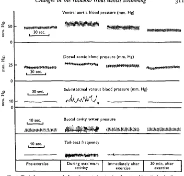
Changes in Blood Pressure, Heart Rate and Breathing Rate During Moderate Swimming Activity in Rainbow Trout
Full text
Figure
Related documents
First, we identify the macroeconomic and financial variables (GDP growth, interest rate maturity spread, stock market’s volatility) and bank-specific variables (size, capital
(c) To discuss extensively the regulations and codes of conduct put in place by financial business regulators in Nigerian to ensure ethical practices in the governance of
In this paper, a Chebyshev finite difference method has been proposed in order to solve nonlinear two-point boundary value problems for second order nonlinear differential
Voltage stability, voltage regulation and power system stability, damping can be improved by using these devices and their proper control [4] .There are various forms of FACTS
The microcapsules were characterized by FTIR, DSC, particle size, SEM, encapsulation efficiency, swelling index, in vitro wash off test and in vitro drug release..
exothermic and a large amount of heat is released in the process.It follows therefore that curing concrete in lime/water solution will result in heating of the
The threat of crime and theft from customers of the Private Member’s Club or Casino is a key motive for CCTV installation, but it is the often internal crime carried out by
Boyle’s introducing his model straightforwardly as if making factual claims would, in analogy to fictionalization, have the undesirable result of forcing his readers to believe
