tinted in Great Britain
METABOLIC AND TISSUE SOLUTE CHANGES
ASSOCIATED WITH CHANGES IN THE FREEZING
TOLERANCE OF THE BIVALVE MOLLUSC
MODIOLUS DEMISSUS
BY DENNIS J. MURPHY*
Department of Zoology, University of Maryland, College Park, Maryland 2074a
(Received 27 July 1976)
SUMMARY
1. A physiological mechanism responsible for increasing the freezing tolerance of the bivalve Modiolus demissus (Dillwyn) following low-temperature acclimation was demonstrated.
2. The rates of oxygen consumption of M. demissus acclimated to temperatures between o and 24 °C were presented as an Arrhenius plot. A change in slope occurred at 10 °C, suggesting that temperature alone was not responsible for the increased decline in the rate of oxygen consumption below 10 °C.
3. Low-temperature acclimation had no effect on blood Na+ or K+ con-centrations but did reduce the concentration of blood Mg2+ and, in addition, resulted in the accumulation of end-products characteristic of anaerobic metabolism - tissue alanine and proline, and blood Ca*"1". Furthermore, maintenance of M. demissus under anaerobic conditions increased freezing tolerance.
4. Taken together, these data indicate that the increased freezing tolerance of M. demissus acclimated to low temperatures involves a con-version to anaerobic metabolism.
5. The increase in blood Ca2+ following low-temperature acclimation was associated with the increased freezing tolerance. Finally, Mg2* simulated the effect of Ca2+ on freezing tolerance, but was only 20 % as effective.
6. These results suggest that a Cas+-dependent mechanism responsible for increasing the freezing tolerance of M. demissus exists, and that the increase in blood Cal+ is due to a conversion to anaerobic metabolism.
INTRODUCTION
A variety of intertidal molluscs are able to tolerate internal ice formation (Kan-wisher, 1955, 1966). Ice forms only extracellularly in these animals and grows at the expense of both intracellular and extracellular water; freezing injury occurs when a critical amount of tissue water is converted to ice (Kanwisher, 1959; Williams, 1970; Murphy & Pierce, 1975). The freezing tolerances of many intertidal molluscs vary seasonally; freezing tolerances increase during the colder winter months (Somme,
1966). Murphy & Pierce (1975) demonstrated that the degree of freezing tolerand exhibited by the intertidal mussel Modiolus demissus depends on the acclimation temperature: lower acclimation temperatures result in greater freezing tolerances. Furthermore, the physiological basis for the increased freezing tolerance of M. demissus acclimated to low temperatures is due to an increased tolerance of greater amounts of tissue ice formation (Murphy, 1974; Murphy & Pierce, 1975).
The conversion of extracellular water to ice leads to an increased concentration of solutes in the unfrozen portion of the extracellular space. As a result, intracellular water diffuses down its concentration gradient into the extracellular space to maintain a vapour pressure equilibrium between the intracellular and extracellular fluids (Mazur, 1963). Extracellular ice formation, therefore, results in cellular dehydration with cell injury occurring when a critical level of cellular dehydration is attained (Lovelock, 1953; Karow &Webb, 1965). Indeed, physiological mechanisms responsible for increasing the freezing tolerance of many overwintering insects and plants to extracellular ice formation involve the accumulation of specific tissue solutes such as sugars, organic acids, proteins, alcohols and ions which reduce the harmful effects produced by freezing dehydration (for review see Heber & Santarius, 1973).
Although much is known about the physiological mechanisms responsible for increasing the freezing tolerance of plants and insects to extracellular ice formation, the mechanisms responsible for increasing the freezing tolerance of molluscs remain unknown. In order to define a physiological mechanism responsible for changes in the freezing tolerance of intertidal molluscs, I have characterized and experi-mentally modified several metabolic and tissue solute changes associated with the increased freezing tolerance of the intertidal, marsh-dwelling mussel Modiolus demissus following low-temperature acclimation. The results of these experiments indicate that a transition from an aerobic to a partially anaerobic metabolism occurs at low temperatures. As a result of anaerobic metabolism, blood Caa+ concentrations increase and greater freezing tolerances are achieved.
MATERIALS AND METHODS Animals
Modiolus demissus were collected periodically between June 1974 and August 1975 from either a salt marsh on the bay side of Assateaque Island, Maryland, or from Little Sippewissett Marsh, Woods Hole, Massachusetts. All mussels ranged from 7 to 9 cm in length and no distinction as to sex was made. Maryland animals were held in aerated artificial sea water (Instant Ocean; salinity = 34%,,) in a constant-temperature room (15 °C) at College Park, Maryland, and Massachusetts animals were held on sea tables in continuously running sea water (32%,,) at approximately 20 °C. The salinity of all sea-water solutions was determined by measuring the osmotic concentration of sea water samples with a freezing point depression osmometer (Osmette, Precision Systems). Osmotic concentrations were converted to parts per thousand (%„) using the formula:
Freezing-injury determinations (A) Whole animals
Intact M. demissus were frozen in air to various subfreezing temperatures for 12 h in an insulated chamber as previously described (Murphy & Pierce, 1975). The animals were then thawed for 1 h at 15 °C in 3 1 of sea water at the salinity of accli-mation. Following the thawing period, mortality was determined and exact LD50 values with 95% confidence limits calculated according to the method of Bliss (1938). A mussel was considered dead when the valves failed to adduct completely either upon direct mechanical stimulation of the posterior adductor muscle or as a result of stroking the shell with a plastic rod.
(B) Isolated tissue preparation -foot muscle
Strips of foot muscle were prepared by cutting the foot longitudinally along the midline into segments with wet weights of approximately 0-15 g. A freezing sample consisted of 10 tissue strips placed in i-oml of incubation medium in a 15 ml polystyrene test tube. These samples were then placed in the freezing chamber and frozen for 12 h at various subfreezing temperatures. The tissue was subsequently thawed by immersing the test tube in a water bath at 25 °C. Under these conditions, the rates of cooling and warming were approximately o-i °C/min and 5-5 °C/min respectively, as determined by an iron-constantan thermocouple (Grass Instrument Co., Model TCT-i).
The degree of tissue freezing injury was determined by measuring the contractility of muscle following a 1 h thawing period according to the following procedure. Each tissue strip was suspended with silk suture vertically in air between a fixed metal hook and a force-displacement transducer (Grass Instrument Co., Model FT 03C). Platinum pin electrodes (Grass Instrument Co., Model E2B) were inserted into the tissue and held in place with a micromanipulator. The muscle was electrically stimulated through the electrodes with square-wave monopolar impulses (10 msec) controlled by a Grass S9 stimulator (Grass Instrument Co.). The output of the transducer was displayed on an oscillograph (Grass Instrument Co., Model 79C). Changes in freezing tolerance were expressed as changes in the minimum voltage stimulus necessary to elicit a twitch contraction.
Influence of temperature acclimation and anaerobiosis on tissue solutes
Mussels were acclimated to temperatures between o and 25 °C in Instant Ocean (32%o) under constant illumination for a period of at least 2 weeks. A constant-temperature room was used to maintain a 12 °C constant-temperature (±0-5 °C) while temperatures below i 2 ° C ( ± i ° C ) were maintained with portable cooling units (Blue M Co.) and temperatures above 12 CC (±o*i °C) with constant-temperature circulating heaters (Haake, Model FE). An additional group of mussels was acclimated at 13 °C for 2 weeks and, subsequently, held under anaerobic conditions by clamping the valves shut with rubber bands and then coating the valves with a layer of paraffin. These animals were held under anaerobic conditions for 14 days.
(A) Blood osmotic pressure
Blood was obtained from M. demissus by slashing the mantle tissue and collecting the drainage from the cut surfaces according to the method of Pierce (1970). The blood was centrifuged at 30000$ for 15 min and the osmotic pressure of the super-natant measured with the freezing point depression osmometer.
(B) Blood cations
The concentrations of Caa+, Mg*+, K+ and Na+ were determined in the blood supernatant using atomic absorption spectrophotometry (Perkin Elmer, Model 503). All ions were measured in an appropriate dilution of a strontium chloride solution (0-25%). The blood was diluted 1:100 for Ca2+ and K+ measurements, 1:50 for Mg2+, and 1:200 for Na+. Strontium chloride prevents ionic bonding of Ca1"1" and Mg2* with any phosphates or sulphates present in the blood and also avoids inter-ference with K+ measurements as occurs when LaC^ is used (Paschen & Fuchs, 1971). The absorbance values were converted to concentrations (jig/ml) by com-parison with standard curves. Standard solutions were made from A.C.S. certified CaCl2, MgCl2> KC1 and NaCl dissolved in double-glass-distilled water. The standard
curves for CaClj, MgClj, KC1 and NaCl were linear over the concentration ranges used with regression coefficients {(}) and 95% confidence limits of + 0*0485 (± 0-0031),
+ 0-0158 (±0-0014), +0-0392 (±0-0063) and +0-0061 (±0-0013) respectively.
Blood Caa+ concentrations were also measured using the murexide colorimetric assay (Williams & Moser, 1953). A working murexide solution was made by dis-solving 8-o mg murexide (ammonium puparate) into 15 ml of double-glass-distilled water and, subsequently, adding 35 ml of 100% ethanol. Blood supernatant was diluted 1:40 with tris buffer (005 M) at pH 6-8 and 2 0 ml of this solution mixed 1:1 with the murexide solution. The absorbance of the red colour produced was read on a double-beam spectrophotometer (Varian Techtron, Model 635) at 475 nm using a murexide-buffer solution (1:1) as the reference. The CaCl, standard curve was linear up to i8/igCa1 +/ml with a regression coefficient and 9 5 % confidence limit of + 0-0283 (± 0-0009). Finally, a statistical comparison of the murexide assay values with those obtained by atomic absorption showed that the blood Ca2+ values obtained by these two techniques did not differ significantly (P > 0-5).
(C) Tissue-free amino acids
Influence of temperature acclimation on oxygen consumption
M. demissus (shell lengths = 7 cm) were acclimated in 33%,, Instant Ocean at 0, 4, 14 and 24 °C for a period of 2-3 weeks under constant illumination. The 14 °C temperature was maintained by a constant-temperature room and temperatures above and below 14 °C maintained as previously described. Following acclimation, individual animals were each placed in a 500 ml clear glass jar filled with sea water from the acclimation tank. The jars were then sealed with screw-on plastic caps coated with petroleum jelly and submersed in the acclimation tanks for either 2-3 h (14 and 24 °C acclimation groups) or 24 h (o and 4 °C acclimation groups). Following the required incubation period, the caps were removed and the dissolved oxygen concentrations rapidly measured with a self-stirring oxygen electrode (YSI, Model 5420A) attached to an oxygen meter (YSI, Model 54). Continuous recordings of oxygen consumption during these incubation periods showed that the rates of oxygen consumption were always linear. In addition, the oxygen consumptions of isolated valves were measured under identical conditions to control for any bacterial or algal contamination.
Influence of divalent cations on freezing tolerance (A) Whole animals
Since M. demissus is an osmo- and ionconformer (Pierce, 1970, 1971), the osmotic pressure and ion concentrations of M. demissus blood can be manipulated simply by altering these factors in the acclimation sea water. Five groups of 60 animals were each placed into 12 gal (45 1) of 3i-5%o artificial sea water (ASW). The ion con-centrations of this sea water were the same as those described by Wilkins (1972) except that Caa+ concentrations were varied from 37 to 16-5 mM. The mussels were acclimated to these solutions for 10-14 days a t 24 °C- Following acclimation, sea water and blood Cas+ concentrations were determined using the murexide assay as described above. Mussels from each group were then frozen to various temperatures for 12 h and LD50 values calculated.
(B) Isolated tissue
The influence of Ca2+ and Mg24" on freezing tolerance was also determined by incubating strips of foot tissue in either Ca2+-free ASW, Mg*+-free ASW, or Ca2+ -and Mga+-free ASW (3i-9%o) (recipes from Wilkins (1972)) for 6 h at 25 °C and then freezing the tissue to various temperatures for 12 h. The tissue was thawed in ASW (3i-9%o) for 1 h at 25 °C and changes in threshold voltages measured. In addition, threshold voltages of non-frozen controls - tissue transferred directly from incubation media to thawing media - were determined for each test solution. The freezing tolerance values of the freeze-thawed muscle were then expressed as responses relative to control, and calculated by the formula:
- 0 - 4
e oo
T5
O 0-4
60 e o
a 0-8
8
d
oe
CO
.3
1-21-6
3-35 3-45 3-55 3-65 Acclimation temperature (1/Tx 103 °K)
Fig. i. Rates of oxygen consumption of M. demusus acclimated to temperatures between o and 34 °C for a weeks. Data are presented as an Arrhenius plot; the change in slope occurs at i o °C. Error bars indicate ± i x standard error of the mean, n = ia.
RESULTS
Influence of temperature acclimation on oxygen consumption
The oxygen consumptions of M. demissus acclimated to temperatures of 24, 14, 4 and o °C progressively declined with the temperature. Furthermore, in an Arrhenius plot, the oxygen consumption data deviated significantly from linearity (F < o-ooi) (Fig. 1). In Fig. 1, therefore, the data are represented as two straight lines inter-secting at 10 °C. The calculated Q10 values for oxygen consumption between 24 and 14 °C was 2-23 and between 4 and o °C was 22-1. These results indicate that the change in the rate of oxygen consumption of M. demissus below 10 °C may represent a qualitative change in metabolism.
Influence of temperature acclimation and anaerobiosis on the concentration of specific tissue solutes
Table i. The effects of macrobiosis and temperature acclimation on the concentrations
of specific tissue solutes and the freezing tolerance of M. demissus
Acclimation conditions
Sea water Ca1+ (miu), n = 3 Blood Ca*+ (miu), n = 10 Blood Cat +-sea water Ca1+ Tissue total free amino acids
0*M/g dry wt.), n = 5 Tissue alanine (% of total), n = Tissue proline (% of Total),
n = 5
Sea water osmotic pressure (m-osmol/kg H,O), n •=• 3 Blood osmotic pressure
(m-osmol/kg HaO), n = 10 Blood osmotic pressure —
sea-water osmotic pressure
LDso(°C)
Group A anaerobic 13 °C
14 days 8-9 194 (±0-7)* io-5»(±o-7)» 562-7 (±7-9)* S ai-83«(±o-87)»
o-8s«(±o-oi)»
9 3 i
957 (±3-4)*
a6«(±3-4)#
— 12 06 (±0-90)**
Group B aerobic 0 °C
14 days
I I - O
138 (io-3) a-8b(±o-3) 4735 (±"-3) i8i7a(±o-6i)
o-68' (±009) 928
94i (±37) i3E(±3-7) -1253 (±041)
Group C aerobic 13 °C
14 days
8-6
93 (±0-2) 07c (±0-2) 465-0(131),
n-6sb(±o-57) o-aob(±o-o3)
938
9S3 (±22)
IS* (±2-*)
- 9 7 8 (±076) • Error values are standard errors.
• • Error values are 95 % confidence limits. l b c
Means with identical superscripts are not significantly different (P > 005). (Non-parametric multiple comparisons by STP was used to determine statistical differences.) (See Sokal & Rohlf, 1969)
to low temperature (o °C) under aerobic conditions were significantly higher than the blood Ca2+ concentration of control mussels acclimated under aerobic conditions at 13 °C (Table 1). Both forced anaerobiosis and low-temperature acclimation also increased the concentration of tissue alanine above that of the controls. Furthermore, the alanine increases were the same under both acclimation conditions (Table 1). Similarly, the concentration of tissue proline was elevated above that of the control mussels by both acclimation conditions, and again by similar amounts (Table 1).
14
13
1 2
11
5 5
5 1
4 7
2 2
1 8
1 4
1 0
4 9 0
4 7 0
4 5 0
-sw
Blood
Ca2
Na+
5 10 15 20 Acclimation temperature (°C)
[image:8.451.102.325.42.373.2]25
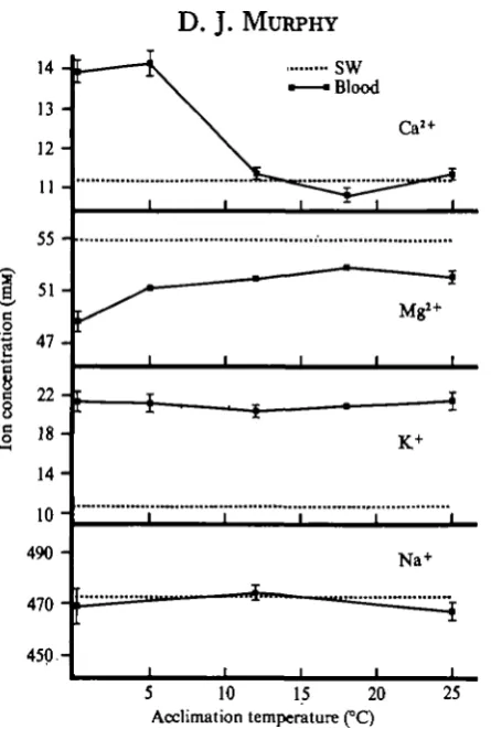
Fig. a. Influence of the temperature of acclimation on the concentration of the major blood cations of M. demistus. Error bars indicate ± i x standard error of the mean, n = io.
Effects of Cai+ and Mg*+ on freezing tolerance
- 1 2
- 1 0
8
- 6
10 12 Blood Ca2+(mM))
[image:9.451.80.367.39.410.2]14 16
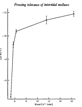
Fig. 3. Relationship between blood C a ^ concentrations and freezing tolerance of M. demutut. Mussels were acclimated in 31-5 % ASW (14 °C) with varying Ca'+ concentration* for 10-14 days prior to freezing. Each point is an L D 5 0 value calculated from 50-80 animals. Error bars indicate 95 % confidence limits.
D I S C U S S I O N
o-o
0-4
0-8
1-2
C a ^ + M g ^ A S W Mg^-freeASW Ca2+-freeASW
+ Mg2+-freeASW
- 4 - 5 - 6 - 7
Freezing temperature (°C)
Fig. 4. Effects of Ca1+ and Mg*+ on the changes in the contractile response of freeze-thawed foot muscle of M. demissus relative to non-frozen controls. Animals used in this experiment were collected during the winter from the salt marshes of Assateague Island, Maryland, and were held in artificial sea water (Instant Ocean; salinity = 34%o) at 15 °C for 1 to 2 weeks prior to use. Increasing values represent increases in the relative amount of voltage necessary to elicit a twitch contraction, or increases in freezing injury. Error bars indicate f i x standard error of the mean, n = 20 (for further details see Materials and Methods section).
temperatures below 10 °C was 22-1. This Q10 value, approximately 10-fold greater than that found in aerobically respiring bivalves, does not indicate a simple temperature-dependent reduction in aerobic metabolism.
More convincing evidence of a switch to anaerobic metabolism at low temperatures is provided from the blood and tissue solute experiments. Solutes which character-istically accumulate during anaerobic metabolism in bivalves such as blood Cas+ (Crenshaw & Neff, 1969) and tissue alanine and proline (Hochachka & Mustafa, 1973) also accumulated in M. demissus following exposure to both low-temperature and anaerobic conditions. Finally, the association between anaerobic metabolism and increased freezing tolerance is further supported by the finding that M. demissus held anaerobically had greater freezing tolerances than aerobic animals acclimated at the same temperature (13 °C).
acclimation had no effect on the concentrations of either blood Na+ or K+. In contrast, M. demissus acclimated to temperatures below 5 °C had blood Ca2+ concentrations 25 % greater than, and blood Mg?+ concentrations 7 % less than, those of mussels acclimated to temperatures between 12 and 25 °C. Furthermore, increasing the blood Ca2+ concentrations of M. demissus acclimated at 24 °C from 5-28 to 11-6i mM caused a simultaneous increase in freezing tolerance. There was no further increase in freezing tolerance when the blood Ca2+ concentration exceeded I I - 6 I mM. It appears, therefore, that increases in blood Ca2+ concentrations can increase the freezing tolerance of M. demissus up to a point, but above that point no further increases occur. In addition, both Ca2+ and Mg*+ increased the freezing tolerance of foot muscle isolated from M. demissus. Incubation of the isolated tissue in either Caa+- or Mg2+-free ASW prior to freezing resulted in the same degree of freezing injury. However, since the Ca2+-free ASW contained approximately 50 mM-Mg!+ whereas the Mg2+-free ASW contained only 10 mM-Ca2+, Mg2+ is only about 20% as effective as Caa+ in altering the freezing tolerance.
In conclusion, a mechanism for increasing the freezing tolerance of M. demissus following low-temperature acclimation involves an increase in the concentration of blood Ca2+. Moreover, Mg2"1" simulates the effect of Ca2+ on freezing tolerance, but is only 20% as effective. Therefore, if Mg2+ is competing with Ca2+, then the drop in blood Mg*+ following low-temperature acclimation may also increase freezing tolerance by increasing the proportion of Caa+ affecting freezing tolerance. Whether or not this is true, however, will depend on determining the mechanism of the Ca2+ effect on freezing tolerance. This Ca2+-dependent mechanism for increasing the freezing tolerance of M. demissus is being investigated.
I wish to thank Dr S. K. Pierce Jr. for his critical reading of the manuscript. This work was supported by NSF Grant BMS 72-02465-A01 to Dr S. K. Pierce Jr. and a Chesapeake Bay Funds Grant, University of Maryland.
Contribution number 59 from the Tallahassee, Sopchoppy and Gulf Coast Marine Biological Association.
REFERENCES
BLISS, C. I. (1938). Determination of a dose-mortality curve for small numbers. J. Pharm. Pharmae. ii, 193-216.
CRKNSHAW, M. A. & NEFF, J. M. (1969). Decalcification at the mantle-shell interface in molluscs.
Am. Zool. 9, 881-5.
DEAL, P. H. (1974). Effects of freezing and thawing on a moderately halophilic bacterium as a function of Na, K and Mg concentration. Cryobiology II, 13-22.
HKBER, U. & SANTARIUS, K. A. (1973). Cell death by cold and heat, and resistance to extreme tem-peratures. Mechanisms of hardening and dehardening. In Temperature and Life (eds H. Precht, J. Christophersen, H. Hensel and W. Larcher), pp. 244-59. New York: Springer-Verlag.
HOCHACHKA, P. W. & MUSTAFA, T. (1973). Animal life without oxygen: Basic biochemical mechanisms.
Am. Zool. 13, 543-55.
KANWISHER, J. (1955). Freezing in intertidal animals. Biol. Bull. mar. biol. Lab., Woods Hole 109, 56-63.
KANWISHER, J. (1959). Histology and metabolism of frozen intertidal animals. Biol. Bull. mar. biol.
Lab., Woods Hole 116, 258-64.
KANWISHER, J. (1966). Freezing in intertidal animals. In Cryobiology (ed. H. T. Meryman), pp. 479-94. New York: Academic Press.
LOVELOCK, J. E. (1953). The mechanism of the protective action of glycerol against hemolysis by, freezing and thawing. Biockim. biopkyt. Acta n , 38-36.
MAZUR, P. (1963). Kinetics of water loss from cells at subzero temperatures and the likelihood or intracellular freezing. J. gen. Physiol. 47, 347-69.
MEINCKE, K. F. (1975). The influence of extreme temperatures on metabolic substances in hemorymph and foot muscle of Helix pomatia. Comp. Biochem. Physiol. 51 A, 373-6.
MURPHY, D. J. (1974). Freezing tolerance of Modiohu demusut: Dependence on tolerance to cell dehydration. Am. Zool. 14, 1250.
MURPHY, D. J. & PIERCE, S. K. JR. (1975). The physiological basis for changes in freezing tolerance of intertidal molluscs. I. Response to subfreezing temperatures and the influence of salinity and temperature acclimation. J. exp. Zool. 193, 313-23.
NEWELL, R. C. (1973). Factors affecting the respiration of intertidal invertebrates. Am. Zool. 13, 513-38.
PASCHEN, K. & FUCHS, C. (1971). A new micro-method for Na, K, Ca, and Mg determinations in a single serum dilution by atomic-absorption spectrophotometry. Climca CMm. Acta 35, 401-8. PIERCE, S. K. JR. (1970). The water balance of Modiolus (Mollusca: Bivalvia: Mytilidae): Osmotic
concentrations in changing salinities. Comp. Biochem. Physiol. 36, 531-33.
PIERCE, S. K. JR. (1971). A source of solute for volume regulation in marine mussels. Comp. Biochem.
Physiol. 38A, 618-35.
READ, K. (1962). Respiration of the bivalved molluscs Mytihu edulis L. and Brachidontes demiinu
plicatulus L. as a function of size and temperature. Comp. Biochem. Physiol. 7, 89-101.
SANTARIUS, K. A. (1971). The effect of freezing on thylakoid membranes in the presence of organic acids. PL Physiol. 48, is6-6a.
SOKAL, R. R. & ROHLF, F. J. (1969). Biometry, pp. 396-97. San Francisco: W. H. Freeman.
SOMME, L. (1966). Seasonal changes in the freezing-tolerance of some intertidal animals. Nytt Mag.
Zool. 13, 53-5.
TERUMOTO, I. (1967). Frost resistance in algae cells. In Cellular Injury and Resistance in Freezing
Organisms (ed. E. Asahina), pp. 191-209. Hakkaido University Press, Japan.
WILKINS, L. A. (1972). Electrophysiological studies in the heart of the bivalve mollusc, Modiolus
demissus. I. Ionic basis of the membrane potential. J. exp. Biol. 56, 273-91.
WILLIAMS, M. B. & MOSER, J. H. (1953). Colorimetric determination of calcium with ammonium purpurate. Analyt. Chem. 35, 1414-17.