Richard A. Suss' Kenneth R. Maravilla' James Thompson2
Received May 7, 1985; accepted after revision August 6, 1985.
Presented at the annual meeting of the American Society of Neuroradiology, New Orleans, February 1985.
1 Department of Radiology, University of Texas Health Science Center, 5323 Harry Hines Blvd., Dallas, TX 75235. Address reprint requests to R. A. Suss.
2 Department of Neurology, University of Texas Health Science Center, Dallas, TX 75235. AJNR 7:235-242, March/April 1986 0195-6108/86/0702-0235
© American Society of Neuroradiology
MR Imaging of Intracranial
Cysticercosis:
Comparison with
CT and Anatomopathologic Features
235
Eight patients with neurocysticercosis were studied with CT and magnetic resonance (MR) imaging. Two cysts were shown better with MR than with CT. A conspicuous, high-intensity mural nodule containing the scolex allowed specific identification of intraven-tricular and parenchymal cysticerci. CT evidence of calcification and metrizamide enhancement in the nodule was also noted in one case. Racemose cysts were seen in the cerebellopontine angle and under the anterior septum pellucidum. Fluid in apparently live cysticerci and in racemose cysts had MR signal properties closely paralleling CSF. A thin subependymal or subpial rim of high signal intensity around the intraventricular and one of the racemose cysts was consistent with tissue reaction and aided diagnosis. While MR showed only one of numerous calcifications, it may be more sensitive than CT in the recognition of perifocal edema and of parenchymal and subarachnoid cysts, may replace invasive ventriculography in the diagnosis of intraventricular cysts, and may be useful in determining the viability of cysts and their response to therapy.
Neurocysticercosis is an infestation of the central nervous system by larvae of Taenia solium, the pork tapeworm. Fecal shedding of eggs by the definitive host, man, contaminates the soil, fingers, etc., leading to ingestion of eggs by an intermediate host such as the pig, or man. The primary larvae, oncospheres, hatch in the intestine and are distributed hematogenously to brain and other tissues, where they develop into secondary larvae, cysticerci [1].
Significant human morbidity caused by dead larvae includes seizures, intracranial hypertension, basal arachnoiditis, focal neurologic deficits, and dementia [2-7]. Cysticercosis is the most common identifiable cause of seizures in young adults in endemic areas [8]. Intracranial hypertension can be caused by degenerating larvae that provoke extensive cerebral edema or cause cerebrospinal fluid (CSF)-pathway obstruction due to arachnoiditis or ependymitis, as well as by live larvae that block the ventricular system or are present in large numbers in the brain parenchyma [7].
Immunologic studies on CSF or serum and abnormal CSF findings such as protein elevation, glucose depression, or eosinophilia may support the diagnosis of cysticercosis, but these tests are often insensitive and nonspecific [1-3, 5, 8-10]. Radiologic studies are the most sensitive and commonly most specific diagnostic tests [11-14], especially with the availability of computed tomography (CT) of the brain, the most commonly affected organ [1, 2]. Accurate diagnosis, especially the recognition of live cysts, has acquired new significance with the possibility of effective chemotherapy using praziquantel [15]. The cysticercus with its spherical wall, fluid contents, and nodular "scolex," is potentially recognizable by magnetic resonance (MR) [16]. This study was undertaken to compare CT and MR findings and to investigate the potential for MR diagnosis of cysticercosis.
Subjects and Methods
236
SUSS ET AL. AJNR:7, March/April 1986TABLE 1: Summary of Clinical, Laboratory, and Imaging Data
Case No.
2 3 4 5 6 7 8
Demographic data:
Age 42 23 41 45 63 50 53 17
Time in USA (months) . N 48 3 12 120 2 60 60 Clinical findings:
Seizures . . . . . . . . . . + + + + + Headaches . . . . . . . . + + + + Other neurologic
complaints + + + +
Papilledema or
ele-vated ICP. + + +
Laboratory findings:
CSF eosinophilia N + + + N +
CSF cysticercus titer
>1:4 N + N N
Serum cysticercus titer
>1:32 N + N N
Imaging findings:
Cerebral Ca++ on CT + + + + +
Cerebral Ca++ (black)
on MR . +
Edema on initial CT + + + +
Edema on initial MR . + + + +
Enhancement (CT) . + + + + +
Hydrocephalus (CT
and MR). + + +
Cyst on initial CT 7 +/7' +
Cyst on initial MR . + +/+' +
Note. -N = information not available; ICP = intracranial pressure: CSF = cerebrospinal fluid: ? = doubtful (case 1) or not recognized prospectively (case 2).
• Two cysts.
cysticercosis were studied with MR. Clinical, laboratory, and imaging findings supporting the diagnosis of cysticercosis are summarized in table 1. Serology consisted of indirect hemagglutination (IHA) titers analyzed at the Centers for Disease Control in Atlanta. Significant additional or corroborative findings were made with MR in five pa-tients (cases 1-5).
Spin-echo (SE) MR imaging was performed on a Diasonics MT/S 0.35 T superconducting system [17]. Multislice sequences with short (0.5 sec) and long (1.5 or 2.0 sec) repetition times (TR) were per-formed using 28 and 56 msec echoes. Axial, coronal, and sometimes sagittal images were obtained. The slice thickness was 7 mm with 3 mm interslice gaps. Images were acquired on a 128 x 128 matrix with a 1.7 mm pixel size and were interpolated to a 256 x 256 matrix for display. MR findings were compared with CT findings from GE 8800 scanners.
Representative Case Reports
Case 1
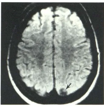
A 42-year-old woman was examined because of a seizure disorder. CT showed several 3-6 mm calcifications in the left cerebral hemi-sphere. MR (fig. 1) showed a round cyst 1 cm in diameter near the left parietal vertex, having signal slightly greater than CSF and con-taining a 3 mm high-signal mural nodule. In retrospect, an arcuate density seen on contrast-enhanced CT may have represented part of the cyst wall, but was indistinguishable from a cortical vein.
Case 2
Fig. 1.-Case 1. SE 1500/28 image. Cysticercus is 1 cm round low-signal area with 3 mm eccentric high-signal mural nodule (arrow) containing scolex.
A 23-year-old woman had a 1 day history of headache, lethargy, confusion, nausea, and vomiting. She was afebrile, with slight nuchal rigidity, no papilledema, and a 3% peripheral eosinophilia. Serum IHA titers for cysticercosis were 1 :128 and 1 :256. Lumbar opening pres-sure was normal. There was a CSF pleocytosis with 95% mononu-clear cells and 1 %-4% eosinophils. A technetium brain scan showed focal uptake in the right parietal vertex.
Intravenously enhanced CT after unenhanced CT showed a 13 x 8 mm thin ring enhancement with a 2 mm dense, enhancing mural nodule. The contents of the ring were otherwise of approximately CSF density, and there was no evidence of surrounding edema (fig. 2A). The third and lateral ventricles, including the temporal horns, were mildly to moderately enlarged. The anterior part of the third ventricle was disproportionately widened. Just behind the middle of the third ventricle and closer to the right lateral wall there was a 2 mm nodule whose apparent density on a 10 mm unenhanced cut was similar to gray matter (fig. 2B). This nodule was less easily appreciated on the contrast-enhanced scan, probably because of adjacent blood vessels and choroid plexus.
MR 7 days later showed a high-signal, long-T2 abnormality (edema) in a 2-3 cm diameter volume of right parietal white matter. A paren-chymal cysticercus within this region was imaged only on the short-TR sequences as a 7 mm cyst with a 2 mm mural nodule (fig. 2C). A 10 x 8 mm oval cyst containing a 4 mm mural nodule was shown in the third ventricle (figs. 2D and 2E). The ventricular cyst fluid had slightly higher signal than did CSF. The mural nodule had a longer T2 than did gray or white matter. Surrounding the third and lateral ventricles there was a high-signal, 3 mm rim with prolonged T2 compared with normal brain tissue. Confirmation of the third ventric-ular cyst was obtained by metrizamide ventriculography (fig. 2F).
[image:2.612.387.558.87.268.2] [image:2.612.56.296.103.385.2]AJNR:7, March/April 1986 MRI OF CYSTICERCOSIS
237
o
E
Fig. 2.-Case 2. A, Intravenously enhanced CT scan shows 1 cm parietal lucency with thin, enhanced rim and mural nodule (arrowhead). e, Unenhanced CT scan. Mild ventriculomegaly, disproportionate enlargement of anterior third ventricle, and 2 mm nodule within third ventricle (arrow). C-E, MR 7 days later but before praziquantel therapy. C, Sagittal SE 500/56 scan. Cyst, nodule
normal and no parenchymal cyst could be detected on long-or short-TR sequences. CT the next day showed a nearly solid, 6 x 8 mm
enhancement in the right parietal lobe without surrounding edema.
Case 3
A 41-year-old woman had a 1.5 year history of tinnitus, vertigo, decreased hearing in the left ear, and intermittent paresthesias of the
left side of the tongue; a 10 month history of total left facial paralysis; and recent onset of headache. She had a complete peripheral left
facial nerve paralysis, decreased sensation in all three divisions of the left trigeminal nerve, and bilateral papilledema with retinal flame
hemorrhages. There was an unexplained secondary (low erythropoi-etin) erythrocytosis with hematocrits of 60%-65%. CT (figs. 3A and 3B) showed a widened left internal auditory canal (lAC) without evidence of a mass but with apparent widening of the
cerebellopon-tine (CP) angle and medullary cisterns. A linear density crossing the
CP angle cistern (fig. 3B) was thought to represent a stretched blood
F
(arrow), and edema in right parietal lobe. Axial SE 2000/28 (0) and coronal SE 1500/56 (E) images show nodule (arrowheads) lying eccentrically in cyst in inferior third ventricle. Above interthalamic adhesion, third ventricle is narrow.
F, Metrizamide ventriculogram confirms nonobstructing cyst within third ve n-tricle. Nodule appeared denser than in A, suggesting metrizamide penetration.
vessel or enhancement of the seventh and eighth nerve bundle stretched slightly cephalad to the lAC. There was no abnormal
contrast enhancement of the brainstem or cerebellum. The third and
lateral ventricles were moderately to markedly enlarged but the fourth
ventricle was normal. MR (figs. 3C and 3D) confirmed the widened
lAC and showed a high-signal focus laterally within it. Cisternal
widening was also confirmed, with signal properties identical to CSF and no high-signal abnormality in the margins. At surgery the seventh and eighth nerve bundle was densely fibrotic. Beneath it a free-floating cystic mass was found in the CP angle and medullary cisterns
that was identified pathologically as a "degenerated larval cyst of Taenia solium."
Case 4
238
SUSS ET AL. AJNR:7, March/April 1986A
B
c
D
A
B
similar to the present one occurred 6 years earlier. She also
com-plained of occasional right hand and face paresthesias. Physical
examination was normal. Lumbar opening pressure was 47 cm H20. CSF glucose was 33 mg/dl (serum 103); protein was 61-81 mg/dl; red blood cell count. 10-350/mm3
; white blood cell count, 40-1165/ mm3. with 4%-14% neutrophils. 44%-48% lymphocytes, 17%-29%
monocytes. and 4%-39% eosinophils (lumbar puncture performed
twice). CT showed a 2.5 x 1.5 cm cyst apparently in the inferior part
Fig. 3.-Case 3. A and B, Intravenously en-hanced CT scans, 1.5 mm slice thickness. Widened lAC and CP angle cistern. Stretched blood vessel or VII and VIII cranial nerve bundle (arrowheads) slightly above level of lAC. Axial SE 500/56 (C) and coronal SE 1500/56 (0) images show widened left CP angle and medullary cisterns and widened inter-nal auditory canal containing high-signal focus (ar -row). Signal properties of fluid within widened cis-tern are identical to those of CSF. No evidence of mass or tissue reaction. Surgically proven race-mose cyst.
Fig. 4.-Case 4. Axial SE 2000/56 (A) and co r-onal SE 500/28 (B) images. Cyst originating in cistern of lamina terminalis and displacing septum pellucidum upward. Prominent high-signal rim in A represents tissue reaction to cyst.
of the septum pellucidum, without edema or abnormal contrast enhancement. MR 5 days later (fig. 4) showed that the cyst lay under
the septum pellucidum, originating in the cistern of the lamina
termi-nalis and pushing the septum up. The cyst fluid had signal properties
identical to CSF and contained no mural nodule. Long-TR sequences
showed the cyst surrounded by a high-signal, 3 mm rim, with
AJNR:7, March/April1986 MRI OF CYSTICERCOSIS 239
Fig. 5.-Case 5. A, Unenhanced CT scan shows
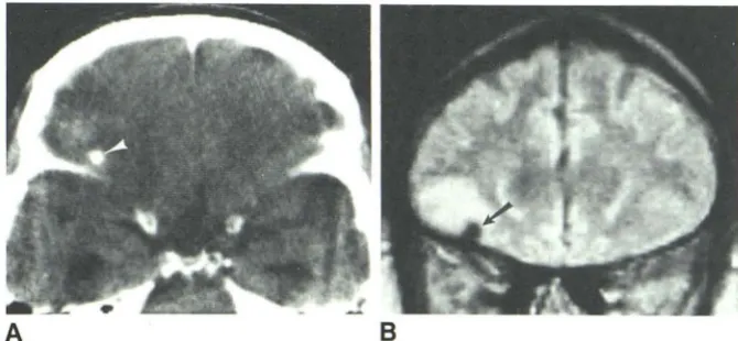
4 mm calcification (arrowhead) just above right sphenoid wing. Higher section (not shown) showed white matter edema. B, SE 1500/56 scan 4 weeks later shows 6 mm area of absent signal (arrow)
corresponding in location to CT calcification. Sur
-rounding zone of abnormal high signal represents edema corresponding predominantly to orbitofron
-tal gray matter.
Case 5
A
A 63-year-old man presented with new onset of seizures, having focal onset in the left arm and subsequent generalization. Physical examination was remarkable only for postictal effects and for papil -ledema. CT showed moderate hydrocephalus, numerous calcifi ca-tions in the choroid plexus or the walls of the third and left lateral
ventricles, and scattered parenchymal and sulcal calcifications. The largest calcification measured 4 mm and was located just above the right lesser sphenoid wing, surrounded by poorly defined lucency (edema) in the inferior frontal white matter (fig. SA). There was no
abnormal contrast enhancement. MR 4 weeks later (fig. 58) showed abnormally high signal (edema) in the right orbitofrontal gray matter. Within this area was a 6 mm zone of absent signal on all sequences, corresponding to the location of the calcification. None of the other
calcifications could be appreciated.
Discussion
Cysticerci
The cysticercus is a vesicular (bladder) larva ranging from
5 mm to 2 cm, less commonly up to 4 em, in diameter [18,
19]. The most typical size is about 1 cm. It develops from the
oncosphere by the envelopment of the solid larva within a
cyst growing from the caudal end of the organism (hence,
cysticercus, meaning bladder tail). Thus the parenchymatous region containing the scolex is transformed into an
invagina-tion of the cyst wall. In the fully developed cysticercus this
region forms a round nodule measuring 2-3 mm, sometimes
up to 5 mm, in diameter. The nodule contains the scolex at
the inner end of a spiral canal that joins with a vestibule to
reach the cyst's surface through an entrance canal. Numerous
calcareous corpuscles (physiologic structures of calcium
car-bonate unrelated to later pathologic calcification [20]) are
closely packed along the spiral canal in the neck of the
organism [11,21]. The scolex proper is only 1 mm in diameter
[1], although for convenience the entire mural nodule is
some-times called the scolex. The cyst wall is 0.07-0.1 mm thick
[22]. The cyst fluid has been described as transparent in live cysts, turning turbid or "jellylike" with the death of the
orga-nism [23-25]. There would not seem to be much taenial
protein antigen in the cyst fluid because fluid spilled at surgery
B
is not necessarily harmful to the patient [26, 27]. Related
cestode larvae have been shown to take up host albumin and
immunoglobulins while alive [28], but the quantitative
signifi-cance for cysticerci is unknown. Explicit measurements of
the protein content per unit volume of fluid for direct
compar-ison with CSF are not available.
This biologic description provides a basis for several CT
and MR observations. The parenchymal and ventricular
cys-ticerci (as opposed to the cisternal racemose cysts) were all
about 1 cm in diameter and each contained a 2-4 mm mural
nodule, producing a characteristic "hole-" or "
ring-with-eccen-tric-dot." Metrizamide may have penetrated the channels in
the nodule of the intraventricular cysticercus (fig. 2F). The
thin wall of the cyst was not imaged per se on CT or MR,
and could only be inferred from the sharp, smooth outline of
the cyst fluid or from the pattern of ventricular or cisternal
distortion. The configuration of a ring or arcuate calcification
around a 2-5 mm solid calcification on skull films has long
been recognized as highly suggestive if not conclusive
evi-dence of cysticercosis [3, 12, 18, 29, 30]; the analogous
configuration of live and early degenerating cysticerci now
imaged with CT [7, 10, 16] and MR should prove at least as
specific.
The significance of the calcareous corpuscles is suggested
by two observations. Considering the effect of
volume-aver-aging, the nodules of the cysticerci seen on unenhanced CT
in this study (case 2) were surprisingly dense. Their intrinsic
density may have been greater than that of brain because of
these calcifications. Moreover, in the one apparently live
cysticercus that was well seen on both CT and MR, the
nodule appeared smaller on CT (about 2 mm in fig. 28) than
on MR (4 mm in fig. 2D). This difference in the imaged sizes
may have resulted from a difference in what is imaged by the
two methods. CT shows calcifications to best advantage, emphasizing the central region of calcareous corpuscles
con-centrated along the spiral canal, but poorly distinguishing the
more peripheral part of the nodule from the surrounding fluid.
MR sensitivity to the difference between fluid water and tissue
water causes the entire volume of the nodule to be imaged clearly, making it larger, more conspicuous, and more
[image:5.615.225.560.90.245.2]240 SUSS ET AL. AJNR:7, March/April 1986
The fluid of the two probably live cystit:;erci appeared to have a slightly higher signal than CSF on long-TR images, while reliable comparison was not possible on the available
short-TR images. This impression was confirmed by signal
intensity measurements and relaxation time calculations in case 2. It becomes difficult, however, to obtain a valid CT density number or MR signal level for a lesion as its diameter
perpendicular to the slice decreases below twice the slice
thickness, and impossible below the slice thickness.
Volume-averaging could explain the slightly increased MR signal as
well as the reported slightly greater CT density [6] of
cysti-cercal fluid compared with CSF. W!lile we cannot exclude a
slight increase in inherent signal level, it was not markedly increased compared with CSF.
If the reported turbidity and gelling of the fluid after cyst death [23-25] correlate with MR signal changes and if reliable measurements on cyst fluid car. be made reasonably free of volume-averaging, signal measurements may help discrimi-nate live from dying cysticerci. One cysticercus, thought to be dying as indicated by CT findings of marked contrast enhancement initially and subsequent solidification, was visi-ble on short-but not long-TR sequences on the initial MR (fig. 2C). This MR finding may have resulted from increased signal intensity (compared with CSF) of the cyst fluid on long-TR
sequences, caused by elevated or otherwise altered protein
content, bringing the cysticercus sufficiently close to
isoin-tensity with brain on these images so that it was not discern-ible. Thus, while the best contrast between high-signal nodule
and low-signal fluid in a live cyst may be found on a long-TR,
short-TE image (e.g., SE 1500/28), a dying cyst's fluid may
contrast very poorly against nodule and brain on long-TR
images and may be perceived best or solely on a short-TR
image. Such MR-derived information indicating cyst death would be useful in patient selection for and monitoring of therapy.
Parenchymal Cysticerci
Our own experience and that of others [10, 13, 14, 31] has indicated that the parenchyrr.a is the mOSt common location of CNS cysticerci. Many of those that appear intracortical on
sectional images actually lie within the sulcal subarachnoid
space, but the sum of sulcal and parenchymal cysts
consti-tutes a majority of the organisms [19]. The propensity to involve the cortical (as well as deep) gray matter [4,12,25,
31, 32] and overlying leptomeninges places many cysts near
the skull, which is a difficult area to examine by CT, with its attendant bone artifacts. The failure to detect by CT a
super-ficial cyst that was easily identified by MR demonstrated an
advantage for MR in detecting convexity cysts, particularly
those near the vertex.
Host Reaction
Microscopically there is a thin host capsule of compressed
tissue with slight gliosis and inflammation and sometimes a
localized endarteritis around live cysticerci in the brain [1, 12,
19, 20, 32], but there is little or no radiologic evidence of host
response [13-16, 33]. The lack of demonstrated response around cysts can produce a "Gruyere (Swiss) cheese" [13] or punched-out appearance in the brain parenchyma and sur-prisingly little distortion of neighboring structures [34]. This inert appearance may be found on MR (fig. 1) as well as CT. Paradoxically, a thin subependymal rim was seen on MR around the intraventricular cyst in case 2 when the organism was thought to be alive and was reduced during praziquantel therapy.
At the death of the organism, a more marked inflammatory response is found [1, 2, 33]. This accelerated inflammation may be the cause of the death of the organism [19], although it has usually been assumed to be the result. The dying cysticercus is ordinarily associated with contrast enhance-ment and edema on CT [15, 16, 33]. MR is at least as sensitive as CT for the detection of brain edema, optimally using SE sequences with long TR and long TE [35], and MR demonstration of cysticercotic edema has previously been illustrated [16]. For most of the patients in this study, too much time elapsed between CT and MR to permit a reliable comparison of detection of inflammatory phenomena. An MR contrast agent such as gadolinium-DTPA [36] would be useful to fill the role of CT contrast enhancement both in the recog-nition of early parasite death and in the detection of organisms in more advanced stages of involution such as the parenchy-mal parasite in case 2 that was invisible on the second MR examination.
The final development in the dead organism is calcification, usually beginning in the nodule [2, 3, 29] (among extracellular
fibers, not the calcareous corpuscles [20]). Calcification has
in our experience and that of others [37] been the most common CT manifestation of cysticercosis, but poor detection of calcification is a known weakness of MR. In case 5, only one of several calcifications was appreciable (fig. 5), while MR failed to detect any of the calcifications in cases 6-8.
Intraventricular Cysticerci
Detection of potentially life-threatening intraventricular cysts is important in the workup of neurocysticercosis. In the Los Angeles County series 46 patients (17%) had intraven-tricular cysts, and six patients died from acute venintraven-tricular obstruction [38]. Plain CT has been considered inadequate for diagnosis of intraventricular cysts [14, 16, 33, 37-44], although a lateral ventricular cyst has been shown directly without intraventricular contrast medium [45] and cysticer-cosis has been suspected on the basis of distortion or dispro-portionate enlargement of the third [38] or fourth [9, 16, 26,
44] ventricle. Ventricular synechiae caused by intraventricular
cysts have been observed by pneumoencephalography [18] and need to be differentiated from the cysts themselves if surgical removal is contemplated.
con-AJNR:7, March/April 1986 MRI OF CYSTICERCOSIS 241
spicuity of the scolex and from the ease of coronal imaging to better appreciate the nature of the ventricular distortion. The potential of MR to replace metrizamide CT ventriculog-raphy in selected cases is important not only for primary diagnosis but also because it may be necessary to reconfirm preoperatively the ventricular location of a known cyst, as these can migrate between ventricles [38, 40, 45].
Definitive cyst identification in the fourth ventricle, where most intraventricular cysticerci occur [4, 5, 23, 26, 38, 43], may prove difficult, especially on CT, because the ballooned ventricle is likely to conform to the cyst wall. On MR the mural nodule and a reactive rim of high signal may indicate the presence of a cyst. Because of the small size of the nodule, contiguous MR sections may be important to detect it. Fourth ventricular cysts can be racemose [19, 43], lacking a scolex, but would probably produce the high-signal rim. Fourth ven-tricular enlargement can also be produced by cysticercal arachnoiditis without a cyst within the ventricle [18], in which case no periventricular high-signal zone would be anticipated.
Racemose Cysts
The racemose cyst is a multilobular, nonviable cyst often several centimeters in size, lacking a scolex. It is the degen-erated form to which a cysticercus in the basal subarachnoid space is transformed [1, 19, 32]. Although sterile, it may be
"alive" in that it can grow by proliferation of its wall [19], and may therefore be amenable to praziquantel therapy. Evidence of chronic meningitis typically accompanies racemose cysts [1 ].
The CP angle and the suprasellar region are locations known to harbor racemose cysts. Several examples of cysti-cercal CP angle cysts, some with lAC erosion and with clinical presentations suggesting acoustic schwan noma, have been
reported [4, 14, 16, 46-48]. Multiple cranial nerves in the vicinity of a cisternal cysticercal cyst may have inflammatory infiltrates among their fibers [19], which can explain the
contrast-enhancement observed (fig. 3B). Cysticercal cysts have been reported beneath [42] and within [49] the septum
pellucidum. In case 4 a high-signal rim surrounded the cyst,
and strong clinical and laboratory evidence for a parasitic
meningitis supported the diagnosis. The differential diagnosis for a cyst in the septal area includes ependymal, arachnoid, and neuroepithelial cysts and cystic tumors including cranio-pharyngioma [49]. Because the racemose cyst's fluid appears identical to CSF on CT and MR, its diagnosis may be sug-gested only by apparent focal cisternal widening, but a high-signal rim caused by adjacent tissue reaction, when present, will be an additional clue.
Conclusions
Cysticerci have a characteristic configuration on MR
imag-ing, consisting of a low-intensity focus about 1 em in diameter
with signal properties closely paralleling ventricular CSF, and a high-intensity, 2-4 mm mural nodule containing the scolex. Racemose cysts are distinguished by their larger size and lack of a mural nodule. Dying or degenerated cysts and
possibly some live cysts may be surrounded by a high-intensity rim of tissue reaction. MR and CT are currently
complementary in detection of these and other features of
cysticercosis. MR shows some cysts (convexity, ventricular)
better than CT does, is probably more sensitive than CT to
surrounding edema, and may show internal changes indicative of cyst death. CT is superior in demonstration of calcification
and, until MR contrast agents are available, blood-brain barrier
breakdown. Positive-contrast CT cisternography and CT
ven-triculography remain the most sensitive detectors of cysts in
the CSF pathways, but are invasive and may not be necessary when MR is positive. MR may prove to be the optimal primary imaging technique for diagnosis of suspected cysticercosis
and for monitoring therapy of cysts and edema.
ACKNOWLEDGMENTS
We thank Jacob Rosenstein and Fred G. Silva for surgical and
pathologic correlation in case 3 and Virginia Vaughn and Jerry Cheek
for photographic assistance.
REFERENCES
1. Marquez-Monter H. Cysticercosis. In: Marcial-Rojas R, ed. P
a-thology of protozoal and helminthic diseases with clinical corr
e-lation. Baltimore: Williams & Wilkins, 1971: 592-617
2. MacArthur WP. Cysticercosis as seen in the British army, with
special reference to the production of epilepsy. Trans R Soc Trop
Med Hyg 1933;27:343-363
3. Dixon HBF, Hargreaves WHo Cysticercosis (Taenia solium): a
further ten years' clinical study, covering 284 cases. Q J Med
1944;13: 1 07-121
4. Obrador S. Clinical aspects of cerebral cysticercosis. Arch Neurol Psychiatry 1948;59: 457 -468.
5. Obrador S. Cysticercosis cerebri. Acta Neurochir (Wien) 1962;10:320-364
6. Schnur JA, Richardson EP. Case records of the Massachusetts
General Hospital: cysticercosis cerebri. N Engl J Med 1977;297:
773-780
7. Torrealba G, Del Villar S, Tagle P, Arriagada P, Kase CS. Cysti
-cercosis of the central nervous system: clinical and therapeutic
considerations. J Neurol Neurosurg Psychiatry 1984;47:784
-790
8. McCormick GF, Zee C-S, Heiden J. Cysticercosis cerebri: review
of 127 cases. Arch Neurol 1982;39: 534-539
9. Bentson JR, Wilson GH, Helmer E, Winter J. Computed tom
og-raphy in intracranial cysticercosis. J Comput Assist Tomogr 1977;1 :464-471
10. Byrd SE, Locke GE, Biggers S, Percy AK. The computed
tomo-graphic appearance of cerebral cysticercosis in adults and
chil-dren. Radiology 1982;144:819-823
11. Brailsford JF. Cysticercus cellulosae-its radiographic detection
in the musculature and the central nervous system. Br J Radial
1941;14:79-93
12. Cardenas-y-Cardenas J. Cysticercosis of the nervous system. II.
Pathologic and radiologic findings. J Neurosurg 1962; 19: 63 5-640
13. Rodriguez-Carbajal J, Boleaga-Duran B. Neuroradiology of
hu-man cysticercosis. In: Flisser A, Willms K, Laclette JP, Larralde
242 SUSS ET AL. AJNR:7, March/April 1986
14. Rodriguez-Carbajal J, Palacios E, Zee C-S. Neuroradiology of cysticercosis of the central nervous system. In: Palacios E, Rodriguez-Carbajal J, Taveras JM, eds. Cysticercosis of the central nervous system. Springfield, IL: Thomas, 1983: 1 01-143 15. Sotelo J, Escobedo F, Rodriguez-Carbajal J, Torres B,
Rubio-Donnadieu F. Therapy of parenchymal brain cysticercosis with
praziquantel. N Engl J Med 1984;310: 1 001-1 007
16. Enzmann DR. Imaging of infections and inflammations of the central nervous system: computed tomography, ultrasound, and
nuclear magnetic resonance. New York: Raven, 1984: 1 03-127 17. Crooks L, Arakawa M, Hoenninger J, et al. Nuclear magnetic
resonance whole-body imager operating at 3.5 kGauss. Radiol
-ogy 1982;143: 169-174
18. Santin G, Vargas J. Roentgen study of cysticercosis of the central nervous system. Radiology 1966;86:520-528
19. Rabiela-Cervantes MT, Rivas-Hernandez A, Rodriguez-Ibarra J, Castillo-Medina S, Cancino F. Anatomopathological aspects of
human brain cysticercosis. In: Flisser A, Willms K, Laclette JP, Larralde C, Ridaura C, Beltran F, eds. Cysticercosis: present state of knowledge and perspectives. New York: Academic, 1982:179-200
20. Grau E, Garrido F, Canedo L. Calcification of the cysticerci of Taenia solium in the human brain. In: Flisser A, Willms K, Laclette JP, Larralde C, Ridaura C, Beltran F, eds. Cysticercosis: present state of knowledge and perspectives. New York: Academic, 1982:499-515
21. Siais J. Morphology of the scolex of cysticercus cellulosae in
brain cysticercosis. In: Flisser A, Willms K, Laclette JP, Larralde
C, Ridaura C, Beltran F, eds. Cysticercosis: present state of knowledge and perspectives. New York: Academic, 1982 :235-259
22. Ramirez-Bon E, Merchant MT, Gonzalez-del Pliego M, Canedo L. Ultrastructure of the bladder wall of the metacestode of Taenia solium. In: Flisser A, Willms K, Laclette JP, Larralde C, Ridaura
C, Beltran F, eds. Cysticercosis: present state of knowledge and perspectives. New York: Academic, 1982:261-280
23. Martinez A. Anatomia patologica de la cisticercosis cerebral.
Neurocirugia (Santiago) 1961;19: 173-190
24. Escobar A, Nieto D. Parasitic diseases. In: Minckler J, ed. P a-thology of the nervous system, vol 3. New York: McGraw-Hili, 1972: 2503-2521
25. Trelles JO, Trelles L. Cysticercosis of the nervous system. In: Vinken PJ, Bruyn GW, eds. Handbook of clinical neurology, vol 35. Amsterdam: North-Holland, 1978: 291-320
26. Loyo M, Kleriga E, Estanol B. Fourth ventricular cysticercosis. Neurosurgery 1980;7: 456-458
27. Gonzalez-Cornejo S. Comment on [43]. Neurosurgery 1983; 12: 151-152
28. Willms K. Host-parasite interface in the metacestode of Taenia solium. In: Flisser A, Willms K, Laclette JP, Larralde C, Ridaura C, Beltran F, eds. Cysticercosis: present state of knowledge and perspectives. New York: Academic, 1982:397-411
29. Dorfsman J. The radiologic aspects of cerebral cysticercosis. Acta Radiol [Diagn] (Stockh) 1963;1 :836-842
30. Taveras JM, Wood EH. Diagnostic neuroradiology, 2d ed. Balti
-more: Williams & Wilkins, 1976:222, 224
31. Martinez B (cited in [18]). Consideraciones sobre la cisticercosis humana (tesis). Mexico, D.F.: U. of Mexico Medical Library, School of Medicine, 1961
32. Escobar A. The pathology of neurocysticercosis. In: Palacios E, Rodriguez-Carbajal J, Taveras JM, eds. Cysticercosis of the central nervous system. Springfield, IL: Thomas, 1983:27-54 33. Handler LC, Mervis B. Cerebral cysticercosis with reference to
the natural history of parenchymal lesions. AJNR 1983;4:709-712
34. Schultz TS, Ascherl GF Jr. Cerebral cysticercosis: occurrence in
the immigrant population. Neurosurgery 1978;3: 164-169
35. Brant-Zawadzki M, Bartkowski HM, Pitts LH, et al. NMR imaging
of experimental and clinical cerebral edema. Noninvas Med Imag 1984;1 :43-47
36. Brasch RC, Weinmann H-J, Wesbey GE. Contrast-enhanced NMR imaging: animal studies using gadolinium-DTPA complex. AJR 1984;142:625-630
37. Mazer S, Antoniuk A, Ditzel LFS, Araujo JC. The computed tomographic spectrum of cerebral cysticercosis. Comput Radiol 1983;7:373-378
38. Zee C-S, Segall HD, Apuzzo MLJ, Ahmadi J, Dobkin WR. Intr a-ventricular cysticercal cysts: further neuroradiologic observations and neurosurgical implications. AJNR 1984;5: 727 -730 39. Rodriguez-Carbajal J, Palacios E, Azar-Kia B, Churchill R.
Ra-diology of cysticercosis of the central nervous system including computed tomography. Radiology 1977;125: 127-131
40. Zee C-S, Segall HD, Miller C, et al. Unusual neuroradiological
features of intracranial cysticercosis. Radiology 1980;137:397 -407
41. Zee C-S, Tsai FY, Segall HD, Teal JS, Ahmadi J. Entrance of
metrizamide into an intraventricular cysticercosis cyst. AJNR 1981;2: 189-191
42. Madrazo I, Garcia-Renteria JA, Paredes G, Olhagaray B.
Diag-nosis of intraventricular and cisternal cysticercosis by comput-erized tomography with positive intraventricular contrast
me-dium. J Neurosurg 1981;55:947-951
43. Madrazo I, Garcia-Renteria JA, Sandoval M, Lopez-Vega FJ.
Intraventricular cysticercosis. Neurosurgery 1983; 12: 148-152
44. Estanol B, Kleriga E, Loyo M, et al. Mechanisms of hydroce
ph-alus in cerebral cysticercosis: implications for therapy. Neurosur -gery 1983;13:119-123
45. Apuzzo MLJ, Dobkin WR, Zee C-S, Chan JC, Giannotta SL,
Weiss MH. Surgical considerations in treatment of intraventric-ular cysticercosis: an analysis of 45 cases. J Neurosurg
1984; 60: 400-407
46. Guevara-Oropeza M. Tumor del angulo pontocerebeloso. Arch Neurol Psiquiat 1938;1 :369-377
47. Bickerstaff ER, Small JM, Woolf AL. Cysticercosis of the poste-rior fossa. Brain 1956;79:622-634
48. Bustamante A. Lesions of the cerebellopontine angle (excluding neurinomas). Rev Inst Nal Neurol Mex 1977;11 :76-81