RESEARCH ARTICLE
COMPUTATIONAL STUDY ON THE EFFECT OF PATTERNED ELECTRICAL STIMULATION IN
NERVE CELLS
*1
Padma Priya, K.,
2Krishnan, J. and
2Malathi, R.
1
Department of Electrical Engineering in Annamalai University, India
2
Department of Electronics and Instrumentation Engineering in Annamalai University, India
ARTICLE INFO ABSTRACT
A neuron is a cell that processes and transmits information through electrical and chemical signals. Dispensed with the neurotransmitter in chemical synapses, few synapses connect both the presynaptic and postsynaptic cells directly. Compared to chemical synapses, nerve impulse conduction by electrical synapses is faster. Nervous messages are mostly associated with an electrical change known as the action potential. An all active Fohlmeister – Coleman – Miller (FCM) model with five nonlinear ion channels was modeled for the neuron cell. The neuron cells are stimulated with a constant dc current and are also analyzed with a patterned biphase current stimulation with Inter Phase Gap.
Copyright © 2014 Padma Priya, et al. This is an open access article distributed under the Creative Commons Attribution License, which permits unrestricted use, distribution, and reproduction in any medium, provided the original work is properly cited.
INTRODUCTION
A Neuron is an electrically excitable cell which processes and transmits the information through electrical and chemical signals. The neurons communicate through the space between the neurons which are called synapses. These synapses play a major role in the propagation of information. Nerve cells communicate via a combination of electrical and chemical signals. Individually neurons are completely separated from one another by their outer cell membranes and cannot directly share the electrical or chemical signals (David M. Lovinger 2008). The exceptional are the electrical synapses, in which the ion-conducting pores made from proteins called connexins connect the intracellular compartments of adjacent neurons, allowing direct ion flow from cell to cell (Kandel et al., 2000). Within the neuron, electrical signals driven by the charged particles allow rapid conduction from one end of the cell to the other end.
Dispensed with the neurotransmitter few synapses connect both the presynaptic and postsynaptic cells directly (Zoidl and Dermietzel 2002). When an action potential reaches such a synapse, the ionic currents flowing into the presynaptic cell can cross the barrier of the two cell membranes through pores called connexons and enter the postsynaptic cell directly stimulating it (Brink et al., 1996). Electrical synapses transmit faster since there is no slow neurotransmitters diffusion across
*Corresponding author: Padma Priya, K.
Department of Electrical Engineering in Annamalai University, India.
the synaptic cleft. They are used whenever fast response and coordination of timing are crucial, like in neural systems that require the fastest possible response, such as defensive reflexes, escape reflexes, the retina of vertebrates, and the heart. Compared to chemical synapses, nerve impulse conduction by electrical synapses is faster, but do not gain much either it remains the same or smaller. So in this proposed method, the electrical synapses are alone considered for the stimulation of the retinal ganglion cells.
Action potential
Nervous messages are mostly associated with an electrical change known as the action potential. This potential arises at a membrane which is situated between the axoplasm, medium inside the axon and the external medium of the neurons (AL Hodgkin and AF Huxley 1939). The description of electrical phenomena in nerves was problematic and was first analyzed. Galvani (1791) noticed that the legs of dissected frogs made active movements when their nerves were connected to a battery and this phenomenon is called as “animal electricity”. Later, Volta (1900) stated that the nerve pulses are electrical conduction phenomena. Helmholtz (1852) performed the first measurements of the propagation velocity of nerves. Ostwald (1890) and others developed the theory of osmosis and electrochemistry, and attempts for relating the flux of ions through the nerve membranes to the propagating action potential (1912). This finally resulted in the model by Hodgkin and Huxley (1952) from 1952 that is the presently accepted
ISSN: 0975-833X
International Journal of Current Research
Vol. 6, Issue, 04, pp.6197-6203, April, 2014
INTERNATIONAL JOURNAL OF CURRENT RESEARCH
Article History:
Received 26thJanuary, 2013
Received in revised form 24thFebruary, 2014
Accepted 10thMarch, 2014
Published online 23rdApril, 2014
Key words:
Action potential, Biphasic stimulation current, FCM model, Inter phase gap, Neuron.
RESEARCH ARTICLE
COMPUTATIONAL STUDY ON THE EFFECT OF PATTERNED ELECTRICAL STIMULATION IN
NERVE CELLS
*1
Padma Priya, K.,
2Krishnan, J. and
2Malathi, R.
1
Department of Electrical Engineering in Annamalai University, India
2
Department of Electronics and Instrumentation Engineering in Annamalai University, India
ARTICLE INFO ABSTRACT
A neuron is a cell that processes and transmits information through electrical and chemical signals. Dispensed with the neurotransmitter in chemical synapses, few synapses connect both the presynaptic and postsynaptic cells directly. Compared to chemical synapses, nerve impulse conduction by electrical synapses is faster. Nervous messages are mostly associated with an electrical change known as the action potential. An all active Fohlmeister – Coleman – Miller (FCM) model with five nonlinear ion channels was modeled for the neuron cell. The neuron cells are stimulated with a constant dc current and are also analyzed with a patterned biphase current stimulation with Inter Phase Gap.
Copyright © 2014 Padma Priya, et al. This is an open access article distributed under the Creative Commons Attribution License, which permits unrestricted use, distribution, and reproduction in any medium, provided the original work is properly cited.
INTRODUCTION
A Neuron is an electrically excitable cell which processes and transmits the information through electrical and chemical signals. The neurons communicate through the space between the neurons which are called synapses. These synapses play a major role in the propagation of information. Nerve cells communicate via a combination of electrical and chemical signals. Individually neurons are completely separated from one another by their outer cell membranes and cannot directly share the electrical or chemical signals (David M. Lovinger 2008). The exceptional are the electrical synapses, in which the ion-conducting pores made from proteins called connexins connect the intracellular compartments of adjacent neurons, allowing direct ion flow from cell to cell (Kandel et al., 2000). Within the neuron, electrical signals driven by the charged particles allow rapid conduction from one end of the cell to the other end.
Dispensed with the neurotransmitter few synapses connect both the presynaptic and postsynaptic cells directly (Zoidl and Dermietzel 2002). When an action potential reaches such a synapse, the ionic currents flowing into the presynaptic cell can cross the barrier of the two cell membranes through pores called connexons and enter the postsynaptic cell directly stimulating it (Brink et al., 1996). Electrical synapses transmit faster since there is no slow neurotransmitters diffusion across
*Corresponding author: Padma Priya, K.
Department of Electrical Engineering in Annamalai University, India.
the synaptic cleft. They are used whenever fast response and coordination of timing are crucial, like in neural systems that require the fastest possible response, such as defensive reflexes, escape reflexes, the retina of vertebrates, and the heart. Compared to chemical synapses, nerve impulse conduction by electrical synapses is faster, but do not gain much either it remains the same or smaller. So in this proposed method, the electrical synapses are alone considered for the stimulation of the retinal ganglion cells.
Action potential
Nervous messages are mostly associated with an electrical change known as the action potential. This potential arises at a membrane which is situated between the axoplasm, medium inside the axon and the external medium of the neurons (AL Hodgkin and AF Huxley 1939). The description of electrical phenomena in nerves was problematic and was first analyzed. Galvani (1791) noticed that the legs of dissected frogs made active movements when their nerves were connected to a battery and this phenomenon is called as “animal electricity”. Later, Volta (1900) stated that the nerve pulses are electrical conduction phenomena. Helmholtz (1852) performed the first measurements of the propagation velocity of nerves. Ostwald (1890) and others developed the theory of osmosis and electrochemistry, and attempts for relating the flux of ions through the nerve membranes to the propagating action potential (1912). This finally resulted in the model by Hodgkin and Huxley (1952) from 1952 that is the presently accepted
ISSN: 0975-833X
International Journal of Current Research
Vol. 6, Issue, 04, pp.6197-6203, April, 2014
INTERNATIONAL JOURNAL OF CURRENT RESEARCH
Article History:
Received 26thJanuary, 2013
Received in revised form 24thFebruary, 2014
Accepted 10thMarch, 2014
Published online 23rdApril, 2014
Key words:
model for the nerve pulse which relies on ionic currents and the membrane capacitor. In the context of their model, the conductance displays rather complex voltage and time dependences that enter the differential equation via a set of empirical parameters. In 1976, Neher and Sakmann described these channels microscopically (Neher and Sakmann 1976). In 1998, MacKinnon and collaborators crystallized the potassium channel and suggested a pathway for the potassium through a pore within the protein (Doyle et al., 1998). Thus, the Hodgkin-Huxley model seemingly finds support in independent experiments. The model by Hodgkin and Huxley is a purely electrical description based on conductors (ion channels and the cytosol of the nerve axon) and on a capacitor, which is the lipid membrane.
Mathematical modeling of action potential
Hudgkin-Huxley model
In the 1950s Alan Hodgkin and Andrew Huxley found the ionic basis of the action potential and developed a mathematical model predicting the speed of spike propagation. Ion channels (pores) are proteins that span the cell membrane allowing the flow of ions through the membrane. Ion channels have three important properties: They conduct ions. They recognize and select specific ions, e.g. sodium Na+ ions. They open and close in response to specific electrical or chemical signals.
FCM models
Neuron was written using a fully implicit method of integration i.e. backward Euler method of integration. Each compartment in the simulation was modeled as an intracellular resistance (Ra) and a membrane mechanism in parallel with a membrane
capacitance and a gap junction conductance in between the compartments. Several constants were specified based on whole-cell recording data which included the value for membrane capacitance (1µF/cm2), membrane resistance (50,000Ωcm2) (Coleman and Miller 1989), and cytoplasmic resistance (110Ωcm)(Coleman and Miller 1989). These values are assumed to be uniform throughout the cell. The simulations were modeled at room temperature (22°C) (Rattay 1990).
Each compartment is modeled with an intracellular resistance (Ra) and a membrane mechanism in parallel with a membrane
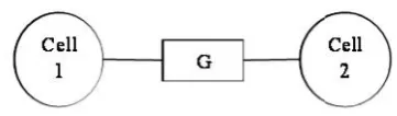
capacitance and a gap junction conductance (G) in between the compartments as shown in Fig. 1. An all active model (FCM) with five nonlinear ion channels was modeled. The linear passive mechanism reduces each cell membrane to a simple parallel RC circuit with a leak. The leak conductance was modeled as a battery at -70mV in series with a conductance of 20µS/cm2. The membrane potential everywhere was initialized
to a resting potential of -70 mV. The HH mechanism is the classic nonlinear description of unmyelinated axons by Hodgkin and Huxley (Hodgkin and Huxley 1952) — a leak
conductance, sodium and potassium channels (
g
Na=120mS/cm2, ENa= 50 mV, ̅ = 36mS/cm
2
, EK= -77mV,
g
l= 0.3mS/cm2, El= 54.3 mV). The FCM model is a complex five channel model based on work by Fohlmeister et al. (1990,1995, 1997). It includes the following conductances:
g
Na (asodium conductance),
g
Ca (a calcium conductance),g
K (adelayed rectifier potassium conductance),
g
A (an inactivatingpotassium conductance), and
g
K,Ca (a non inactivatingcalcium activated potassium conductance) (Fohlmeister et al., 1990). All channels are modeled as simple voltage-gated conductances except
g
K,Ca, which is modeled as acalcium-gated conductance. It was this unique combination of channel kinetics which best emulated the firing pattern of ganglion cells (Fohlmeister et al., 1990). The calcium and potassium conductances served to shape the finer properties of the action potential including the ability to produce slow repetitive firing which is impossible using the Hodgkin–Huxley channels completely. The model for membrane potential takes the familiar Hodgkin/Huxley form (Fohlmeister et al., 1990):
)
(
)
(
)
(
)
(
)
(
, 3 4 3 3 K Ca K K A A K K Ca Ca Na Na mE
E
g
E
E
h
a
g
E
E
n
g
E
E
c
g
E
E
h
m
g
dt
dE
C
(1)Where the rate constants for m, h, c, n, a, and hAall solve the first order kinetic equation (Plonsey and Barr 1988):
x x
x
x
dt
dx
)
(
(2)Electrotonic current
The instantaneous membrane potential varies from each compartment to the other and also the electrotonic current across the boundaries between the neighboring compartments. Except the injected stimulus current into the compartment, there are no current sources or sinks within the compartments. So in order to maintain the conservation of current, the sum of all currents flowing across the entire boundary of any individual compartment must always be zero or equal to Istim.
Each compartment’s boundary consistsof its plasma membrane and its intercompartmental interfaces i.e. the cytoplasmic cross sections across which all the currents are electrotonic. So the net electrotonic current is the residual current that equals in magnitude of the sum of all instantaneous membrane currents, including ion channel, capacitive, and leak deducing Istim. With
regard to the algebraic sign, we define positive electrotonic current as the net positive charge flowing into the compartment
[image:2.595.67.248.410.463.2]from its neighboring compartments. The positive
Na
ionsFig. 1. Model representing connections between two neuron cells
model for the nerve pulse which relies on ionic currents and the membrane capacitor. In the context of their model, the conductance displays rather complex voltage and time dependences that enter the differential equation via a set of empirical parameters. In 1976, Neher and Sakmann described these channels microscopically (Neher and Sakmann 1976). In 1998, MacKinnon and collaborators crystallized the potassium channel and suggested a pathway for the potassium through a pore within the protein (Doyle et al., 1998). Thus, the Hodgkin-Huxley model seemingly finds support in independent experiments. The model by Hodgkin and Huxley is a purely electrical description based on conductors (ion channels and the cytosol of the nerve axon) and on a capacitor, which is the lipid membrane.
Mathematical modeling of action potential
Hudgkin-Huxley model
In the 1950s Alan Hodgkin and Andrew Huxley found the ionic basis of the action potential and developed a mathematical model predicting the speed of spike propagation. Ion channels (pores) are proteins that span the cell membrane allowing the flow of ions through the membrane. Ion channels have three important properties: They conduct ions. They recognize and select specific ions, e.g. sodium Na+ ions. They open and close in response to specific electrical or chemical signals.
FCM models
Neuron was written using a fully implicit method of integration i.e. backward Euler method of integration. Each compartment in the simulation was modeled as an intracellular resistance (Ra) and a membrane mechanism in parallel with a membrane
capacitance and a gap junction conductance in between the compartments. Several constants were specified based on whole-cell recording data which included the value for membrane capacitance (1µF/cm2), membrane resistance (50,000Ωcm2) (Coleman and Miller 1989), and cytoplasmic resistance (110Ωcm)(Coleman and Miller 1989). These values are assumed to be uniform throughout the cell. The simulations were modeled at room temperature (22°C) (Rattay 1990).
Each compartment is modeled with an intracellular resistance (Ra) and a membrane mechanism in parallel with a membrane
capacitance and a gap junction conductance (G) in between the compartments as shown in Fig. 1. An all active model (FCM) with five nonlinear ion channels was modeled. The linear passive mechanism reduces each cell membrane to a simple parallel RC circuit with a leak. The leak conductance was modeled as a battery at -70mV in series with a conductance of 20µS/cm2. The membrane potential everywhere was initialized
to a resting potential of -70 mV. The HH mechanism is the classic nonlinear description of unmyelinated axons by Hodgkin and Huxley (Hodgkin and Huxley 1952) — a leak
conductance, sodium and potassium channels (
g
Na=120mS/cm2, ENa= 50 mV, ̅ = 36mS/cm
2
, EK= -77mV,
g
l= 0.3mS/cm2, El= 54.3 mV). The FCM model is a complex five channel model based on work by Fohlmeister et al. (1990,1995, 1997). It includes the following conductances:
g
Na (asodium conductance),
g
Ca (a calcium conductance),g
K (adelayed rectifier potassium conductance),
g
A (an inactivatingpotassium conductance), and
g
K,Ca (a non inactivatingcalcium activated potassium conductance) (Fohlmeister et al., 1990). All channels are modeled as simple voltage-gated conductances except
g
K,Ca, which is modeled as acalcium-gated conductance. It was this unique combination of channel kinetics which best emulated the firing pattern of ganglion cells (Fohlmeister et al., 1990). The calcium and potassium conductances served to shape the finer properties of the action potential including the ability to produce slow repetitive firing which is impossible using the Hodgkin–Huxley channels completely. The model for membrane potential takes the familiar Hodgkin/Huxley form (Fohlmeister et al., 1990):
)
(
)
(
)
(
)
(
)
(
, 3 4 3 3 K Ca K K A A K K Ca Ca Na Na mE
E
g
E
E
h
a
g
E
E
n
g
E
E
c
g
E
E
h
m
g
dt
dE
C
(1)Where the rate constants for m, h, c, n, a, and hAall solve the first order kinetic equation (Plonsey and Barr 1988):
x x
x
x
dt
dx
)
(
(2)Electrotonic current
The instantaneous membrane potential varies from each compartment to the other and also the electrotonic current across the boundaries between the neighboring compartments. Except the injected stimulus current into the compartment, there are no current sources or sinks within the compartments. So in order to maintain the conservation of current, the sum of all currents flowing across the entire boundary of any individual compartment must always be zero or equal to Istim.
Each compartment’s boundary consistsof its plasma membrane and its intercompartmental interfaces i.e. the cytoplasmic cross sections across which all the currents are electrotonic. So the net electrotonic current is the residual current that equals in magnitude of the sum of all instantaneous membrane currents, including ion channel, capacitive, and leak deducing Istim. With
regard to the algebraic sign, we define positive electrotonic current as the net positive charge flowing into the compartment
from its neighboring compartments. The positive
Na
ionsflowing into the compartment produce the negative Na current which is counter to the convention for membrane currents.
Stimulation Current
The neuron cells are stimulated with a constant dc current and are also analyzed with a patterned biphase current stimulation. An intracellular stimulation current, IStim, was a bi-phasic
current injection as shown in Figure 2. The pattern stimulation current consists of two phases: the cathodic phase and the anodic phase. The durations of the cathodic and anodic phases are ω-and ω+ respectively. The intraphase gap (IPG) delays; separates the pulses and also avoids the reversal of the earlier physiological effect of the previous pulse (Mark et al., 1999). An increasing interphase gap leads to a decrease in the charge required to cause a neuron to spike (Craig O. Savage et al., 2012).
RESULTS
Fig. 3. (a) Action potential of a neuron cell (b) Electrotonic current flowing across the boundaries between the neighboring
[image:3.595.319.553.49.234.2]compartments
[image:3.595.50.276.274.458.2]Fig. 2. The biphasic stimulation current waveform
Fig. 4. Action potential of a cell and Electrotonic current flowing across the boundaries between the neighboring compartments for different values of stimulating current
Istim
(a) Istim= (5 3 1), and G1, G2= 0.001.
(b) Istim= (6 4 2), and G1, G2= 0.001.
flowing into the compartment produce the negative Na current which is counter to the convention for membrane currents.
Stimulation Current
The neuron cells are stimulated with a constant dc current and are also analyzed with a patterned biphase current stimulation. An intracellular stimulation current, IStim, was a bi-phasic
current injection as shown in Figure 2. The pattern stimulation current consists of two phases: the cathodic phase and the anodic phase. The durations of the cathodic and anodic phases are ω- and ω+ respectively. The intraphase gap (IPG) delays; separates the pulses and also avoids the reversal of the earlier physiological effect of the previous pulse (Mark et al., 1999). An increasing interphase gap leads to a decrease in the charge required to cause a neuron to spike (Craig O. Savage et al., 2012).
RESULTS
Fig. 3. (a) Action potential of a neuron cell (b) Electrotonic current flowing across the boundaries between the neighboring
compartments
[image:3.595.330.545.287.700.2]Fig. 2. The biphasic stimulation current waveform
Fig. 4. Action potential of a cell and Electrotonic current flowing across the boundaries between the neighboring compartments for different values of stimulating current
Istim
(a) Istim= (5 3 1), and G1, G2= 0.001.
(b) Istim= (6 4 2), and G1, G2= 0.001.
flowing into the compartment produce the negative Na current which is counter to the convention for membrane currents.
Stimulation Current
The neuron cells are stimulated with a constant dc current and are also analyzed with a patterned biphase current stimulation. An intracellular stimulation current, IStim, was a bi-phasic
current injection as shown in Figure 2. The pattern stimulation current consists of two phases: the cathodic phase and the anodic phase. The durations of the cathodic and anodic phases are ω- and ω+ respectively. The intraphase gap (IPG) delays; separates the pulses and also avoids the reversal of the earlier physiological effect of the previous pulse (Mark et al., 1999). An increasing interphase gap leads to a decrease in the charge required to cause a neuron to spike (Craig O. Savage et al., 2012).
RESULTS
Fig. 3. (a) Action potential of a neuron cell (b) Electrotonic current flowing across the boundaries between the neighboring
compartments
Fig. 2. The biphasic stimulation current waveform
Fig. 4. Action potential of a cell and Electrotonic current flowing across the boundaries between the neighboring compartments for different values of stimulating current
Istim
(a) Istim= (5 3 1), and G1, G2= 0.001.
Fig. 6. Biphasic stimulating current and the action potential evoked by varying the IPG (a)∆ = 1ms (b) ∆ = 2ms(c) ∆ = 3ms.
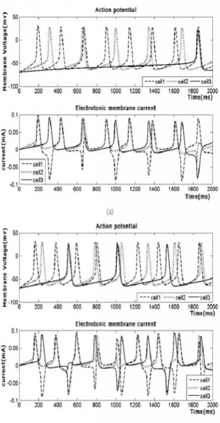
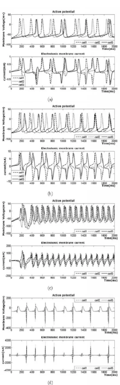
Fig. 5. Action potentials of three cell compartments and the electrotonic current flowing through their cell membranes for the given values of stimulating current Istim= (6 4 2) and different
values of gap conductances with G1= G2
(a)G1, G2= 0.01, (b) G1, G2= 0.1, (c) G1, G2= 1, (d) G1, G2= 10.
Fig. 6. Biphasic stimulating current and the action potential evoked by varying the IPG (a)∆ = 1ms (b) ∆ = 2ms(c) ∆ = 3ms.
Fig. 5. Action potentials of three cell compartments and the electrotonic current flowing through their cell membranes for the given values of stimulating current Istim= (6 4 2) and different
values of gap conductances with G1= G2
Fig. 7. Biphasic stimulating current and the action potential produced with non-uniform pulse duration.
Fig. 8. Biphasic stimulating current and the action potential originated by increasing the pulse duration, cathodic pulse amplitude respectively.
Fig. 9. Biphasic stimulating current and the action potential developed by increasing stimulating frequency
Fig. 10. Biphasic stimulating current and the action potential initiated by increasing stimulating amplitude and IPG for 50Hz frequency
pulse Fig. 7. Biphasic stimulating current and the action potential
produced with non-uniform pulse duration.
Fig. 8. Biphasic stimulating current and the action potential originated by increasing the pulse duration, cathodic pulse amplitude respectively.
Fig. 9. Biphasic stimulating current and the action potential developed by increasing stimulating frequency
Fig. 10. Biphasic stimulating current and the action potential initiated by increasing stimulating amplitude and IPG for 50Hz frequency
DISCUSSION
The action potential of the designed neuron cell and the electrotonic current flows across the boundaries between the neighboring compartments were analysed for the constant dc stimulus current bounding up to 20mA (Padma Priya et al., 2013). Though the stimulating current is applied continuously the action potential is provoked in the neurons only when the RGC crosses the threshold and it is not generated in the refractory period. The spike potentials for the cells were inspected for different values of gap conductances (G) ranging from 1x10-30mS/cm2to 1.4mS/cm2and stimulating current Istim
up to 10mA which leads to the observation, that when the amplitude of Istim is increased the spiking rate of the neuron
increases (Padma Priya et al., 2013). The increasing conductance of the cells increases the spiking rate of the neuron and gets collapsed for higher values of G greater than 1mS/cm2 and the current through the membrane increases (Padma Priya et al., 2013). The action potential of the designed neuron cell and the electrotonic current flows across the boundaries between the neighboring compartments is
illustrated in Fig.3. Though the stimulating current is applied continuously the action potential is provoked in the neurons only when the nerve cell crosses the threshold and it is not generated in the refractory period. The spike potentials for three cells are depicted in Fig.4 for equal gap conductances G1,
and G2= 0.001 between the cells and for different values of
stimulating current Istim= (5 3 1), and Istim= (6 4 2) in Fig. 4(a)
and Fig. 4(b) which leads to the observation that when the amplitude of Istim is increased the spiking rate of the neuron increases. In Fig. 5, the conductances of the membrane are increased and the action potential and the membrane currents are sketched. The increasing conductance of the cells increases the spiking rate of the neuron and gets collapsed for higher values of G greater than 1 and the current through the membrane increases. The Fig.6 manifests the symmetric biphasic stimulation current with the pulse frequency of 20Hz i.e. a pulse for each 50ms and the corresponding action potential developed. The anodic and cathodic phase duration ω+ and ω- is 3ms. The Anodic amplitude, A+ of 10mA and Cathodic amplitude, A- of -5mA is applied with an inter phase delay,∆ as 0ms. The action potential originated with such a biphasic stimulation current with the IPG for 1ms, 2ms and 3ms is presented in Fig. 6(a), 6(b), and 6(c) respectively. The fig.7 narrates the action potential produced with the biphasic stimulating current with non-uniform pulse duration i.e. Anodic phase duration, ω+ = 3ms, and Cathodic phase duration, ω- = 1ms in fig. 7(a) and the Cathodic phase duration, ω- = 2ms in fig. 7(b). The Fig. 8 pictures the action potential initiated and the biphasic stimulating current by increasing both the anodic and cathodic pulse duration to 4ms in 8(a), and by increasing the Anodic pulse amplitude to 15mA in Fig. 8(b). The Fig. 9 characterizes the spiking of the neuron for the pulse frequency 50Hz and 100Hz in Fig. 9(a) and 9(b) respectively. The spiking of the neuron with the IPG as 2ms and 3 ms are presented in Fig. 10(a) and 10(b) respectively. The Fig. 10 reveals that the action potential is not initiated for certain pulses in the biphasic current waveform with 50Hz frequency, anodic phase duration, ω+ = 3ms, anodic pulse amplitude, A+ = 10mA, cathodic phase duration, ω- = 3ms, and cathodic pulse amplitude, A- = -5mA. These parameters of the biphasic stimulation current could be adjusted to develop the action potential. The stimulating current parameters are adapted as, Cathodic phase duration, ω -= 2.5ms, 2ms, 1ms, and Inter Phase Delay, ∆ = 1.5ms, 2ms, 3ms, so as to provoke the action potential and is represented in Fig. 11(a), 11(b) and 11(c) respectively. The stimulation pattern can be changed and the observation studies may be extended and is in current progress.
REFERENCES
AL Hodgkin, AF Huxley, Action Potentials Recorded from Inside a Nerve Fibre, Nature 144, 710-711, 1939.
Bernstein, J., 1912. Elektrobiologie. Die Lehre von den elektrischen Vorg¨angen im Organismus auf moderner Grundlage dargestellt. Vieweg & Sohn.
Brink PR, Cronin K, Ramanan SV (1996). "Gap junctions in excitable cells". J. Bioenerg. Biomembr. 28 (4): 351–8. Chen W. K., Linear Networks and Systems. Belmont, C A :
Wadsworth, 1993, pp. 123–135.
Coleman and R. F. Miller, “Measurement of passive membrane parameters with whole-cell recording from
Fig. 11. Biphasic stimulating current and the action potential generated by increasing stimulating amplitude and IPG
DISCUSSION
The action potential of the designed neuron cell and the electrotonic current flows across the boundaries between the neighboring compartments were analysed for the constant dc stimulus current bounding up to 20mA (Padma Priya et al., 2013). Though the stimulating current is applied continuously the action potential is provoked in the neurons only when the RGC crosses the threshold and it is not generated in the refractory period. The spike potentials for the cells were inspected for different values of gap conductances (G) ranging from 1x10-30mS/cm2to 1.4mS/cm2and stimulating current Istim
up to 10mA which leads to the observation, that when the amplitude of Istim is increased the spiking rate of the neuron
increases (Padma Priya et al., 2013). The increasing conductance of the cells increases the spiking rate of the neuron and gets collapsed for higher values of G greater than 1mS/cm2 and the current through the membrane increases (Padma Priya et al., 2013). The action potential of the designed neuron cell and the electrotonic current flows across the boundaries between the neighboring compartments is
illustrated in Fig.3. Though the stimulating current is applied continuously the action potential is provoked in the neurons only when the nerve cell crosses the threshold and it is not generated in the refractory period. The spike potentials for three cells are depicted in Fig.4 for equal gap conductances G1,
and G2= 0.001 between the cells and for different values of
stimulating current Istim= (5 3 1), and Istim= (6 4 2) in Fig. 4(a)
and Fig. 4(b) which leads to the observation that when the amplitude of Istim is increased the spiking rate of the neuron increases. In Fig. 5, the conductances of the membrane are increased and the action potential and the membrane currents are sketched. The increasing conductance of the cells increases the spiking rate of the neuron and gets collapsed for higher values of G greater than 1 and the current through the membrane increases. The Fig.6 manifests the symmetric biphasic stimulation current with the pulse frequency of 20Hz i.e. a pulse for each 50ms and the corresponding action potential developed. The anodic and cathodic phase duration ω+ and ω- is 3ms. The Anodic amplitude, A+ of 10mA and Cathodic amplitude, A- of -5mA is applied with an inter phase delay, ∆ as 0ms. The action potential originated with such a biphasic stimulation current with the IPG for 1ms, 2ms and 3ms is presented in Fig. 6(a), 6(b), and 6(c) respectively. The fig.7 narrates the action potential produced with the biphasic stimulating current with non-uniform pulse duration i.e. Anodic phase duration, ω+ = 3ms, and Cathodic phase duration, ω- = 1ms in fig. 7(a) and the Cathodic phase duration, ω- = 2ms in fig. 7(b). The Fig. 8 pictures the action potential initiated and the biphasic stimulating current by increasing both the anodic and cathodic pulse duration to 4ms in 8(a), and by increasing the Anodic pulse amplitude to 15mA in Fig. 8(b). The Fig. 9 characterizes the spiking of the neuron for the pulse frequency 50Hz and 100Hz in Fig. 9(a) and 9(b) respectively. The spiking of the neuron with the IPG as 2ms and 3 ms are presented in Fig. 10(a) and 10(b) respectively. The Fig. 10 reveals that the action potential is not initiated for certain pulses in the biphasic current waveform with 50Hz frequency, anodic phase duration, ω+ = 3ms, anodic pulse amplitude, A+ = 10mA, cathodic phase duration, ω- = 3ms, and cathodic pulse amplitude, A- = -5mA. These parameters of the biphasic stimulation current could be adjusted to develop the action potential. The stimulating current parameters are adapted as, Cathodic phase duration, ω -= 2.5ms, 2ms, 1ms, and Inter Phase Delay, ∆ = 1.5ms, 2ms, 3ms, so as to provoke the action potential and is represented in Fig. 11(a), 11(b) and 11(c) respectively. The stimulation pattern can be changed and the observation studies may be extended and is in current progress.
REFERENCES
AL Hodgkin, AF Huxley, Action Potentials Recorded from Inside a Nerve Fibre, Nature 144, 710-711, 1939.
Bernstein, J., 1912. Elektrobiologie. Die Lehre von den elektrischen Vorg¨angen im Organismus auf moderner Grundlage dargestellt. Vieweg & Sohn.
Brink PR, Cronin K, Ramanan SV (1996). "Gap junctions in excitable cells". J. Bioenerg. Biomembr. 28 (4): 351–8. Chen W. K., Linear Networks and Systems. Belmont, C A :
Wadsworth, 1993, pp. 123–135.
Coleman and R. F. Miller, “Measurement of passive membrane parameters with whole-cell recording from
neurons in the intact amphibian retina,” J. Neurophysiol., vol. 61, pp. 218–230, 1989.
Craig O. Savage, Tatiana Kameneva, David B. Grayden, Hamish Meffin, Anthony N. Burkitt, Minimisation of Required Charge for Desired Neuronal Spike Rate, 34th Annual International Conference of the IEEE EMBS, San Diego, California USA, 28 August - 1 September, 2012. David M. Lovinger, Communication Networks in the Brain,
Alcohol Research & Health, Vol. 31, No. 3, 196-214, 2008. Doyle P. A., D. A., J. Morais, J. M. Gulbis, A. L. Cohen, B. T.
Chait, and R. MacKinnon. 1998. The structure of the potassium channel: Molecular basis of k+ conduction and selectivity. Science 280:69.
Fohlmeister J. F. and R. F. Miller, “A comprehensive model of repetitive impulse encoding in retinal ganglion cells,” Invest. Ophthalmol. Vis. Sci. Suppl., vol. 36, no. 4, pp. S-9301995, Abstract.
Fohlmeister J. F. and R. F. Miller “Mechanisms by which cell geometry controls repetitive impulse firing in retinal ganglion cells,” J. Neurophysiol., vol. 78, pp. 1948–1964, 1997.
FohlmeisterJ. F., P. A. Coleman, and R. F. Miller, “Modeling the repetitive firing of retinal ganglion cells,” Brain Res., vol. 510, pp. 343–345, 1990.
Galvani, A., 1791. Abhandlung ¨uber die Kr¨afte der Electricit¨at bei der Muskelbewegung, volume 52 of
Ostwald’s Klassiker der exakten Wissenschaften (1894).
Hodgkin A. L. and A. F. Huxley, “A quantitative description of membrane current and its application to conduction and excitation in nerve,” J. Physiol., London, U.K., vol. 117, pp. 500–544, 1952.
Hodgkin, A. L., and A. F. Huxley. 1952. A quantitative description of membrane current and its application to conduction and excitation in nerve. J. Physiol. 117:500– 544.
Kandel, E. R.; Schwartz, J. H.; and Jessell, T. M., Principles of Neural Science. New York: McGraw-Hill, 2000.
Mark S. Humayun, Eugene de Juan Jr., James D. Weiland, Gislin Dagnelie, Steve Katona, Robert Greenberg, Satoshi Suzuki, Pattern electrical stimulation of the human retina, Vision Research 39 (1999) 2569–2576.
Neher, E., and B. Sakmann. 1976. Single-channel currents recorded from membrane of denervated frog muscle fibres. Nature 260:779–802.
Ostwald, W. 1890. Elektrische Eigenschaften halbdurchl¨assiger Scheidew ¨ande. Z. Phys. Chem. 6:71– 82.
Padma priya. K, Krishnan. J, Malathi. R, Computational study on the effect of patterned electrical stimulation in retinal ganglion cells, International Journal of Recent Scientific Research Vol. 4, Issue-10, pp.1538-1544, October, 2013. Plonsey R. and R. C. Barr, Bioelectricity, A Quantitative
Approach. New York: Plenum, 1988, pp. 285–286.
Rattay F., Electrical Nerve Stimulation: Theory, Experiments and Applications. New York: Springer-Verlag, 1990. Volta, A., Untersuchungen ¨uber den Galvanismus, volume
118 of Ostwald’s Klassiker der exakten Wissenschaften
(1900), 1796-1800.
Von Helmholtz. 1852. Messungen ¨uber fortpflanzungsgeschwindigkeit derreizung in den nerven. Arch. Anat. Physiol. wiss. Med. 199–216.
Zoidl G, Dermietzel R (2002). "On the search for the electrical synapse: a glimpse at the future". Cell Tissue Res. 310 (2): 137–42.