University of Warwick institutional repository: http://go.warwick.ac.uk/wrap
A Thesis Submitted for the Degree of PhD at the University of Warwick
http://go.warwick.ac.uk/wrap/66592
This thesis is made available online and is protected by original copyright.
Please scroll down to view the document itself.
in
Xenopus laevis.
Subreena Lutchmy Simrick, B.Sc. (Hons.).
A thesis submitted to the University of Warwick
for the degree of Doctor of Philosophy.
Molecular Physiology,
Department of Biological Sciences.
University of Warwick,
Coventry,
CV47AL.
Contents
Preface
Contents 11
List of Figures VI
List of Tables IX
Acknowledgements X
Declarations XI
Summary xii
Abbreviations Xlll
Chapter I: Introduction
1.1 The pronephros; the simplest of the 3 kidney forms
1.1.1 Xenopus pronephros: Overview of structure and function 1
1.1.2 Xenopus pronephric development 7
1.1.2.1 The pronephric anlagen 7
1.1.2.2 The glomus anlagen and development of the nephrocoel 13
1.1.2.3 The Pronephric Tubules 14
1.1.2.4 The Duct 17
1.2 Developmental congenital kidney disease 18
1.2.1 Wilms' Tumour J 19
1.2.2 Paired box 2 22
1.2.3 Nephrin 25
1.2.4 PKDJ 26
1.3 Aims of thesis 28
Chapter 2: Materials and Methods
2.1 Materials 29
2.2 Media and stock solutions 29
2.3 Plasmids 29
2.4 Bacterial strains (E. coli) 30
2.5 DNA Techniques 30
2.5.1 Agarose gel electrophoresis 30
2.5.2 Restriction digests 30
2.5.3 Ligation of DNA into plasmid vectors 31
2.5.4 PCR 31
2.5.5 Primer Design 34
2.5.6 DNA probe labelling 34
2.6 RNA Techniques 34
2.6.1 RNA extraction from embryos and animal caps 34
2.6.2 Reverse Transcription PCR 35
2.6.3 Preparation of in vitro transcription of mRNA 35
2.6.4 Wholemount in situ hybridisation 36
2.7 Protein Techniques 36
2.7.1 In vivo translation 36
2.7.2 In vitro translation 36
2.7.3 Immunostaining 37
2.8 2.8.1 2.8.2
2.8.3
2.9 2.9.1 2.9.2 2.9.3 2.9.4 2.10 2.10.1 2.10.2 2.10.3 2.10.4 2.10.5 2.10.6 2.10.7 2.10.8 Microbiological TechniquesDNA minipreps and glycerol stock formation Transformation
Competent cells
Phage Library Screening
Infection of XLI-Blue MRF' with phage In vivo excision
Southern blotting Southern hybridisation Embryo culture
In vitro fertilisation Staging
Micro-injection Fixing
Animal cap dissections Wax-sectioning
Measurement of tissue area Pronephric Index
Results Part 1
Identification of Genes Involved in the Pronephric Development of
Xenopus laevis
Results Part 1: Introduction
38
38 3838
39 39 39 39 41 41 41 42 42 42 42 44 44 4447
Chapter 3: Screening of a stage 13 cDNA library with a subtracted probe.
3.1 Introduction 49
3.2 Results 53
3.2.1 Preparation of probes and initial screening 53 3.2.2 Selection of clones within positive plaques 59
3.2.3 Characterisation of identified clones 65
3.3 Discussion: Screening of a stage 13 cDNA phagemid library with a 84 subtracted probe
Chapter 4: Analysis of potential pronephric UniGene cluster genes
4.1 Introduction 91
4.2 Results: Temporal and Spatial expression patterns of UniGene 92 cluster genes
4.3 Discussion: Potential future analysis of UniGene cluster genes 97
Chapter 5:
Pod
J and
Darmin r
are expressed in the developing pronephros
5.1 Introduction 104
5.1.1 Pod I 104
5.1.2 Darmin r 108
5.2 Results 109
Contents
Results Part 1: Conclusion
119
Results Part 2
Developmental role of
Pod
J
in Xenopus laevis
Results Part 2: Introduction
121
Chapter 6: Pronephric lineage targeted Pod lover-expression results in a
reduced glomus
6.1 Introduction 122
6.2 Results 123
6.2.1 Pod I is evolutionarily conserved through a number of vertebrate 123 6.2.2 6.2.3 6.2.4 6.2.4.1 6.2.4.2 6.2.4.3 6.2.5 6.2.5.1 6.3 species
Pod 1 expression is concentrated in the pronephric glomus Pod 1 is highly expressed in a number of adult tissues
Pronephric lineage targeted Pod lover-expression causes a phenotype
Pronephric lineage targeted Pod lover-expression results in a reduced glomus phenotype as indicated by WTl and xlmxlb in situ analysis
Pronephric lineage targeted Pod lover-expression does not effect the pronephric tubules and duct as indicated by 3G8 and 4A6 antibody staining
Pronephric lineage targeted Pod lover-expression does not effect the pro nephric anlagen as indicated by Xliml in situ hybridisation Pod lover-expression has no clear effect on heart development Pod lover-expression fails to result in a clear heart phenotype although may result in an altered heart position.
Discussion
Chapter 7:
Pod 1 expression is required for glomus development
123 126 126 129 138 138 141 141 149
7.1 Introduction 153
7.2 Results 153
7.2.1 Pod 1 knock-down 153
7.2.1.1 Pod 1 expression is required for glomus development 159 7.2.1.2 Pod 1 knock-down disrupts tubule morphology 167
7.2.1.3 Pod 1 knock-down has no obvious effect on Xliml expression, but 170 abrogates
Pax
8 expression in the pronephric anlagen7.2.2 Pod 1 expression was initiated at the same stage as other pronephric 174 development genes
7.2.3 Pod 1 expression in animal caps can up-regulated other pronephric 174 development genes
7.2.4 Co-injection of Pod 1 with class A basic helix-loop-helix 176 transcription factors E12, E47, HEB and lTF-2 fails to indicate any
potential association
7.3 Discussion 181
Results Part 2: Conclusion
188
Results Part 3
The role of
Darmin r
in the development of the pronephros
Results Part 3: Introduction
190Chapter 8: Pronephric lineage targeted
Darmin r
over-expression disrupts
pronephric tubule formation
8.1 Introduction 8.2 Results
8.2.1 Dannin r amino acid sequence is evolutionarily conserved
8.2.2 Darmin r is expressed in the pronephric tubules
8.2.3 Dannin r is expressed ubiquitously in adult Xenopus tissues
191 192 192 192 196 8.2.4 Retinoic acid and activin A do not have an effect on Dannin r 196 8.2.5 8.2.5.1 8.2.5.2 8.2.5.3 8.2.5.4 8.2.6 8.3
expression in animal caps
Darmin r over-expression gives a pronephric phenotype Darmin r over-expression disrupts tubule development
Darmin r over-expression does not effect the size of the glomus Darmin r over-expression may effect nephrostome distribution Darmin r over-expression may reduce the size of the pronephric
anlagen
Darmin r knock-down experiments
Discussion 196 201 204 204 206 208 208
Results Part 3: Conclusion
213
Chapter 9:
Discussion
9.1 Potential target gene identification 214
9.1.1 Screen of a stage 13 cDNA library with a subtractive hybridisation 214 probe
9.1.2 UniGene cluster genes 215
9.1.3 Data mining 215
9.2 Pod I 216
9.3 Darmin r 219
9.4 Future Work 219
9.5 Conclusion 220
References
221Appendix
Statistical data 237
Vector maps 260
List of Figures
Chapter 1
1.1
1.21.3
1.4
1.5
Chapter 2
2.1
2.2
2.3
2.4
Chapter 3
3.1
3.2
3.3
3.4
3.5
3.6
3.7
3.8
3.9
3.10
3.11
3.12
3.13
Chapter 4
4.1
4.2
4.3
4.4
Chapter 5
5.1
5.2
5.3
Introduction
The pronephros, mesonephros and the metanephros
Integrated and non-integrated nephrons
Structure of the Pronephros
Segregation of the
Xenopuspronephric anlagen from the
intermediate mesoderm
Formation of the pronephric tubules in
Xenopusas observed by
immunohistochemistry using the tubule specific monoclonal
antibody 3G8 and Darmin r wholemount in situ hybridisation
Materials and Methods
Arrangement of Southern blotting apparatus
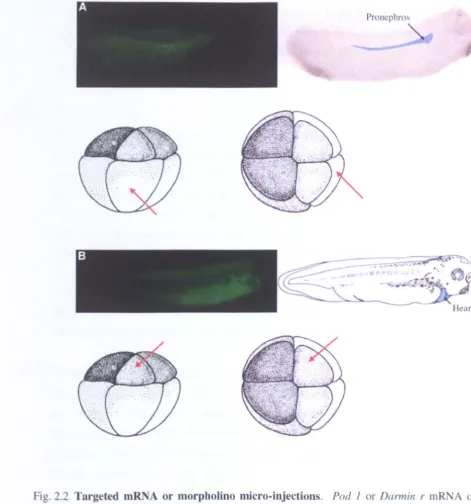
Targeted mRNA or morpholino micro-injections
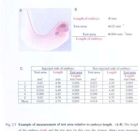
Example of measurement of test area relative to embryo length
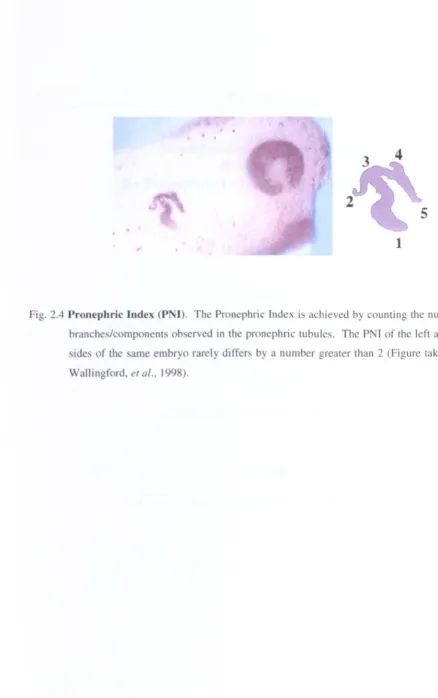
Pronephric Index (PNI)
Screening of a stage 13 cDNA library with a subtracted probe.
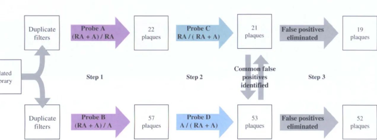
Clontech PCR-Select™ cDNA Subtraction Hybridisation
In vitro induction of pronephric tubules
Probe synthesis from treated animal cap mRNA
Stage 13 phagemid cDNA library screening
First round peR of positive samples identified in initial screen
Southern hybridisation of first round PCR of positive samples
identified in initial screen
Hybridisation of plaque lift showing typical clones chosen
Temporal expression patterns of clones identified in screens
Comparison of whole embryo and pronephric temporal expression
patterns of clones identified in screens
In situ hybridisation analysis of clones ,2.f6 and §a4 In situ hybridisation analysis of clones 12al and l.2.b2 In situ hybridisation analysis of clones l.2.c7 and 41 b2 In situ hybridisation analysis of clones 6C5 and 18B 1
Analysis of potential pronephric UniGene cluster genes
Temporal expression patterns of selected UniGene cluster genes
Temporal expression patterns of selected UniGene clusters genes
in dissected kidney anlagen
In situ
hybridisation analysis of UniGene cluster genes xl.5110
and xl.5983
In situ
hybridisation analysis of UniGene cluster genes xl.12848
and xl. 16795
Pod 1
and
Darmin
r are expressed in the developing pronephros
Temporal expression patterns of
PodJ
and Darmin r
Pod 1 is expressed in the developing pronephros Darmin r
is expressed in the developing pronephros
Chapter 6
6.1
6.2
6.3
6.4
6.5
6.6
6.7
6.8
6.96.10
6.11
6.12
Chapter 77.1
7.2
7.3
7.4
7.5
7.6
7.77.8
7.9
7.10 7.117.12
7.13
ChapterS 8.1Pronephric lineage targeted Pod lover-expression results in a reduced glomus
Alignment of homologous Pod 1 amino acid sequences Spatial pronephric expression of Pod 1 in stage 42 X laevis Expression pattern of Pod 1 in the adult organs of X laevis Oocyte translation of Xenopus and mouse Pod 1 mRNA
Xenopus and mouse Pod 1 show the same over-expression phenotype demonstrated by WTI in situ indicating the size of the glomus
Sections of Xenopus Pod lover-expression analysis in embryos confirms a reduction in glomus size
Xenopus Pod lover-expression glomus phenotype confirmed by xlmxl b expression analysis
Histochemical analysis of the Xenopus Pod J over expression phenotype showed no observable effect on either pronephric tubule or duct
Xenopus Pod lover expression analysis with Xlim 1 does not indicate a pronephric anlagen phenotype
Analysis of the Xenopus Pod J heart expression pattern by wholemount in situ hybridisation and sectioning
Xenopus Pod J over expression does not have an effect on the heart, as indicated with Cardiac troponin I expression
Transverse sections of wax embedded embryos over-expressing Pod J suggest a possible heart phenotype
Pod 1 expression is required for glomus development
Morpholino oligonucleotides bind to mRNA inhibiting translation Pod J morpholino specifically knocks-down Xenopus Pod 1 mRNA translation in vivo
Pod 1 expression is required for WTI expression in the glomus Pod 1 expression is required for glomus development
Pod 1 knock-down may reduce survival of glomus cells Pod J knock-down disrupts tubule morphology
Pod
1
knock-down does not have an effect on early Xlim 1 expressionPod 1 expression is required for Pax 8 expression
Pod 1 temporal expression compared to a selection of pronephric development genes and class A basic helix-loop-helix transcription factors
Pod J pronephric gene up-regulation was not enhanced by the presence of class A basic helix-loop-helix transcription factors E12, E47, HEB and ITF2b
Pod 1 may up-regulate other pronephric development genes
Pod J expression in animal caps treated with retinoic acid and activin
Hypothetical Pod
1
gene networkPronephric lineage targeted
Darmin r
over-expression disrupts pronephric tubule formationAlignment of homologous Darmin r amino acid sequences
List of figures
8.2
Spatial pronephric expression of
Darmin r in stage 42 X laevis 1958.3
Expression pattern of
Darmin r in the adult organs of X laevis 197 8.4 Darmin rexpression in animal caps treated with retinoic acid and
198
activin A
8.5
Oocyte translation of
Darmin r mRNA200
8.6 Darmin r over-expression disrupts tubule formation
202
8.7 Darmin r over-expression does not have an effect on the glomus205
8.8 Darmin rover-expression reduces the size of the pronephric
207
anlagen
Chapter 2 2.1
Chapter 3
3.1
3.2
3.3
3.4
3.5
Chapter 4
4.1
Chapter 7 7.1
Materials and Methods Primers used in this study
Screening of a stage 13 cDNA library with a subtracted probe. Relative concentrations of Activin A used to induce different tissues in animal cap assays
Summary of results of PCR and Southern hybridisation of phage pick and sequencing of PCR products
Preparation of antisense DIGlabelled RNA probe s for initial whole mount in situ hybridisation
First round in situ hybridisation of the 19 clones chosen in screening
Complete sequencing of chosen clones
Analysis of potential pronephric UniGene cluster genes
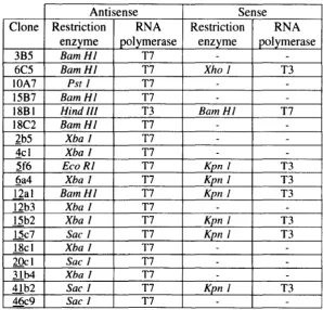
IMAGE consortium data and preparation of antisense and sense RNA DIG labelled probes for initial wholemount in situ hybridisation
Pod 1 expression is required for glomus development mRNA sources used in animal cap assays
32
51
67
70
71
73
96
Acknowledgements
A huge thank you goes to my supervisor Liz Oliver-Jones, for her tireless enthusiasm, guidance and drive. I really wouldn't be here if it wasn't for you. Also Karine Masse,
who has always been the first port of call for advice or a good chat!
Thank you to everyone who has been in the lab during my stretch, specifically Rob and Steph for their kind friendship. Not forgetting Surinder who is always at hand with a
random joke or two.
I would like to thank my family, namely, "Have you done it yet?" Jim, "Just do it" Didi and "Have you got a job yet ... " Mum, who have put up with and supported me.
Finally, I dedicate this thesis to Matthew John Paul, who has been there for me during all the happy and the not so happy moments of this PhD. Thank you for sharing this
The results presented in this thesis are the work of the author unless specified.
Microinjection and dissections were carried out by Professor Elizabeth Oliver-Jones.
Wax-embedding and sectioning were performed by Mr Surinder Bhamra. Hormone
injections to
Xenopus laevis
females were given by Mr Robert Taylor and Paul Jarrett.Sources of information have been acknowledged by reference.
None of this work has been previously used to apply for a degree.
Summary
The fonnation of the kidney in vertebrates proceeds through a succession of up to three structures. the pronephros. metanephros and mesonephros. This project aimed to identify a suitable target gene involved in the development of the pronephros and study its role in development. By analysing the functional role of a target gene. this thesis ultimately aims to further elucidate the developmental regulation of pronephric organogenesis.
The candidate target genes were identified from 3 sources. Firstly. a Xenopus laevis
stage 13 cDNA library was screened using a probe made from retinoic acid and activin A treated animal caps. Secondly. four UniGene cluster genes were investigated. after initial analysis indicated they were highly pronephros specific (Personal correspondence Pollet. N). Finally, data mining identified Xenopus homologues of important kidney development genes identified in higher vertebrates or newly identified genes expressed in the pronephros. On the basis of preliminary data. Darmin r and Pod J were taken
forward for functional studies.
Pod J, a basic-helix-Ioop-helix transcription factor. was found to be expressed from pronephric initiation with enhanced expression in the pronephros. The expression patterns of Pod J strongly matched that of the mouse homologue. implying a conserved evolutionary role. Pronephros targeted over-expression resulted in the reduction of glomus tissue, with no alteration in pronephric tubule or duct morphology. In addition. targeted knock-down resulted in the absence of cells in the glomus region.
Preliminary analysis of Darmin r. a cytosolic non-specific dipeptidase. indicated a role
later in pronephric tubule development. Targeted over-expression experiments disrupted tubule morphology and reduced the size of the pronephric anlagen.
Initial promising data clearly placed Pod J and Darmin r as important pronephric
AP
alkaline phosphataseAPS
ammonium persulphateATP
adenosine triphosphatebFGF
basic fibroblast growth factorbHLH
basic helix-loop-helixbp
base pairsBSA
bovine serum albumineDNA
complementary deoxyribonucleic acidCi
CurieC-terminal
carboxyl-terminaldATP
deoxyadenosine triphosphatedCTP
deoxycytidine triphosphatedGTP
deoxyguanosine triphosphatedHlO
distilled waterDIG
digoxygeninDNA
deoxyribonucleic acidDNase
deoxyribonucleasedNTPs
de ox yri bon ucl eosi de tri phos phatesdpe
days postcoitumE. coli Escherichia coli
EDTA
ethylene diamine tetra acidEST
expressed sequence tagFGF
fibroblast growth factorg gram
GFP
green fluorescent proteinHCI
hydrochloric acidH2O
waterH20
2 hydrogen peroxideI
litreLBroth
Luria Brothkb
kilobaseskDa
kilo DaltonM
molarMBT
mid-blastula transitionmg
milligramml
millilitremM millimolar
MMLV
moloney murine leukaemia virusMOPS
3-N-morpholinoJpropane sui phonic acidMO
morpholino oligonucleotidemRNA
messenger ribonucleic acidAbbreviations ng nl nM N-terminal PAGE
PBS
PBT
PBST
PCR
pM pmolPMSF
PNI
RA RNA RNase rpmRT-PCR
SDS
ssDNATBE
TBS
TdT
TEMED Tris Tris-CITUNEL
UTR
V v/v w/v U JLgJLI
J.LM
nanogram nanolitre nanomolar amino-terminalpolyacrylamide gel electrophoresis phosphate buffered saline
phosphate buffered tris phosphate buffered tris polymerase chain reaction picomolar picomoles phenylmethylsulphonyl fluoride Pronephric Index retinoic acid ribonucleic acid ribonuclease
revolutions per minute reverse transcription PCR sodium dodecylsulphate single stranded DNA tris buffered EDT A tris buffered saline
Terminal deoxynucleotidyl transferase N. N. N' -Tetramethylethylenediamine tris (hydroxymethyl) aminomethane
tris (hydroxymethyl) aminomethane. pH adjusted with HCI
Introduction
1.1
The pronephros; the simplest of the 3 kidney forms
Kidney organogenesis in vertebrates follows a succession of kidney structures, which are formed, degraded, and reformed with increasing structural complexity (Saxen, 1987). Induction of each successive kidney structure requires the previous temporal form for its own induction from the intermediate mesoderm (Brandli, 1999: Vize, et al.,
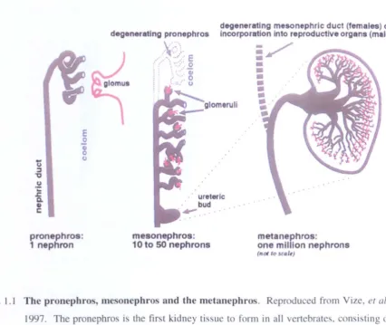
1997). The 3 kidney structures are the pronephros, mesonephros and metanephros (Fig. 1.1, reproduced from Vize, et al., 1997). When functional, the kidney structures all have the same principle role of osmoregulation and waste disposal. This role is achieved by their basic functional unit, the nephron. The pronephros, comprising only one nephron, is the simplest and also the earliest in evolutionary terms. The mesonephros is next, consisting of 10-50 nephron-like structures and finally, the metanephros, with approximately I million nephrons is the most sophisticated. The final structural complexity of the kidney is correlated to the complexity of organism. In lower vertebrates, amphibia and fish, the progression is from the pronephros to the mesonephros only, where the mesonephros is the functioning kidney of the adult. In higher vertebrates, mammals and birds, all three kidney stages are created, with the metanephros being the functioning kidney of the adult and the pronephros being vestigial. This thesis will focus on the simplest kidney structure, the pronephros, and its development in Xenopus laevis.
1.1.1
Xenopus
Pronephros: Overview of Structure and Function
The Xenopus pronephros, comprising one nephron, has 3 distinct structures: a glomus, the blood capillary network responsible for filtration of the blood: pronephric tubules, used for the selective reabsorption of water and nutrients from the filtrate; and the pronephric duct, which channels waste to the exterior.
The Glomus
E o
"8
(.)pronephros: 1 nephron
degenerating pronephros
V.·,
•.. ~: ... .
:>-\; __
;
i
)
glomus
mesonephros: 10 to SO nephrons
degenerating mesonephric duct (females) or incorporation into reproductive organs (males)
eruli
: /
•
metanephros:
one million nephrons (not 10 seQ/,)
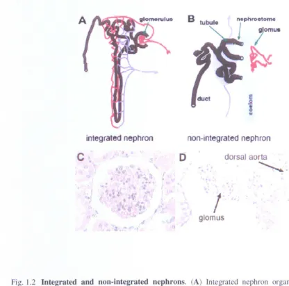
[image:17.515.40.466.79.438.2]Instead, the glomus and tubules are separated by the coelom and the nephrostomes (Fig. 1.2). Integrated nephrons can be found in the mesonephros and the metanephros.
The glomus is composed of a high pressured capillary network, derived from the dorsal aorta, which projects into the nephrocoel space within the pronephric capsule (Fig. 1.3). Filtration from the high pressured capillary network of the glomus through to the nephrocoel is both passive, through the slit diaphragms, and active, through the podocyte foot processes. The surface of the glomus vasculature 'bulb' is covered by podocytes with foot processed protruding into the basement membrane of the capillaries. The foot processes of metanephric kidneys have been found to have characteristics consistent with high levels of endocytosis (Mundel and Kriz, 1995). Between the podocytes are slit diaphragms which are the dominant size selection element during filtration (Abrahamson, 1986). The matrix of the glomus vasculature 'bulb' is high in podocalyxin and nephrin. Podocalyxin helps maintain foot process structure and function (Schnabel, et al., 1989 and Horvat, et al., 1986) and nephrin is a major component of slit diaphragms (Tryggvason, 1999).
The Pronephric Tubules
The pronephric tubules are joined to the nephrocoel through nephrostomes (Fig. 1.3). Nephrostomes are thin epithelial tubes lined with cilia along their length. The epithelial cells of the nephrostomes are distinct from the pronephric tubule cells by their lack of brush borders and convoluted membranes. The cilia draw fluid from the nephrocoel into the pronephric tubules by ciliary action. The pronephric tubules possess two morphologically distinct segments called the proximal tubules and the distal tubule. The proximal tubules are connected to the nephrostomes and are consistently arranged (Vize, et aI., 1997). The epithelial cells forming the proximal tubules have a dense mat
of short microvilli on the apical surface (M~bjerg, et al., 2000). This is known as the
integrated nephron
c
o
(
.
nophroetomo
i
omu •e
o ti o"
non-integrated nephron
dorsal aor1a
• I .
~
~
,
"
glomus
Fig. 1.2 Integrated and non-integrated nephrons. (A) Integrated nephron organisation. A
blood vessel (red) enters the Bowman's capsule where wastes are filtered into the Bowman's space. The blood vessel then exits the capsule and surrounds the distal (near the capsule) and proximal tubules. Resorbed nutrients, salts, and water are then returned
to the blood stream via the venous system () JrIJ1l). (B) Organisation of a non-integrated nephron. Wastes filtered into the coelom are collected by tubules. Resorbed
[image:19.514.7.423.56.468.2]Endothel ial cells
0
Podocyte
~
Matrix
Basement membrane
•••••••
••••••••••••••••
Coelom
Nephrostome
+
-Proximal Tubules
Pnml'llhric sinus
""-'p • • • • • • • • •
J)ud
....,
... .
\ \
nutrient uptake. The pronephric tubules are surrounded by a capillary bed called the blood sinus. The blood sinus returns the reabsorbed water, salt and nutrients, to the blood stream (Brandli, 1999).
The Duct
The epithelial cells of the pronephric duct resemble those of the distal tubule, but have a smooth basal membrane (M~bjerg, et al., 2000). The pronephric duct is fused with a cloacal horn, which extends the structure to the cloaca (Fig. 1.3). The resulting waste after the filtration through the glomus and reabsorbtion through the pronephric tubules is channelled out of the organism though the pronephric duct and cloaca.
1.1.2
Xenopus
Pronephric Development
The pronephros is derived from the intermediate mesoderm, located between the somites and the lateral plate. The developing pronephros is therefore exposed to signals from the somites, lateral plate and ectoderm. However, the complete picture of the molecular control of pronephros formation is still unclear, although, certain factors have been identified to playa role. The genes actively involved in pronephric development have been reviewed, although this does not include genes expressed by terminally differentiated tissues.
1.1.2.1 The Pronephric anlagen
Morphological development of the pronephric anlagen
i
I )I
I
~ I
f
~;
.
-~ .)
r
cc.lom/ pronepllrOt
Ii
,
10t'WIic I ... ~clWtlc~.
I
.~...
..
_ ...
.ra ...Stage 21 Stage 27 Stage 27
Fig. 1.4 Segregation of the Xenopus pronephric anlagen from the intermediate mesoderm. At
stage 2 J slight thickning of the somatic portion 0 f the lateral mesodenn is caused by
pronephric precursor cells becoming more columnar. By stage 24 this fonns a compact
aggregate, referred to a the pronephric swelling. At stage 27 the pronephric anlage is a
distinct mass, lying distal to the lateral plate. The tubule, duct and glomus anlagen can be
distinguished at this stage. Cells are arranged radially, which leads to a tiny lumen forming
pronephric swelling. Cells from the pronephric anlagen then become radial and gradually form a lumen at stages 27 through to 32 (Vize, et al., 1997).
Gene expression and regulation in the pronephric anlagen
Xliml, a LIM family transcription factor, is one of the earliest markers of pronephric specification and is visible at stage 12.5 by wholemount in situ hybridisation (Taira, et aI., 1994). Expression, however, is not restricted to the pronephric anlagen and can be seen in the prechordal mesoderm and forming notochord before the pronephric expression. Pronephric expression is seen in the lateral mesoderm and continues throughout condensation of the somatic lateral mesoderm, labelling the entire pronephric anlagen. This expression ceases in the glomus primordium around the time that it divides from the pronephric tubule and duct primordium. Xliml expression then continues in the pronephric tubule and duct primordium until late tail bud stages. This loss of expression is concurrent with the initiation of xWTI. Wilms' Tumour I, expression in the extreme anterior of the pronephric anlagen. which may indicate an exclusion of Xliml expression by xWTI (Carroll. et al., 1999). Indeed, targeted xWTI over-expression reduces Xliml expression in the pronephric anlagen (Wallingford, et al., 1998). However, this exclusion is not complete and a small proportion of cells in the extreme anterior of the pronephric tubule and duct primordium do express both transcription factors.
Chapter I
xPax 8, paired box transcription factor, is another early marker of pronephric specification, and like Xliml, is observed by wholemount in situ hybridisation during late gastula (Carroll and Vize, 1999). The expression pattern of xPax 8 in the developing gastula, differs from that of Xliml. xPax 8 expression appears more diffuse over the lateral mesoderm and initially, is only expressed in the presumptive glomus and pronephric tubule tissues. Later, between stages 20 to 25, the elongating neurula, Xliml and xPax 8 expression co-localise to the pronephric anlagen. Over-expression of either
Xliml or xPax 8 in Xenopus laevis leads to an enlargement of the pronephros or ectopic pronephroi, indicated by antibody staining for the pronephric tubules. Furthermore, co-injection of the transcription factors has a synergistic effect on this phenotype (Carroll and Vize, 1999). Retinoic acid and activin treatment of dissected animal caps can produce pronephric tissues (Reviewed Chapter in 3). Interestingly, this treatment of dissected animal caps also induces expression of Xliml and xPax 8 (Heller and Brandli, 1999 and Chan, et al., 2000). In fact, Xliml expression was found to be required for the induction of pronephric tissues in treated animal caps. These data suggests that Xliml and xPax 8 are involved in pronephric specification and patterning, possibly in collaboration, although, observation of their early expression patterns suggest that they may be regulated by different mechanisms.
X/db-I, a LIM domain binding protein, has been shown to bind to Xliml and synergize with it to activate the Spemann organizer and neural markers when co-injected (Breen,
et al., 1998). Expression is observed in the pronephros and central nervous system in the elongating neural a, stages 23 to 28 (Chan, et al., 2(00). However, there is no evidence of their co-activity in the pronephros.
In Xenopus, two homologs of the cold-inducible RNA binding protein have been identified, XCIRP and XCIRP-l (Uochi and Asashima, 1998 and Peng, et al., 2(00).
activin treated animal caps. The role of XCIRP-l is proposed to be required for marking genes or gene transcripts for future activation. where it might change the conformation of the DNA or RNA to establish competence for differentiation programs.
Xwnt-4 and xPax 2. another paired box transcription factor. have similar expression patterns to Xliml. although they are observed later in the elongating neurula (Heller and Brandli. 1997 and Carroll, et al., 1999). Xliml. Xwnt-4 and xPax 2 expression in the developing pronephric anlagen is entirely co-localised. even as expression is restricted to the pronephric tubule and duct primordium. with a slight over-lap with xWT I. The co-expression of Xliml, Xwnt-4 and xPax 2 with xWT 1 in the extreme anterior portion of the pronephric tubule and duct primordium is transient and by stage 30. xWTl expression is exclusive to the glomus primordium.
NDRG 1, N-myc downstream-regulated gene I, expression is initiated between stages 12 and 15 and is observed in the pronephric anlagen at stage 26 by wholemount in situ hybridisation (Kyuno, et al., 2003). Over-expression of NDRGI resulted in a reduction and disorganisation of pronephric structures and somites. Morpholino oligonucleotide knock-down of NDRG 1 caused a complete oblation of pronephric structures. NDRG 1 is therefore involved in pronephric development. possibly. pronephric anlagen specification events due to the timing of pronephric expression and mis-expression effects on the whole pronephros.
Bone morphogenetic proteins, BMP. are secreted TGF-p superfamily members and have been implicated in a diverse range of biological processes. BMP inhibitors are associated with axis patterning and are believed to be one of the dorsal ising factors diffused from the organizer of the blastula (Dale and Wardle. 1999). BM P4 is expressed in the lateral mesoderm and in the pronephros, although detailed analysis of pronephric expression has not been published (Fainsod. et al .• 1994). BMP4 acts as a morphogen in the specification of ventral tissues, such as blood and epidermis with high concentrations and lateral tissues, such as pronephros and muscle with lower concentrations, in a gradient across the embryo (Dale. et al., 1992 and Dale. 2000).
Chapter 1
Xwnt-ll, expressed in pro nephric tubules at stage 28 (Ku and Melton, 1993), has been
suggested to provide an important signal during the earliest stages of pronephric development, from preliminary experiments from our laboratory (Tetelin and Jones, unpublished).
Notch- 1. Serrate- 1. and Delta- 1 are believed to be involved in a feedback loop within the pronephric anlagen that controls the division of glomus. tubule and duct primordia (McLaughlin, ef al .• 2000). Notch- 1 and Delta- 1 expression is visible at stage 21/22 in the anterior region of the pronephric anlagen. Serrate-l expression is observed in a similar area. a stage later at 23124. From this similar origin of expression the expression patterns dynamically change, so that at stage 30. Notch- 1 is expressed in the duct.
Serrate-l is expressed in the pronephric tubules. and Delfa-l is expressed in the glomus to pronephric tubule border. The role of Notch- 1 in pronephric development was investigated using targeted injection of activated or dominant negative forms of the transcription factor SuCH) to mimic or inhibit Notch signaling. Activation of Notch signaling inhibited Delta-l and enhanced Serrate-l expression and inhibition of Notch signaling inhibited Serrate-land enhanced Delta- 1 expression at stage 30. Surprisingly. activation of Notch leads to an enlargement in tubule and glomus structures and a reduction of duct structures. In addition. inhibition of Notch signaling enhanced duct formation. In fact, activation of Notch promoted Xliml expression in the anterior pronephric anlagen. the area of the presumptive glomus and pronephric tubules. and completely reduced expression in the presumptive duct region at stage 30. Notch- 1
signaling is therefore believed to partition the fates of the pronephric anlagen by a feedback loop with Delta- 1 and Serrate- 1.
There are three members of the Hepatocyte Nuclear Factor (HNF) transcription factor family that are expressed in the Xenopus pronephros. HNF-la, HNF-IP and HNF-4. HNF-l
P
transcripts are detected in the somatic layer of the intermediate mesoderm at stage 15 and in the pronephric anlage at stage 23. where it is expressed in both the tubules and duct later in development (Demartis. et al .• 1994). HNF1-aand HNF-4 are expressed from stage 20 onwards and later in development are detected in the pronephric tubules (Holewa, et al., 1996, Weber, et al., 1996a and Weber. et al.,leads to the activation of HNF-J
a
via an activin A response element in the promoter to establish tissue-specific gene expression (Weber, et al., 1996a). Expression of a human gain-of-function HNF-JP
mutant in Xenopus, leads to defective development andagenesis of the pronephros. This phenotype is similar to HNF-IP mRNA
over-expression and demonstrates a conserved role in renal development (Wild, et al., 2000).
1.1.2.2 The Glomus anlagen and development of the nephrocoel
Morphological development of the glomus and nephrocoel
The capillary network of the glomus is derived from the dorsal aorta and is visible at stage 29-30 (Nieuwkoop and Faber, 1994). Although the blood supply to the glomus does not commence until stage 35/36. The nephrocoel and the glomus 'bulb' are formed from the pronephric anlagen containing condensing intermediate mesoderm. During the early tail bud stages, the nephrocoel and the glomus 'bulb' primordium segregate from the pronephric anlagen (Jones, 2005). The area of intermediate mesoderm (within the nephrocoel and the glomus 'bulb' primordium) lying adjacent to the developing glomus capillary network forms a visceral epidermal layer which protrudes into the nephrocoel and forms the glomus 'bulb' (Nieuwkoop and Faber, 1994). The area not lying adjacent to the glomus capillary network is the parietal epidermal layer which forms the lining of the nephrocoel.
Gene expression and regulation in the glomus and nephrocoel
WIl, a zinc-finger transcription factor, is the earliest glomus-specific gene expression
in the pronephric anlagen (described in 1.1.2.1 Gene expression and regulation in the pronephric anlagen, Carroll, et al., 1999). Expression is observed in the lining of the nephrocoel and glomus of late tail bud stage embryos and at stage 38 is also detected in the heart (Carroll and Vize, 1996 and Semba. et al., 1996). xWII over-expression disrupts glomus formation and inhibits pronephric tubule development (Wallingford, et
al., 1998). It is hypothesised that the function of xWIJ may be to reserve the fate of
pronephric competent cells for the formation of the glomus, possibly by preventing
xPax 2, Xliml or xWnt-4 expression, and thus tubule specification, in the glomus
Chapter 1
development genes from the glomus primordium shortly after the onset of xWfl expression.
The other early glomus marker is the LIM domain binding protein xlmx I b, which is expressed from stage 10.5 in neural and presumptive pronephric regions (Haldin, et al., 2(03). It is expressed in the glomus primordium at stage 27 and expression continues throughout development. Xlmxl b morpholino oligonucleotide knock-down reduces the size of the glomus and disrupts tubule morphology (Haldin and Jones, unpublished). Like xWT1, xlmxlb has been implicated in early glomus specification, although has yet been shown to inhibit the expression of pronephric tubule development genes.
Nephrin is a trans-membrane protein, expressed in the developing podocytes in the glomus from stage 25 (Gerth, et al., 2005). A component of slit diaphragms, nephrin has been used to study the 3 dimensional structure of the glomus.
VEGF, Vascular Endothelial Growth Factor, and its tyrosine kinase receptor, flk-l expression are observed in juxtaposed tissues through-out the Xenopus vasculature (Cleaver, et al., 1997). Expression of VEGF is seen in the glomus from stage 32. Over-expression analysis reveals an alteration in the vasculature morphogenesis. Ectopic expression in embryos revealed VEGF acts as a chemoattractant for angioblasts, indicating a role in vertebrate vascular development (Cleaver and Krieg, 1998).
1.1.2.3 The Pronephric Tubules
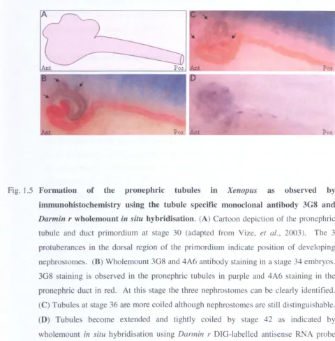
immunohistochemistry using the tubule specific monoclonal antibody 3G8 and Darmin r wholemount in situ hybridisation. (A) Cartoon depiction of the pronephric tubule and duct primordium at stage 30 (adapted from Vize, et al., 2003). The 3
protuberances in the dorsal region of the primordium indicate position of developing
nephrostomes. (B) Wholemount 3G8 and 4A6 antibody staining in a stage 34 embryos.
3G8 staining is ob erved in the pronephric tubules in purple and 4A6 staining in the
pronephric duct in red. At this stage the three nephrostomes can be clearly identified.
(C) Tubules at stage 36 are more coiled although nephrostomes are still distinguishable.
(D) Tubule become extended and tightly coiled by stage 42 as indicated by
wholemount in situ hybridisation using Darmin r DIG-labelled antisense RNA probe
[image:30.523.7.487.57.545.2]Chapter I
visible by 3G8 (pronephric tubules) and 4A6 (pronephric duct) immunohistchemistry. As the tadpole develops, the distal tubule extends and coils, forming a convoluted mass by feeding stages (Vize, et al., 2003).
Gene expression and regulation in the pronephric tubules
As the pronephric tubule and duct primordium develops, Xliml pronephric expression is dramatically restricted to the tubules, before becoming exclusive to the nephrostomes, the regions of increased extension and growth (Carroll, et al., 1999). This is similar to the later expression patterning of xPax 2 but not xPax 8 (Carroll, et al., 1999 and Heller and Brandli, 1997). As described previously, co-injection of Xliml and xPax 8 leads to an enlargement of the pronephros or ectopic pronephroi, indicated by antibody staining for the pronephric tubules (Carroll and Vize, 1999). Interestingly, xPax 2 can also form an enlargement of the pronephros or ectopic pronephroi when co-injected with X/iml. Comparison of xPax 2 and xPax 8 with Xliml expression patterns during pronephric tubule morphogenesis shows a much closer relationship of Xliml with xPax 2. It has been suggested, therefore, that xPax 8 is involved with early pronephric patterning events, and xPax 2 is involved with later pronephric tubule patterning events.
In the developing pronephric tubule and duct primordium, XWnt-4, Xliml and xPax 2 expression coincides with the anterior-dorsal most region of
wn
expression (Carroll and Vize, 1999). It is not clear whether this environment contributes to the formation of the nephrostomes, or is a result of unspecified cells yet to be determined as glomus or tubule tissue. In zebrafish, nephrostomes are formed transiently, and are derived from the coelom before the tubules develop (Drummond, et al., 1998). In Xenopus, however, the nephrostomes are formed from the tubule primordium, but are again formed before tubule morphogenesis (Jones, 2005).XTRAP-y is one of four subunits that make up the translocon-associated protein complex which is involved in intracellular protein transport (Li, et al., 2005).
Expression was observed in the pronephros from stage 25 in the pronephric tubules. Morpholino oligonucleotide knock-down of XTRAP-y disrupted the pronephric tubule formation and expression of xPax 2 and xWnt-4 at stage 30. Conversely for a pronephric tubule promoting gene, Xliml pronephric expression increased in targeted XTRAP-y knock-down embryos. XTRAP-y expression was also found to be required for the induction of pronephric tissues in treated animal caps, which suggests an earlier role in pronephric induction.
Annexin IV is expressed in the pronephric primordium from stage 26 and later on the luminal surface of the pronephric tubules (Seville, et al., 2002). Over-expression experiments resulted in a disruption of WTI expression in the glomus, although no effect on Xliml or xPax 8 expression was observed. Morpholino oligonucleotide
knock-down analysis gave a single shortened, enlarged tubule, rather than the branching tubules seen in control embryos.
PKDI forms part of an ion channel complex with PKD2 and has been implicated in the activation of intracellular signalling pathways (Burtley, et al., 2005). Expression is observed by dissected pronephric RT-PCR from pronephric initiation, stage 14, and continues until stage 40, the last stage tested. xPKDI expression is induced by the treatment of animal caps with retinoic acid and activin to produce pronephric tubules. Inactivation of xPKDJ ion channel properties by blocking calcium flux where xPKDI is expressed, suppresses tubulogenesis.
1.1.2.4 The Duct
Chapter 1
induces the surrounding mesonephric mesenchyme to form the mesonephros, before gradually regressing to form part of the genital system in males.
Gene expression and regulation in the pronephric duct
A detailed previously, Xliml pronephric expression is initiated at stage 12.5 continues to late tail bud stages (Carroll, et al., 1999). Later expression includes a reducing expression in the anterior duct which is maintained in the portion extending towards the cloacal horns, although not in the cloacal horns themselves. Interestingly, similar to the expression in the pronephric tubules, this is also a region of extension and growth. This expression pattern differs from xPax 2 in the pronephric duct, which has been comparable in the other pronephric tissues. xPax 2 expression is ubiquitous over the pronephric duct and includes the cloacal horns.
Xfz8 is detected in the pronephric anlagen at stage 22 and becomes restricted to the pronephric duct by stage 28. Depletion of Xfz8 results in reduced pronephric tubule branching and a reduction in 4A6 duct staining.
XJz8
is hypothesised to be involved in epithelium formation in the developing pronephric tubule and duct after specification (Satow, et ai., 2004).At stage 27, gremlin, a BMP antagonist, is expressed anteriorly in the early developing duct and proceeds posteriorly as the duct develops (Hsu, et al., 1998). Xenopus c-ret has been detected at stage 28 in the caudal tip of the migrating duct primordium and in the tubules (Carroll, et al., 1999).
1.2
Developmental Congenital Kidney Disease
form for its own induction (Brandli, 1999 and Vize, et al., 1997). Structural defects in the development of the earlier structures may therefore effect the formation of later kidney structures.
Xenopus laevis was the model organism used in this study. The pronephros in Xenopus forms just below the epidermal surface, allowing for effortless accessibility. In addition, the morphological development of the pronephros in Xenopus has been well documented. Xenopus, itself is an ideal organism to use due to its external development, large egg and embryo sizes, ease of embryo culture and adult husbandry. The external development and detailed fate mapping of early division allows for targeted micro-injection of mRNA in over-expression experiments, or morpholino oligonucleotides in knock down experiments. The other common model organism to use in kidney development research is the mouse. However, the metanephric kidneys of the mice are complex and their internal development means embryo manipulation is more difficult. Also the knock-out phenotypes of many kidney development genes are embryo lethal, restricting developmental analysis.
To impress the importance of studying kidney development, four important human and
Xenopus development genes will be highlighted. WTl, Pax 2, Nephrin and PKDl have been previously shown to have critical roles in pronephric development (1.1.2 Xenopus Pronephric Development). The roles of these genes in human developmental congenital kidney diseases will now be discussed.
1.2.1
Wilms' Tumour
1
Wilms' tumour is a cancer of the kidney that affects I in 10,000 children (Discenza and Pelletier, 2(04). It is believed to arise when the developing metanephric mesenchyme fails to properly differentiate, forming nephrogenic rests. These rests degenerate, remain dormant, or in the case of Wilms' tumour, proliferate. Human WT 1, a four zinc-fingered transcription factor, was identified from positional cloning identifying genes on the Wilms' tumor locus human chromosome II (p 13) (Call, et al., 1990 and Gressler, et
Chapter I
The earliest observation of human
wn
expression in the kidney is in the developing podocytes of the mesonephros and the metanephros, the condensing metanephric mesenchyme and the tip of the ureteric bud (Discenza and Pelletier, 2004). Expression is also observed in the sex cord region of the urogenital ridge, spleen, epicardium and other epithelial tissues.Denys-Drash syndrome
W[J mutations were found at a higher incidence, 90%, with Denys-Drash Syndrome sufferers (Little and Wells, 1997). Denys-Drash syndrome is associated with nephropathy due to diffuse mesangial sclerosis and intersex (Denys, et al., 1967 and
Drash, et al., 1970). Denys-Drash syndrome kidneys typically show glomerular lesions,
enlarged immature podocytes, thickening of glomerular membranes, reduced capillary lumens and severely damaged tubules. Hypertension is often a symptom, as fibrotic material is produced in the cells lying adjacent to the glomerular, causing the collapse of the arteries (Scharnhorst, et al., 2001). Patients also suffer from a high incidence of
Wilms' Tumours (Little and Wells, 1997). 8 out of 10 unrelated Denys-Drash syndrome patients were found to be heterozygous for point mutations in exon 9 of
wn
(Pelletier, et ai., 1991). These point mutations are believed to alter the structure of thethird zinc-finger and so hinder the binding of WTI to the target DNA binding site (Pelletier, et ai., 1991, Little, et ai., 1993 and Scharnhorst, et at., 200 I). Indeed, WTI
DNA binding has been shown to be interrupted in vitro by point mutations in exon 9 of
the WI'1 gene. Chimeric and heterozygous mice for a mutation that truncates the third zinc finger, coded by exon 9, have been created. Interestingly, the phenotypes observed in the mutant mice were similar to those of Denys-Drash syndrome patients (Patek, et ai., 1999). Also co-incident with the human syndrome, >75% of Denys-Drash syndrome mutant mice developed Wilms' Tumours, further implying a link.
WI'1 has been shown to repress the Pax 2 promoter and inhibit its expression (Ryan, et at., 1995). In fact, Pax 2 down regulation was found to be required for podocyte
maturation, (Quaggin, 2002). Unsurprisingly, abnormal Pax 2 podocyte expression is observed in patients with Denys-Drash syndrome (Yang, et ai., 1999). However, it is
Frasier's syndrome
Related to Denys-Drash syndrome, but less severe, Frasier's syndrome was also found to be associated with
wn
mutations (Barbaux, et al., 1997). Like Denys-Drash syndrome, Frasier's syndrome is characterized by intersex and nephropathy, although, no Wilms' tumours have been detected. The nephropathy, however, is due to focal segmental glomerulosclerosis, where kidneys have fused podocyte foot processes and scar tissue in the glomerulus. The condition is termed focal, as not all nephrons are affected, and segmental due to the localised effect on the filtration barrier of the glomerulus. Frasier's syndrome patients are missing one of the alternatively spliced WTI isoforms, known as the +KTS isoform (Barbaux, et al., 1997). This loss is caused by mutations in the donor splice site in intron 9 of Wf J, loosing the addition of three amino acids (KTS) between WTl zinc-fingers 3 and 4. The 3 additional amino acids are believed to be involved with subnuclear localisation of the WTl protein (Scharnhorst, et al., 200 1).WAGR syndrome QYilms' tumour, Aniridia, Genitourinary defects and Mental Retardation)
WAGR syndrome is characterized by development of Wilms' tumours, iris abnormalities (Aniridia), genital defects and mental retardation. W AGR syndrome has been linked with the deletion of part of the short arm of chromosome II, the region of the Wf I and Pax 6 genes (Riccardi, et al., 1978). The reduction of Wf I expression, due to gene deletion, has been implicated in the unexpected or late-occurring renal failure in some W AGR syndrome patients (Breslow, et al., 2000). The condition of Aniridia has been linked to the deletion of Pax 6 (Discenza and Pelletier, 2004).
The null Wfl mouse does not produce any kidney structures, and heterozygous null
Chapter 1
WTl and other vertebrates
Analysis of
wn
expression in developing quail chick embryos revealed similar expression observed in mouse kidneys, that the mesonephric glomerulus and duct (ureteric bud) and the condensing metanephric mesenchyme (Carmona, et al., 2001). Although no mis-expression experiments have yet been performed, quail expression patterns suggest a link with Slug, which is also expressed in podocytes and is involved in their mesenchymal-epithelial transition (Carmona, et al., 2001 and Davidson, et al., 2(02).The null
wn
mouse lacks kidneys, gonads and adrenal glands (Kreidberg, et al., 1993).Morpholino oligonucleotide knock-down experiments in zebrafish have shown similar phenotypes, with disrupted pronephric and interrenal (adrenal gland) organogenesis.
Ff] b is a functional homologue of SF], which is a sex determining gene in mammals.
wn knock-down in zebrafish was also found to reduce ff] b expression, also effecting
gonad development (Hsu, et ai., 2003).
1.2.2
Paired bo;!
£
Pax 2 is part of the paired box transcription factor family. The highly conserved paired box encodes a bipartite helix-loop-helix DNA-binding domain that enables Pax 2 to bind directly to gene promoters (Sanyanusin, et al., 1996). Pax 2 is positioned at the boundary band of q24 and q25 of chromosome 10 (Gough, et al., 2(03). Comparable with mouse expression, Pax 2 is believed to be expressed in the pronephric tubule, duct and mesonephric tubules of early human developing kidneys (Eccles, 2(02). During induction of the metanephros, expression is observed in the epithelium of the branching ureteric bud and in the condensing mesenchyme. Later expression is observed in the differentiating renal vesicle and the developing nephron, and excluded from the glomerulus. Interestingly, in adulthood, expression has also been observed in regenerating proximal tubule cells following treatment with nephrotoxins. This indicates that Pax 2 may also be involved in regenerative processes (Eccles, 1998). Other areas of mouse expression include the central nervous system, eye and ear (Favor,
Papillorenal syndrome I Renal-coloboma syndrome
Papillorenal syndrome is generally associated with a reduced kidney, resulting from reduced ureteric bud branching and a reduced number of nephrons. Other symptoms include vesicoureteral reflux, optic nerve and disc defects, and a high-frequency of hearing loss. Papillorenal syndrome is caused by a single nucleotide deletion in exon 2 or 5 of Pax 2, creating a frame-shift of the Pax 2 coding region creating a premature stop codon which results in a truncated protein (Sanyanusin, et af., 1995 and Sanyanusin, et af., 1999). Symptoms vary in extremity and are increased with homozygous mutations, leading to the hypothesis of haploinsufficiency, where phenotypes are produced from a reduction of healthy protein to inadequate levels (Eccles, 2002).
A spontaneous mutation, typical of the Papillorenal syndrome mutation in humans, has been studied in mice (Favor, et al., 1996). The mutation creates a frame-shift in the helix-loop-helix DNA-binding domain of the Pax 2 protein, resulting in 27 mis-matched amino acids in the DNA-binding domain and a truncated protein. The truncated mutant protein is therefore unable to recognize its normal target genes and so is ineffectual. The Pax
i
Neu heterozygous mice were reported to have unaffected, reduced or absentkidneys and eye defects, including severe abnormalities of the optic disc region. The developing homozygous Pax
i
Ncu mouse had a reduced metanephric anlagen and a deficiency in tubule formation. Other homozygous phenotypes observed in the developing mouse include defects in the mid-hindbrain region, lack of inner ear structures and severely disrupted eye structures. Not only are these phenotypes largely analogous to the symptoms observed in Papillorenal syndrome, but also they display the same variation in intensity. This is again suggestive of haploinsufficiency and collaborates with the inadequacy of the truncated protein.In mouse metanephric development, the ureteric bud branches into the condensing metanephric mesenchyme, forming a nephron at each ureteric bud tip outgrowth. The number of ureteric bud branches therefore sets the number of nephrons formed for life.
Pax 2 is believed to protect the ureteric bud against apoptosis. Indeed, increased apoptosis was observed in the kidneys of heterozygous Pax l'Neu mice (Porteous, et al .•
Chapter I
et al., 2003). In addition, the inhibition of apoptosis in heterozygous Pax
i
Neu mice, led to a partial rescue of the decreased ureteric bud branching and nephron number.Pax 2 and adult carcinoma
The role of Pax 2 in the protection against apoptosis is not restricted to developmental events. Renal cell carcinoma accounts for 3% of all adult aggressive tumours and is the most common malignancy in the adult kidney, 90-95% (Gnarra and Dressler, 1995). Renal cell carcinoma cells lines were found to express Pax 2. Inhibition of Pax 2 by antisense oligonucleotide resulted in the inhibition of cell proliferation. Pax 2 is therefore a potential anti-apoptotic carcinoma gene. Pax 2 expression was also observed in Kaposi Sarcoma cells (Buttiglieri, et al., 2004). Kaposi Sarcoma is a cancer of the blood vessels associated with immunodepressive conditions, such as HIV-l infection and long term post-transplantation therapy. Again, Pax 2 inhibition, this time by transfection with an antisense Pax 2 gene, resulted in the reduction of cell proliferation and an enhanced sensitivity to apoptosis.
Pax 2 and other vertebrates
Similar to the mouse and human expression, chick Pax 2 expression is found in the early developing pronephros and mesonephros, the developing ear and brain (James and Schultheiss, 2003 and Hutson, et al., 1999). Also, chick Pax 2 has been shown to have a role in early kidney induction, where exogenous mesodermal expression was sufficient to produce ectopic kidney structures (Bouchard, et al., 2(02). The chick homologue of Pax 2 has even been shown to have a role in protection against apoptosis, although in the development of the ear, not the kidney (Li, et al., 2(04). These common characteristics suggest conserved evolutionary properties of Pax 2 in development.
Two Pax 2 zebrafish homologues have been identified, Pax 2.1 and Pax 2.2, however
Pax 2.1 more closely resembles the mammalian Pax 2 based on its expression pattern (Majumdar, et ai., 2000). Pax 2.1 expression is observed in the central nervous system, ear, optic tracts and pronephros, analogous to the mouse (Majumdar, et al., 2000 and Favor, et al., 1996). The zebrafish no isthmus mutant has a truncated form of the Pax
not match the increase in apoptosis observed in the kidneys of "Papillorenal syndrome" mice (Porteous, et ai., 2000).
1.2.3
Nephrin
Nephrin is a major component of the slit-diaphragm of podocytes (Fig. 1.3)
(Tryggvason, 1999 and Ruotsalainen, et ai., 1999). Without slit-diaphragms, the nephrons are unable to stop large proteins, such as albumin, from entering the tubules and resulting in proteinuria. Nephrin was identified using positional cloning to identify
the gene responsible for Congenital Nephrotic Syndrome of the Finnish type (See below) (Kestila, et ai., 1998). Nephrin was found to be located on chromosome
19q13.1.
Congenital Nephrotic Syndrome of the Finnish type
Congenital Nephrotic Syndrome is associated with massive proteinuria at, or shortly after birth (Kestila, et ai., 1998). Congenital Nephrotic Syndrome of the Finnish type is
autosomal-recessive disorder with an frequency of I in 10,000 births in Finland, although significantly less in other European countries. Symptoms of proteinuria and glomerula lesions are observed during embryo development. Structurally, Congenital Nephrotic Syndrome of the Finnish type kidneys have podocytes lacking in foot processes and slit-diaphragms (Fig.I.3), dilated proximal tubules and an increased number of nephrons, resulting in enlarged kidneys. The most frequent cause of Congenital Nephrotic Syndrome of the Finnish type is two nucleotide deletions in exon 2, which result in a frame-shift and a truncated protein. However, many different
Nephrin mutations have been identified (Koziell, et ai., 2002).
Experiments were performed using a human embryonic cell line and 21 mis-sense disease causing Nephrin mutations (Liu, et ai., 200 I). Analysis revealed a localisation of mutant Nephrin to the endoplasmic reticulum, not the cell surface of the podocyte.
This could explain the lack of slit-diaphragms in Congenital Nephrotic Syndrome of the Finnish type kidneys, and the associated proteinuria.
Chapter 1
homozygous and the null Nephrin mouse has enlarged kidneys, dilated tubules, massive proteinuria, edema and the absence of slit-diaphragms. Heterozygous null Nephrin
mice also have similar phenotypes, but these are less severe and proteinuria and edema do not develop until a week after birth.
Nephrin and other vertebrates
In zebrafish, nephrin expression is localised to the podocytes (Kramer-Zucker, et af.,
2(05). Morpholino oligonucleotide mis-translation results in a general edema that spreads throughout the larval body. Embryos die aged I week, with pronephric tubule cysts and aberrant glomerulus morphology. The majority of podocytes lack foot processes and no slit-diaphragms are present. The disrupted morphology of the glomerulus reduces the size discrimination of filtration, and large protein deposits are found in the tubules. These phenotypes exactly match the symptoms of Congenital Nephrotic Syndrome of the Finnish type, showing a conserved evolutionary role of
nephrin in kidney development and a correlation between pronephric and metanephric development.
1.2.4
PKDJ
PKDl encodes Polycystin I, a trans-membrane protein with potential adhesion and protein to protein interactions (Kim, et af., 2000). Polycystin I has been found to be expressed in epithelial tissues, endothelial and vascular smooth muscle cells and at cell to cell junctions. In mice kidneys, expression is observed in mature collecting ducts (Gallagher, et af., 2(02). PKDl has been located chromosome 16 and was identified by its link with Adult polycystic kidney disease.
Autosomal Dominant Polycystic Kidney Disease