Original Article
Elastography for the differentiation of benign and
malignant cervical lymph node: a meta-analysis
Qiong Xie, Yibing Li, Haoping Li, Hongli Ji
Department of Ultrasound, Women & Infants Hospital of Zhengzhou, Zhengzhou, China
Received November 1, 2015; Accepted May 20, 2016; Epub August 15, 2016; Published August 30, 2016
Abstract: Objectives: To assess the diagnostic efficacy of elastography in diagnosis of benign and malignant cervical
lymph node. Methods: After comprehensive search and study selection, a meta-analysis was performed on data from 548 patients pooled from 14 studies for evaluating FLT accuracy, in which data from 351 patients pooled from
ten double-tracer studies were used for direct comparison with FDG. Weighted sensitivity and specificity were used
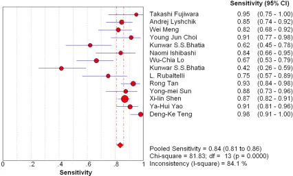
as main indicators of test performance. The data from individual study were extracted and patient subgroup analy-ses were performed. Results: Elastography had a pooled sensitivity (random effect model) of 85% (95% CI,
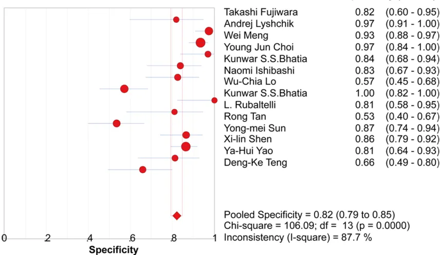
0.76-0.90) in the differential diagnosis of benign and malignant LNs (lymph nodes). The pooled specificity (random-effect
model) was 86% (95% CI, 0.77-0.91) which were showed in Figures 2 and 3. The summary positive LR and negative LR were 5.86 (95% CI, 3.62-9.50) and 0.18 (95% CI, 0.12-0.28), respectively. The summary diagnostic odds ratio (DOR) used to examine the elastography accuracy is 32.641 (95% CI, 16.772-63.525) and the area under the SROC
was 0.92 (95% CI, 0.89-0.94). Significant heterogeneity was found in sensitivity (heterogeneity, chi-square = 95.68,
P = 0.00, I-square = 86.41), specificity (heterogeneity, chi-square = 109.23, P = 0.00, I-square = 88.10), indicat -ing that more than 80% of variance across studies is attributed to heterogeneity rather than chance. Conclusion: Elastograohy has high accuracy in differentiating benign and malignant cervical LNs.
Keywords: Cervical lymph node, elastography, meta-analysis, accuracy
Introduction
A lymph node (LN) is an oval-shaped organ of the lymphatic system, distributed widely throughout the body including the armpit and stomach and linked by lymphatic vessels [1-3]. Lymph nodes are major sites of B, T, and other immune cells. Lymph nodes are important for the proper functioning of the immune system,
acting as filters for foreign particles and cancer
cells. Lymph nodes do not deal with toxicity, which is primarily dealt with by the liver and
kid-neys. Lymph nodes also have clinical signifi
-cance. They become inflamed or enlarged in
various infections and diseases which may range from trivial throat infections, to life-threatening cancers [4-6]. The condition of the lymph nodes is very important in cancer stag-ing, which decides the treatment to be used, and determines the prognosis. When swollen,
inflamed or enlarged, lymph nodes can be hard, firm or tender [7].
Evaluation of LN in patients with various under-lying diseases is important to detect current status, suitable treatment and prognosis of the patients [8]. It is crucial to distinguish malig-nant LNs from benign LNs to follow appropriate therapy. The gold standard for evaluating enlarged LNs is pathologic examination of
obtained tissue. Although fine-needle aspira
-tion (FNA) is considered as the most efficient
method for differentiating benign and malig-nant LNs, it is considered as an invasive meth-od which is prone to sampling errors and ana-lytic uncertainty [9]. Its false negative rate has been reported to be between 12.5% and 25% [10, 11].
Different modalities such as ultrasound com-puted tomography, and magnetic resonance imaging are currently used as imaging
tech-niques for differentiating benign and malignant
malig-cesses like cancer modify the physical charac-teristics of diseased tissues. This principle has already been exploited for diagnostic purposes, but interest in elastography has been increased by the recent development of integrated sys-tems that facilitate the inclusion of sonoelasto-graphic studies in routine practice. Soft tissues show more displacement than stiff ones. It has been applied in the evaluation of different organs such as breast, thyroid, pancreas, liver and LNs [13-16].
Several previous studies had evaluated accu-racy of this modality in differentiating benign
and malignant LNs. Its sensitivity and specifici -ty ranged from 79% to 100% and 50-96%, respectively [17-21]. The aim of present study was to perform a meta-analysis of published information to investigate the overall accuracy of elastography for differentiation of benign and malignant cervical LNs.
Materials and methods
Literature search
A systematic search was conducted for the studies on association between the elastogra-phy and cervical lymph nodes published before October, 2015 in PubMed, Web of Science Embase, Cochrane Central Trials and CNKI (China National Knowledge Infrastructure) databases. The search was performed without any restrictions on language. The search terms were as follows: “lymph nodes” in combination with “strain imaging”, “elasticity imaging
tech-niques”, “elasticity” or “elasticity”. To identify
additional relevant publications and missing
data, the reference lists of identified studies
and review articles were manually searched and study authors were contacted. Two investi-gators (A and B) searched and assessed stud-ies for eligibility independently, and disagree-ments were resolved by discussion. If any
clarification of data was necessary, we contact -ed the authors for detail-ed information.
ration for cytology (FNAC); (3) the outcome data available to reconstruct a diagnostic 2×2 con-tingency table for true positives (TP), false posi-tives (FP), false negaposi-tives (FN) and true nega-tives (TN).
The exclusion criteria were as following: (1) not related to the topic or cannot get the full texts; (2) incomplete data available; (3) publication types such as reviews, editorials, letters, ani-mal experiments and case reports; (4) when publications involved the overlapping data sets, only the study with the largest number of par-ticipants was included, and updated data were excluded directly.
Data extraction and quality assessment
Two authors (A and B) reviewed and extracted data independently. The following data were
extracted: the name of the first author, year of
publication, country, description of the study population (number of patients, median age), number of cervical LNs, malignant LN rate, ref-erence, method and a cutoff value. Additionally, direct data of TP, FP, FN, TN and indirect data of
sensitivity, specificity, positive predictive value
(PPV), negative predictive value (NPV) and accuracy for the 2×2 table could be extracted. Once the data they obtained were different, the third author (C) would check the data. Finally, disagreements were resolved through discus-sion among the authors.
Statistical analysis
The summary sensitivity and specificity were
benign and malignant cervical lymph nodes if AUC approached to 1 approximately.
Heterogeneity was evaluated based Q statistic and I2 test, with P < 0.10 or I2 > 50% considered
statistically significant. Funnel plots are the
most common used tool to detect publication bias. With the drawbacks of funnel plots, a wide range of studies of different sizes and subjec-tive judgments, publication bias was detected by Egger’s test. We calculated indirect data using SPSS version 20.0 for constructing con-tingency table. Other analyses were done using STATA 12.0 and meta-DISC. Two-sided P < 0.05 was considered indicative of statistically signi-
ficant.
Results
Characteristics of included studies
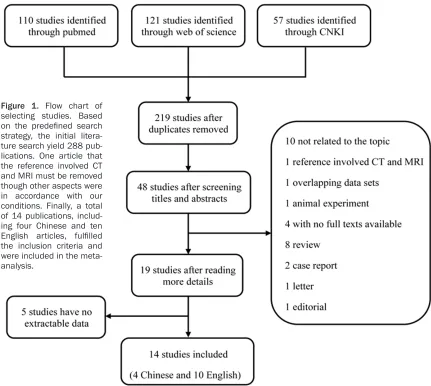
Based on the predefined search strategy, the
One article that the reference involved CT and MRI must be removed though other aspects were in accordance with our conditions. Finally, a total of 14 publications, including four
Chinese and ten English articles, fulfilled the
inclusion criteria and were included in the
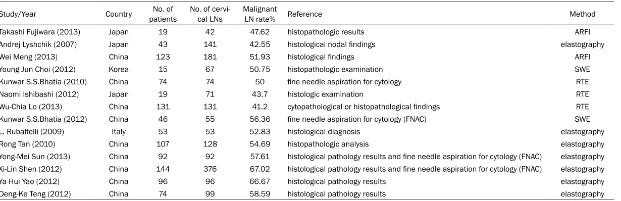
meta-analysis [21-26]. The flow chart that dis -plays the study selection process was shown in Figure 1. 1606 cervical lymph nodes from 1036 patients were diagnosed by histological pathology results and FNAC. In general, the
[image:3.612.89.520.70.456.2]number of cervical lymph nodes had a signifi -cant change in the included studies (Table 1). The median number was 115 nodes, ranging from 42 to 376. The proportion of malignant lymph nodes was 52.96% (range 41.2 to 67.02). The mean age of the included patients was 51.37 years (range 40.8 to 69.9). 52.72 per-cent of the patients were male. In Addition, there existed some variation of the type of elastography used in these fourteen studies. Figure 1. Flow chart of
selecting studies. Based
on the predefined search
strategy, the initial litera-ture search yield 288 pub-lications. One article that the reference involved CT and MRI must be removed though other aspects were in accordance with our conditions. Finally, a total of 14 publications, includ-ing four Chinese and ten
English articles, fulfilled
Naomi Ishibashi (2012) Japan 19 71 43.7 histologic examination RTE Wu-Chia Lo (2013) China 131 131 41.2 cytopathological or histopathological findings RTE Kunwar S.S.Bhatia (2012) China 46 55 56.36 fine needle aspiration for cytology (FNAC) SWE L. Rubaltelli (2009) Italy 53 53 52.83 histological diagnosis elastography Rong Tan (2010) China 107 128 54.69 histopathologic analysis elastography Yong-Mei Sun (2013) China 92 92 57.61 histological pathology results and fine needle aspiration for cytology (FNAC) elastography Xi-Lin Shen (2012) China 144 376 67.02 histological pathology results and fine needle aspiration for cytology (FNAC) elastography Ya-Hui Yao (2012) China 96 96 66.67 histological pathology results elastography Deng-Ke Teng (2012) China 74 99 58.59 histological pathology results elastography
[image:4.792.94.709.83.282.2]impulse elastography (ARFI), while three per-formed ultrasound real-time elastography
(RTE). Two applied the technique of shear wave
elastography (SWE) for checking nodes, and the remaining seven articles did not show read-ers the type of elastography. Missing data that were modeled from the given factors using sto-chastic regression analysis included the mean age (one study), male percentage (one study) and the cutoff value (one study).
Meta-analysis results
Diagnostic accuracy: We display the test results for elastography in differentiating benign and malignant cervical lymph nodes. Elastography had a pooled sensitivity (random effect model) of 85% (95% CI, 0.76-0.90) in the differential diagnosis of benign and malignant LNs. The
pooled specificity (random-effect model) was
86% (95% CI, 0.77-0.91) which were showed in Figures 2 and 3. The summary positive LR and negative LR were 5.86 (95% CI, 3.62-9.50) and 0.18 (95% CI, 0.12-0.28), respectively. The summary diagnostic odds ratio (DOR) used to examine the elastography accuracy is 32.641 (95% CI, 16.772-63.525) and the area under the SROC was 0.92 (95% CI, 0.89-0.94).
Heterogeneity study: Significant heterogeneity
was found in sensitivity (heterogeneity,
chi-square = 95.68, P = 0.00, I-chi-square = 86.41), specificity (heterogeneity, chi-square = 109.23, P = 0.00, I-square = 88.10), indicating that
more than 80% of variance across studies is attributed to heterogeneity rather than chance. To explore the sources of heterogeneity, meta-regression analysis was performed. The out-comes of meta-regression analysis are shown method were associated with a higher RDOR. The other characteristics were not statistically
significant in the regression model.
Publication bias: Funnel spot and test results of the publication bias are shown that there
was no statistically significant publication bias among the 14 included studies (P = 0.942).
Discussion
[image:5.612.92.518.74.331.2]rect differentiation of benign and malignant LNs is essential for clinical decision making.
Elastography is a new technique of sonography
that is noninvasive, available and easy to apply. It evaluates the stiffness of the lesions based on response to the compression and decom-pression. By applying a mechanical force to the target lesion, an elastogram will be obtained. The results of the response of the lesions to mechanical force will appear as red or green indicating softness or blue, indicating hard-ness of the tissue. Cell types of the lesion, the
quantity of the entire types of cells and micro
and macro pathological structures have roles in rate of stiffness. Qualitative elastography scoring method or SR measurement. By means of scoring system, operator should score the target lesion according to the proportion of blue areas in the lesions. It is semi-objective, and it depends on different factors such as the operator’s experience and scoring system (5 or 4 point). Two different methods could be obtained for elastography evaluation. Strain ratio measurement has been considered to be more accurate that scoring method because it could estimate the difference between stiff-ness of the lesions and the surrounding tissue
[28]. One of the advantages of the SR method
is that as it is quantitative, in cases that the
scores are the same visually, the SR could be different. The nature of the tissue was analyzed
either by a qualitative method or a quantitative
method [18, 29, 30].
In this meta-analysis, we evaluated accuracy of elastography in differentiating benign and malignant cervical LNs. We detected high
sen-sitivity and specificity for both elastography
scoring system and SR. We also obtained high DORs for both scoring and SR evaluation. It can show that the odd of obtaining positive results in diseased rather than non-diseased individu-als by means of elastography is high. The other included study by Giovannini et al. [18, 31]
reported 84% sensitivity and 95% specificity.
[image:6.612.92.520.85.333.2]The variation may be produced during the sub-jective interpretation of the color pattern even if the same threshold was used. The reap-praised pooled results still suggested a high accuracy of elastography for differentiating benign and malignant LNs. In a previous sys-tematic review conducted by Ying et al. [32], diagnostic accuracy of SR and scoring system method in differentiating benign and malignant cervical and axillary LNs had been evaluated. Figure 3. Forest plot (random-effect model) of the meta-analysis of specificity for the differentiation of benign and
They found that sensitivity, specificity and diagnostic OR of SR method is significantly
higher than scoring method. We found hetero-geneity in all measurements of both methods (I2 > 50%) but as a limitation, we did not do
meta-regression analysis to find the source of
heterogeneity Ying et al. performed
meta-regression analysis and evaluated 11 specific
covariates of patient and study, but they did not
find the source of heterogeneity in their study
[32]. Our meta-analysis results support conclu-sion Elastograohy has high accuracy in differ-entiating benign and malignant cervical LNs. However, there were some limitations in this study. First, only 14 trials were included in the meta-analysis. As a result, the small num-ber of studies might reduce the power of the tests on publication bias and source of hetero-geneity. Second, different diagnostic standards for elastography were used in the selected studies.
Acknowledgements
Thank all our colleagues working in the Department of Ultrasound, Women & infants Hospital of Zhengzhou.
Disclosure of conflict of interest
None.
Address correspondence to: Dr. Qiong Xie, Depart- ment of Ultrasound, Women & Infants Hospital of Zhengzhou, Zhengzhou 450002, China. Tel: +86-371-63883127; Fax: +86-+86-371-63883127; E-mail: [email protected]
References
[1] Tan PW, Goh T, Nonomura H and Tan BK. Hilar vessels of the submandibular and upper jugu-lar neck lymph nodes: anatomical study for vascularized lymph node transfer to extremity lymphedema. Ann Plast Surg 2016; 76: 117-23
[2] Kato S, Shirai Y, Kanzaki H, Sakamoto M, Mori S and Kodama T. Delivery of molecules to the lymph node via lymphatic vessels using ultra-sound and nano/microbubbles. Ultraultra-sound Med Biol 2015; 41: 1411-1421.
[3] Matsuda T, Iwasaki T, Mitsutsuji M, Hirata K, Maekawa Y, Tsugawa D, Sugita Y, Sumi Y, Shimada E and Kakeji Y. Cranially approached radical lymph node dissection around the mid-dle colic vessels in laparoscopic colon cancer
surgery. Langenbecks Arch Surg 2015; 400: 113-117.
[4] Nowikiewicz T, Srutek E and Zegarski W.
Problems concerning patients’ qualification for
surgical procedures allowing for evaluation of the condition of axillary fossa lymph nodes in the radical treatment of breast cancer. Pol Przegl Chir 2015; 87: 290-294.
[5] Zielinski P, Dyszkiewicz W, Piwkowski CT, Dworacki G and Gasiorowski L. Can the condi-tion of the cell microenvironment of mediasti-nal lymph nodes help predict the risk of metas-tases in non-small cell lung cancer? Cancer Epidemiol 2009; 33: 387-390.
[6] van Lanschot JJ, Tilanus HW and Obertop H. [Transhiatal or transthoracic resection of esophageal carcinoma based on tumor loca-tion, positive high-thoracic lymph nodes and preoperative physical condition]. Ned Tijdschr Geneeskd 2003; 147: 2097-2100.
[7] Katakai T, Hara T, Lee JH, Gonda H, Sugai M and Shimizu A. A novel reticular stromal struc-ture in lymph node cortex: an immuno-platform for interactions among dendritic cells, T cells and B cells. Int Immunol 2004; 16: 1133-1142.
[8] Lecuru F, Mathevet P, Querleu D, Leblanc E, Morice P, Darai E, Marret H, Magaud L, Gillaizeau F, Chatellier G and Dargent D. Bilateral negative sentinel nodes accurately predict absence of lymph node metastasis in early cervical cancer: results of the SENTICOL study. J Clin Oncol 2011; 29: 1686-1691. [9] Ross DS. Nonpalpable thyroid
nodules--man-aging an epidemic. J Clin Endocrinol Metab 2002; 87: 1938-1940.
[10] Eloubeidi MA, Wallace MB, Reed CE, Hadzijahic N, Lewin DN, Van Velse A, Leveen MB, Etemad B, Matsuda K, Patel RS, Hawes RH and Hoffman BJ. The utility of EUS and EUS-guided
fine needle aspiration in detecting celiac lymph
node metastasis in patients with esophageal cancer: a single-center experience. Gastro- intest Endosc 2001; 54: 714-719.
[11] Hernandez LV, Mishra G, George S and Bhutani MS. A descriptive analysis of EUS-FNA for me-diastinal lymphadenopathy: an emphasis on clinical impact and false negative results. Am J Gastroenterol 2004; 99: 249-254.
[12] Xu W, Shi J, Zeng X, Li X, Xie WF, Guo J and Lin Y. EUS elastography for the differentiation of benign and malignant lymph nodes: a meta-analysis. Gastrointest Endosc 2011; 74:
1001-1009; quiz 1115 e1001-1004.
titis C: noninvasive diagnosis by means of real-time tissue elastography--establishment of the method for measurement. Radiology 2011; 258: 610-617.
[16] Kanamoto M, Shimada M, Ikegami T, Uchiyama H, Imura S, Morine Y, Kanemura H, Arakawa Y and Nii A. Real time elastography for
noninva-sive diagnosis of liver fibrosis. J Hepatobiliary
Pancreat Surg 2009; 16: 463-467.
[17] Giovannini M, Hookey LC, Bories E, Pesenti C, Monges G and Delpero JR. Endoscopic
ultra-sound elastography: the first step towards vir -tual biopsy? Preliminary results in 49 patients. Endoscopy 2006; 38: 344-348.
[18] Giovannini M, Thomas B, Erwan B, Christian P, Fabrice C, Benjamin E, Genevieve M, Paolo A, Pierre D, Robert Y, Walter S, Hanz S, Carl S,
Christoph D, Pierre E, Jean-Luc VL, Jacques D,
Peter V and Andrian S. Endoscopic ultrasound elastography for evaluation of lymph nodes and pancreatic masses: a multicenter study. World J Gastroenterol 2009; 15: 1587-1593. [19] Choi YJ, Lee JH and Baek JH. Ultrasound
elastography for evaluation of cervical lymph nodes. Ultrasonography 2015; 34: 157-164. [20] Andreo Garcia F, Centeno Clemente CA, Sanz
Santos J, Barturen Barroso A, Hernandez Gallego A and Ruiz Manzano J. Initial experi-ence with real-time elastography using an ul-trasound bronchoscope for the evaluation of mediastinal lymph nodes. Arch Bronconeumol 2015; 51: e8-11.
[21] Choi YJ, Lee JH, Lim HK, Kim SY, Han MW, Cho KJ and Baek JH. Quantitative shear wave elas-tography in the evaluation of metastatic cervi-cal lymph nodes. Ultrasound Med Biol 2013; 39: 935-940.
[22] Alam F, Naito K, Horiguchi J, Fukuda H, Tachikake T and Ito K. Accuracy of sonographic elastography in the differential diagnosis of en-larged cervical lymph nodes: comparison with conventional B-mode sonography. AJR Am J Roentgenol 2008; 191: 604-610.
[25] Lo WC, Cheng PW, Wang CT and Liao LJ. Real-time ultrasound elastography: an assessment of enlarged cervical lymph nodes. Eur Radiol 2013; 23: 2351-2357.
[26] Lenghel LM, Bolboaca SD, Botar-Jid C, Baciut G and Dudea SM. The value of a new score for sonoelastographic differentiation between be-nign and malignant cervical lymph nodes. Med Ultrason 2012; 14: 271-277.
[27] Snow GB, Patel P, Leemans CR and Tiwari R. Management of cervical lymph nodes in pa-tients with head and neck cancer. Eur Arch Otorhinolaryngol 1992; 249: 187-194.
[28] Teng DK, Wang H, Lin YQ, Sui GQ, Guo F and Sun LN. Value of ultrasound elastography in assessment of enlarged cervical lymph nodes. Asian Pac J Cancer Prev 2012; 13: 2081-2085.
[29] Opacic D, Rustemovic N, Kalauz M, Markos P, Ostojic Z, Majerovic M, Ledinsky I, Visnjic A, Krznaric J and Opacic M. Endoscopic ultra-sound elastography strain histograms in the evaluation of patients with pancreatic masses. World J Gastroenterol 2015; 21: 4014-4019. [30] Hirche TO, Ignee A, Barreiros AP,
Schreiber-Dietrich D, Jungblut S, Ott M, Hirche H and Dietrich CF. Indications and limitations of en-doscopic ultrasound elastography for evalua-tion of focal pancreatic lesions. Endoscopy 2008; 40: 910-917.
[31] Lee YN, Moon JH, Kim HK, Choi HJ, Lee SH, Choi MH, Kim DC, Lee TH, Cha SW, Cho YD and Park SH. A triple approach for diagnostic as-sessment of endoscopic ultrasound-guided
fine needle aspiration in pancreatic solid
masses and lymph nodes. Dig Dis Sci 2014; 59: 2286-2293.
[32] Ying L, Hou Y, Zheng HM, Lin X, Xie ZL and Hu YP. Real-time elastography for the
differentia-tion of benign and malignant superficial lymph