1,1
000-(Ethane-1,2-diyl)bis(1,4,7-triazonane)
James C. Knight* and Ian A. Fallis
Main Building, School of Chemistry, Cardiff University, Park Place, Cardiff CF10 3AT, Wales
Correspondence e-mail: [email protected]
Received 14 May 2010; accepted 25 May 2010
Key indicators: single-crystal X-ray study;T= 150 K; mean(C–C) = 0.003 A˚;
Rfactor = 0.068;wRfactor = 0.208; data-to-parameter ratio = 18.2.
In the centrosymmetric title compound (dtne), C14H32N6, two 1,4,7-triazacyclononane (tacn, or 1,4,7-triazonane) moieties are linked together each at an amino position by a single ethylene spacer. The molecular packing is supported by pairs of intermolecular N—H N hydrogen bonds, which form
R22(22) ring motifs and link the molecules into infinite chains running parallel to theaaxis.
Related literature
For an investigation into the coordination chemistry of dtne derivatives and similarly bridged polyaza macrocyclic frame-works, see: Schro¨der et al. (2000). For dinuclear metal complexes of related ligands, see: Sinnecker et al. (2004); Marlin et al. (2005). For the crystal structure of the related compound 1,4,7-triazacyclononane (tacn), see: Battle et al.
(2005). For the structures of other metal complexes of dtne, see: Liet al.(2009). For hydrogen-bond motifs, see: Bernstein
et al.(1995). For the preparation of a similar compound, see: Burdinskiet al.(2000).
Experimental
Crystal data
C14H32N6 Mr= 284.46 Triclinic,P1 a= 6.2732 (3) A˚ b= 6.4988 (3) A˚
c= 10.7152 (6) A˚ = 99.751 (2) = 93.115 (2) = 110.410 (3)
V= 400.45 (3) A˚3
MoKradiation = 0.08 mm1
0.40.280.28 mm
Data collection
Bruker–Nonius KappaCCD diffractometer
Absorption correction: multi-scan (SORTAV; Blessing, 1995) Tmin= 0.649,Tmax= 0.985
4952 measured reflections 1806 independent reflections 1599 reflections withI> 2(I) Rint= 0.099
Refinement
R[F2> 2(F2)] = 0.068 wR(F2) = 0.208 S= 1.23 1806 reflections 99 parameters
H atoms treated by a mixture of independent and constrained refinement
max= 0.30 e A˚
3
min=0.33 e A˚
3
Table 1
Hydrogen-bond geometry (A˚ ,).
D—H A D—H H A D A D—H A
N2—H2 N1i
0.80 (3) 2.37 (3) 3.129 (3) 159 (2)
Symmetry code: (i)xþ1;y;z.
Data collection: COLLECT (Nonius, 2000); cell refinement:
SCALEPACK (Otwinowski & Minor, 1997); data reduction:
DENZO (Otwinowski & Minor, 1997) and SCALEPACK;
program(s) used to solve structure:SIR92 (Altomare et al., 1993); program(s) used to refine structure:SHELXL97(Sheldrick, 2008); molecular graphics: X-SEED (Barbour, 2001); software used to prepare material for publication:WinGX(Farrugia, 1999).
This project was supported by the EPSRC (research grant No. EP/E030122/1).
Supplementary data and figures for this paper are available from the IUCr electronic archives (Reference: SJ5006).
References
Altomare, A., Cascarano, G., Giacovazzo, C. & Guagliardi, A. (1993).J. Appl. Cryst.26, 343–350.
Barbour, L. J. (2001).J. Supramol. Chem.1, 189–191.
Battle, A. R., Johnson, D. L. & Martin, L. L. (2005).Acta Cryst.E61, o330– o332.
Bernstein, J., Davis, R. E., Shimoni, L. & Chang, N.-L. (1995).Angew. Chem. Int. Ed. Engl.34, 1555–1573.
Blessing, R. H. (1995).Acta Cryst.A51, 33–38.
Burdinski, D., Bothe, E. & Wieghardt, K. (2000).Inorg. Chem.39, 105–116. Farrugia, L. J. (1999).J. Appl. Cryst.32, 837–838.
Li, Q.-X., Wang, X.-F., Cai, L., Li, Q., Meng, X.-G., Xuan, A.-G., Huang, S.-Y. & Ai, J. (2009).Inorg. Chem. Commun.12, 145–147.
Marlin, D. S., Bill, E., Weyhermu¨ller, T., Bothe, E. & Wieghardt, K. (2005).J. Am. Chem. Soc.127, 6095–6108.
Nonius (2000).COLLECT. Nonius BV, Delft, The Netherlands.
Otwinowski, Z. & Minor, W. (1997). Methods in Enzymology, Vol. 276, Macromolecular Crystallography, Part A, edited by C. W. Carter Jr & R. M. Sweet, pp. 307–326. New York: Academic Press.
Schro¨der, M., Blake, A. J., Danks, J. P., Li, W.-S. & Lippolis, V. (2000).J. Chem. Soc. Dalton Trans.pp. 3034–3040.
Sheldrick, G. M. (2008).Acta Cryst.A64, 112–122.
Sinnecker, S., Neese, F., Noodleman, L. & Lubitz, W. (2004).J. Am. Chem. Soc.
126, 2613–2622. Structure Reports
Online
supporting information
Acta Cryst. (2010). E66, o1513 [https://doi.org/10.1107/S1600536810019562]
1,1
′
-(Ethane-1,2-diyl)bis(1,4,7-triazonane)
James C. Knight and Ian A. Fallis
S1. Comment
The coordination chemistry of ligand frameworks which contain two tacn moieties linked by two to six carbon atoms has
been extensively studied (Schröder et al., 2000). The ability of these so-called "earmuff" ligands to form dinuclear metal
complexes, in which two metal centres lie in close proximity, has provided a useful means of investigating the active sites
of various biological systems. For example, the dinuclear manganese complexes of ligands dtne (Sinnecker et al., 2004)
and 1,2-bis(4,7-dimethyl-1,4,7-triaza-1-cyclononyl)ethane (Me4dtne) (Marlin et al., 2005) have received particular
attention as a means of investigating Photosystem II. Whilst crystal structures of several dtne transition metal complexes
have been reported (Li et al., 2009), the structure of the free ligand in the solid state has, until now, remained elusive.
We can report that dtne crystallizes in the triclinic space group P -1 with one molecule in the unit cell. The asymmetric
unit contains one-half molecule with the other half generated by a centre of inversion which lies at the midpoint of the C7
—C7i bond [Symmetry code: (i) = -x, -y, -z] (Figure 1). The bond lengths and angles within each tacn moiety are
comparable to those found in the crystal structure of 1,4,7-triazacyclononane hemihydrate (Battle et al., 2005). The N3—
C7—C7i bond angle is 112.12 (15) ° which indicates no significant stretching or compression of the ethylene bridge. The
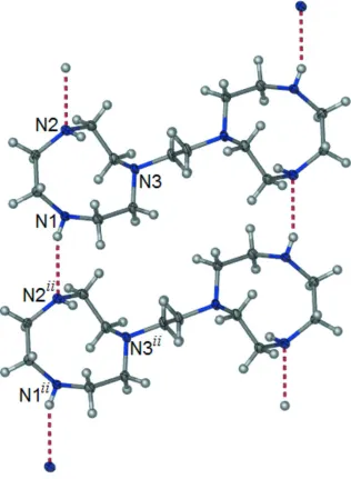
molecular packing (Figure 2) is supported by pairs of N—H···N hydrogen bonds between N1 and N2ii [Symmetry code:
(ii) = x-1, y, z] (Figure 3). These H-bond interactions generate R22(22) ring motifs (Bernstein et al., 1995) and link the
molecules into supramolecular one-dimensional chains which run parallel to the a-axis.
S2. Experimental
1,2-Bis(1,4,7-triaza-1-cyclononyl)ethane, commonly referred to by the abbreviation dtne, was prepared by a modification
of the procedure for that of 1,2-bis(4-methyl-1,4,7-triazacyclononyl)ethane (Me4dtne) reported by Burdinski et al., 2000.
To a stirred solution of 1,4,7-triazatricyclo[5.2.1.04,10]decane (6.96 g, 5 mmol) in dry acetonitrile (25 ml) was added
1,2-dibromoethane (4.51 g, 2.4 mmol). After 5 days an off-white hygroscopic precipitate was collected by filtration and
subsequently dissolved in 6 M hydrochloric acid (100 ml). The resulting solution was heated at reflux for 3 days after
which the solvent was removed by evaporation under reduced pressure. The title compound was isolated by the addition
of 10 M NaOH (20 ml) and subsequent removal of water by azeotropic distillation with toluene and a water collector.
Solvent removal under reduced pressure afforded the title compound as a low melting slightly yellow solid. Crystals
appropriate for data collection were obtained by slow diffusion of diethyl ether into a chloroform solution under an inert
atmosphere.
S3. Refinement
The carbon bound H atoms were placed in calculated positions and subsequently treated as riding with C—H distances of
0.99 Å and Uiso(H) = 1.2Ueq(C). The hydrogen atoms located on N1 and N2 were located on a difference map and freely
Figure 1
Perspective view of the asymmetric unit, showing the atom numbering. Displacement ellipsoids are at the 50%
probablility level. H atoms are represented by circles of arbitrary size. Unlabelled atoms are related to labelled atoms by
the symmetry operation -x, -y, -z.
Figure 2
[image:3.610.131.477.336.625.2]Figure 3
A fragment of the molecular packing, clearly showing H-bond interactions between adjacent molecules. [Symmetry code:
(ii) x-1, y, z.].
1,1′-(Ethane-1,2-diyl)bis(1,4,7-triazonane)
Crystal data
C14H32N6
Mr = 284.46
Triclinic, P1 Hall symbol: -P 1
a = 6.2732 (3) Å
b = 6.4988 (3) Å
c = 10.7152 (6) Å
α = 99.751 (2)°
β = 93.115 (2)°
γ = 110.410 (3)°
V = 400.45 (3) Å3
Z = 1
F(000) = 158
Dx = 1.18 Mg m−3
Mo Kα radiation, λ = 0.71073 Å Cell parameters from 6758 reflections
θ = 1.0–27.5°
Block, colourless
Data collection
Bruker–Nonius KappaCCD diffractometer
Radiation source: fine-focus sealed tube Graphite monochromator
φ and ω scans
Absorption correction: multi-scan (SORTAV; Blessing, 1995)
Tmin = 0.649, Tmax = 0.985
4952 measured reflections 1806 independent reflections 1599 reflections with I > 2σ(I)
Rint = 0.099
θmax = 27.5°, θmin = 1.9°
h = −7→8
k = −8→8
l = −13→13
Refinement
Refinement on F2
Least-squares matrix: full
R[F2 > 2σ(F2)] = 0.068
wR(F2) = 0.208
S = 1.23 1806 reflections 99 parameters 0 restraints
Primary atom site location: structure-invariant direct methods
Secondary atom site location: difference Fourier map
Hydrogen site location: inferred from neighbouring sites
H atoms treated by a mixture of independent and constrained refinement
w = 1/[σ2(F
o2) + (0.0859P)2 + 0.2591P]
where P = (Fo2 + 2Fc2)/3
(Δ/σ)max < 0.001
Δρmax = 0.30 e Å−3
Δρmin = −0.33 e Å−3
Special details
Geometry. All esds (except the esd in the dihedral angle between two l.s. planes) are estimated using the full covariance matrix. The cell esds are taken into account individually in the estimation of esds in distances, angles and torsion angles; correlations between esds in cell parameters are only used when they are defined by crystal symmetry. An approximate (isotropic) treatment of cell esds is used for estimating esds involving l.s. planes.
Refinement. Refinement of F2 against ALL reflections. The weighted R-factor wR and goodness of fit S are based on F2,
conventional R-factors R are based on F, with F set to zero for negative F2. The threshold expression of F2 > σ(F2) is used
only for calculating R-factors(gt) etc. and is not relevant to the choice of reflections for refinement. R-factors based on F2
are statistically about twice as large as those based on F, and R- factors based on ALL data will be even larger.
Fractional atomic coordinates and isotropic or equivalent isotropic displacement parameters (Å2)
x y z Uiso*/Ueq
N1 0.1344 (3) 0.5390 (3) 0.32247 (17) 0.0229 (4)
H1 0.145 (5) 0.533 (5) 0.233 (3) 0.033 (7)*
N2 0.6399 (3) 0.5507 (3) 0.29452 (16) 0.0202 (4)
H2 0.775 (5) 0.583 (4) 0.312 (2) 0.019 (6)*
N3 0.1962 (3) 0.1707 (3) 0.15019 (15) 0.0200 (4) C1 0.3489 (3) 0.6779 (3) 0.4061 (2) 0.0231 (5)
H1A 0.3671 0.5978 0.4744 0.028*
H1B 0.3328 0.819 0.4475 0.028*
C2 0.5687 (3) 0.7378 (3) 0.3420 (2) 0.0234 (5)
H2A 0.5468 0.808 0.2696 0.028*
H2B 0.6945 0.8513 0.404 0.028*
C3 0.5883 (3) 0.4564 (3) 0.15762 (18) 0.0234 (5)
H3B 0.5167 0.5461 0.1168 0.028* C4 0.4308 (3) 0.2106 (3) 0.12080 (18) 0.0225 (5)
H4A 0.4267 0.1575 0.0282 0.027*
H4B 0.4954 0.1211 0.1664 0.027*
C5 0.1748 (3) 0.1640 (3) 0.28616 (18) 0.0211 (5)
H5A 0.329 0.2224 0.3355 0.025*
H5B 0.0966 0.0072 0.2955 0.025*
C6 0.0377 (3) 0.3056 (3) 0.33779 (19) 0.0230 (5)
H6A −0.1207 0.2366 0.2934 0.028*
H6B 0.0293 0.3037 0.4296 0.028*
C7 0.0265 (4) −0.0249 (3) 0.06487 (18) 0.0241 (5)
H7A −0.1166 −0.0741 0.1049 0.029*
H7B 0.0849 −0.1493 0.0528 0.029*
Atomic displacement parameters (Å2)
U11 U22 U33 U12 U13 U23
N1 0.0170 (8) 0.0243 (9) 0.0240 (9) 0.0054 (7) 0.0015 (6) 0.0006 (7) N2 0.0140 (8) 0.0237 (9) 0.0189 (8) 0.0049 (6) 0.0007 (6) −0.0013 (6) N3 0.0168 (8) 0.0211 (8) 0.0152 (8) 0.0016 (6) 0.0000 (6) −0.0021 (6) C1 0.0182 (9) 0.0242 (10) 0.0229 (10) 0.0068 (8) 0.0025 (7) −0.0037 (8) C2 0.0190 (9) 0.0193 (9) 0.0276 (10) 0.0033 (7) 0.0038 (7) 0.0009 (8) C3 0.0199 (9) 0.0266 (10) 0.0183 (9) 0.0033 (8) 0.0038 (7) 0.0010 (8) C4 0.0205 (10) 0.0246 (10) 0.0181 (9) 0.0061 (8) 0.0024 (7) −0.0028 (7) C5 0.0209 (9) 0.0214 (9) 0.0166 (9) 0.0034 (7) 0.0007 (7) 0.0018 (7) C6 0.0174 (9) 0.0243 (10) 0.0226 (10) 0.0028 (7) 0.0043 (7) 0.0016 (8) C7 0.0231 (10) 0.0193 (9) 0.0205 (10) −0.0007 (7) −0.0025 (7) −0.0014 (8)
Geometric parameters (Å, º)
N1—C6 1.466 (3) C3—C4 1.526 (3)
N1—C1 1.475 (2) C3—H3A 0.99
N1—H1 0.96 (3) C3—H3B 0.99
N2—C2 1.460 (3) C4—H4A 0.99
N2—C3 1.462 (2) C4—H4B 0.99
N2—H2 0.80 (3) C5—C6 1.524 (3)
N3—C7 1.464 (2) C5—H5A 0.99
N3—C4 1.464 (2) C5—H5B 0.99
N3—C5 1.477 (2) C6—H6A 0.99
C1—C2 1.531 (3) C6—H6B 0.99
C1—H1A 0.99 C7—C7i 1.525 (4)
C1—H1B 0.99 C7—H7A 0.99
C2—H2A 0.99 C7—H7B 0.99
C2—H2B 0.99
C6—N1—C1 114.96 (17) H3A—C3—H3B 107.5
C6—N1—H1 106.1 (17) N3—C4—C3 113.41 (16)
C2—N2—H2 111.5 (17) N3—C4—H4B 108.9
C3—N2—H2 106.5 (18) C3—C4—H4B 108.9
C7—N3—C4 112.85 (15) H4A—C4—H4B 107.7
C7—N3—C5 112.41 (15) N3—C5—C6 109.75 (16)
C4—N3—C5 112.25 (15) N3—C5—H5A 109.7
N1—C1—C2 116.37 (17) C6—C5—H5A 109.7
N1—C1—H1A 108.2 N3—C5—H5B 109.7
C2—C1—H1A 108.2 C6—C5—H5B 109.7
N1—C1—H1B 108.2 H5A—C5—H5B 108.2
C2—C1—H1B 108.2 N1—C6—C5 113.78 (16)
H1A—C1—H1B 107.3 N1—C6—H6A 108.8
N2—C2—C1 115.49 (16) C5—C6—H6A 108.8
N2—C2—H2A 108.4 N1—C6—H6B 108.8
C1—C2—H2A 108.4 C5—C6—H6B 108.8
N2—C2—H2B 108.4 H6A—C6—H6B 107.7
C1—C2—H2B 108.4 N3—C7—C7i 112.1 (2)
H2A—C2—H2B 107.5 N3—C7—H7A 109.2
N2—C3—C4 115.55 (17) C7i—C7—H7A 109.2
N2—C3—H3A 108.4 N3—C7—H7B 109.2
C4—C3—H3A 108.4 C7i—C7—H7B 109.2
N2—C3—H3B 108.4 H7A—C7—H7B 107.9
C4—C3—H3B 108.4
C6—N1—C1—C2 106.9 (2) C7—N3—C5—C6 −97.75 (19)
C3—N2—C2—C1 101.1 (2) C4—N3—C5—C6 133.75 (17)
N1—C1—C2—N2 −67.9 (2) C1—N1—C6—C5 −71.3 (2)
C2—N2—C3—C4 −118.6 (2) N3—C5—C6—N1 −56.6 (2)
C7—N3—C4—C3 151.78 (17) C4—N3—C7—C7i −77.7 (3)
C5—N3—C4—C3 −79.9 (2) C5—N3—C7—C7i 154.1 (2)
N2—C3—C4—N3 67.4 (2)
Symmetry code: (i) −x, −y, −z.
Hydrogen-bond geometry (Å, º)
D—H···A D—H H···A D···A D—H···A
N2—H2···N1ii 0.80 (3) 2.37 (3) 3.129 (3) 159 (2)