Apoplastic Effector Gene Family of the Potato Cyst
Nematodes
Sebastian Eves-van den Akker1,2, Catherine J. Lilley1, John T. Jones2, Peter E. Urwin1*
1Centre for Plant Sciences, University of Leeds, Leeds, United Kingdom,2Cell and Molecular Sciences Group, Dundee Effector Consortium, James Hutton Institute, Invergowrie, Dundee, United Kingdom
Abstract
Sedentary endoparasitic nematodes are obligate biotrophs that modify host root tissues, using a suite of effector proteins to create and maintain a feeding site that is their sole source of nutrition. Using assumptions about the characteristics of genes involved in plant-nematode biotrophic interactions to inform the identification strategy, we provide a description and characterisation of a novel group of hyper-variable extracellular effectors termed HYP, from the potato cyst nematode
Globodera pallida. HYP effectors comprise a large gene family, with a modular structure, and have unparalleled diversity between individuals of the same population: no two nematodes tested had the same genetic complement of HYP effectors. Individuals vary in the number, size, and type of effector subfamilies. HYP effectors are expressed throughout the biotrophic stages in large secretory cells associated with the amphids of parasitic stage nematodes as confirmed by in situ
hybridisation. The encoded proteins are secreted into the host roots where they are detectable by immunochemistry in the apoplasm, between the anterior end of the nematode and the feeding site. We have identified HYP effectors in three genera of plant parasitic nematodes capable of infecting a broad range of mono- and dicotyledon crop species.In plantaRNAi targeted to all members of the effector family causes a reduction in successful parasitism.
Citation:Eves-van den Akker S, Lilley CJ, Jones JT, Urwin PE (2014) Identification and Characterisation of a Hyper-Variable Apoplastic Effector Gene Family of the Potato Cyst Nematodes. PLoS Pathog 10(9): e1004391. doi:10.1371/journal.ppat.1004391
Editor:David L. Williams, Rush University Medical Center, United States of America
ReceivedApril 3, 2014;AcceptedAugust 11, 2014;PublishedSeptember 25, 2014
Copyright:ß2014 Eves-van den Akker et al. This is an open-access article distributed under the terms of the Creative Commons Attribution License, which permits unrestricted use, distribution, and reproduction in any medium, provided the original author and source are credited.
Data Availability:The authors confirm that all data underlying the findings are fully available without restriction. HYP effectors present in the G. pallida genome assembly (GPLIN_001208400, GPLIN_001025300, GPLIN_001135100, GPLIN_000907700) available at: ftp://ftp.sanger.ac.uk/pub/pathogens/Globodera/pallida/ Gene_Predictions/ Unique genomic sequences for all amplified, cloned G. pallida HYP effectors are available at GenBank under accession numbers KM206198 to KM206272.
Funding:SEvdA was supported by a Ph.D. studentship jointly funded by the UK Biotechnology and Biological Sciences Research Council (BBSRC) and The James Hutton Institute. The James Hutton Institute receives funding from the Scottish Government. The funders had no role in study design, data collection and analysis, decision to publish, or preparation of the manuscript.
Competing Interests:The authors have declared that no competing interests exist. * Email: [email protected]
Introduction
Plant parasitism by nematodes is a major threat to global food security, with at least one nematode species targeting each of the world’s most economically important crops [1]. Damage to crops caused by plant-parasitic nematodes worldwide has been valued at over £75 billion each year [2]. A better understanding of the mechanisms by which these organisms parasitise plants has the potential to make a significant contribution to global food security. Plant parasitic nematodes display a wide range of parasitic strategies, from simple migratory ectoparasites that live in soil and feed on root epidermal cells, to migratory endoparasites that feed destructively as they move through roots. However, the most complex, well-adapted, economically important, and consequently most widely studied are the sedentary endoparasites, including the root-knot and cyst nematodes of Clade 12 of the phylum Nematoda [3]. These biotrophic pathogens invade the host roots as second stage juveniles (J2) and migrate to cells near the vascular cylinder. A suite of ‘‘effector proteins’’, which modify host tissues to create a feeding site, are injected into root cells via a needle-like stylet. The female nematodes will feed from these sites for a period
of 4–6 weeks while they develop and swell into mature, egg producing adults [4]. At the time of induction of the feeding site the nematode becomes sedentary, losing the ability to move. If at any time during these 4–6 weeks the feeding site is compromised the nematode cannot survive. Nematodes, like other biotrophic plant pathogens, have therefore evolved the ability to suppress host defences (reviewed in [5,6]).
Much recent work for both cyst and root-knot nematodes has focused on identification of effectors and their localisation within the host tissue. It is becoming increasingly clear that the apoplasm is an important recipient compartment for effectors during both the migratory and sedentary stages. The CLAVATA-like or CLE peptides of Heterodera schachtii are expressed in the dorsal pharyngeal gland cell throughout infection [9], and are apparently secreted into syncytia then subsequently transported to the apoplasm by existing plant mechanisms [10]. The MAP-1 protein of Meloidogyne incognita, initially identified as a putative avirulence factor is secreted by the amphids [11] and accumulates in the apoplasm [12,13]. Similarly the Mi-ASP2 and Mi-PEL3 proteins are secreted into the apoplasm by the subventral glands during both migration of the juvenile and the sedentary feeding stages of mature females [12]. In addition, electron microscopy studies of feeding cyst nematodes have identified a structure produced by endoparasitic nematodes that is directly associated with the plant-nematode interface; the feeding plug [14,15]. Feeding plugs appear as electron dense material located in the apoplasm between the anterior end of the nematode and the feeding site [14].
Until recently, nematode effector identification was centred on the dorsal or subventral pharyngeal gland cells, either by localisation of sequenced gene expression to these structures or by direct isolation of RNA from the pharyngeal gland cells [16,17]. These have successfully identified various effectors with a range of functions [5,6] however these strategies may fail to identify effectors involved in maintenance and suppression of host defences throughout the biotrophic phases, in particular those that do not originate from the gland cells. Indeed, nematodes have numerous other tissues with the capacity to secrete proteins into their host. The amphids are the primary sense organs and their function was thought to be confined to the migratory stages of sedentary endoparasitic nematodes (reviewed in [18]). However, structural changes occur in the amphids during the transition from migratory juvenile to sedentary feeding stages [19], suggesting distinct roles at each stage. Two previous studies showed the feeding plugs of cyst nematodes are continuous with the amphid openings of sedentary females [14,15], one of which concluded
use of RNAseq data to identify and characterise effectors with continual expression throughout the biotrophic phases of cyst nematodes. We present the first description and characterisation of a hyper-variable extracellular effector gene family, termed HYP effectors.
Results
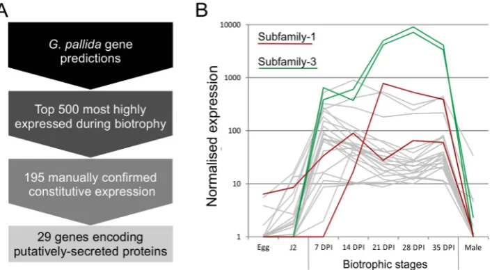
Using the assumption that effectors involved throughout the biotrophic interaction would be specifically and abundantly expressed in all sedentary stages, and would encode secreted proteins, a pipeline was developed to identify the most promising candidate genes (Figure 1). All predicted genes in the recently assembled genome sequence of the potato cyst nematode Globodera pallida[22] were ranked by their ratio of normalised expression during biotrophic stages compared to non-biotrophic stages. The top 2% (<500) of genes were manually verified, and those that were not evenly highly expressed across all biotrophic stages were discarded. The remaining 195 sequences were taken forward for secreted protein prediction. This resulted in 56 proteins with predicted signal peptides, 29 of which had no transmembrane domains. These 29 genes were compared to all published sequences in the non-redundant database, and those that had no significant homologues or no conserved domains were analysed further. One gene family identified, subsequently named HYP, was characterised in detail.
Gp-hypgene family identification and cloning
Based on the genomic sequence of GPLIN_001208400 (ftp://ftp. sanger.ac.uk/pub/pathogens/Globodera/pallida/Gene_Predictions/), primers were designed to amplify the coding region after the predicted cleavage site of the signal peptide to the stop codon (Table S1). This primer pair amplified a range of different sized products, which when sequenced could be grouped into three subfamilies 1 -2 and -3, each sharing considerable stretches of conserved bases at the 59and 39ends of the genes. Each subfamily was compared back to the genome by BLAST. Two complete genes were present, the previously identified GPLIN_001208400 and GPLIN_001025300, corresponding to subfamily -1 and -3 respectively. There were also two gene fragments (either a partial sequence or containing poly-N regions), named GPLIN_001135100 and GPLIN_000907700, corresponding to subfamilies -1 and -3 respectively. Based on the genomic sequence of gene GPLIN_001208400, primers were designed to amplify from the start to the stop codon. These specifically amplified a range of different sized products that could be grouped into subfamily -1 and -3 only.
No subfamily-2 members were present in the assembled genome sequence, however twoGp-hypsequences were present in thede novotranscriptome assembly of early sedentary stage nematodes (7 days post infection), both of which corresponded to subfamily -2. of a large array of effector proteins. This diversity may
Primers designed to amplify from the start to the stop codon amplified a range of products that all corresponded to subfamily -2. Subfamilies -1 and -3 were not separable by PCR of coding regions. 39 RACE was carried out for each of the three subfamilies, which identified a single specific UTR per subfamily (Figure 2). Each single subfamily specific 39UTR corresponded to the expected 39 UTR sequence present for that subfamily from either the assembled genome orde novotranscriptome.
Inter-subfamily variation
Subfamily-specific PCR primers from start codon to 39 UTR (Figure 2) were able to differentiate between subfamilies. In general subfamilies and locations of primer combinations can be described by Figure 2. In total, 75 unique genomic sequences were identified across Gp-hyp-1, -2 and -3, where Gp-hyp-1 is numerically dominant. All cloned Gp-hyp genes, irrespective of subfamily, shared stretches of 410 and 94 nucleotides with.90% identity at the 59 and 39 ends respectively. Between highly
conserved regions, subfamilies are characterised by a series of variable number subfamily-specific tandem repeats, summarised in Figure 2. No Gp-hyp sequences identified to date encode any annotated domains with the exception of a predicted signal peptide at the N-terminus of the protein.
Intra-subfamily variation
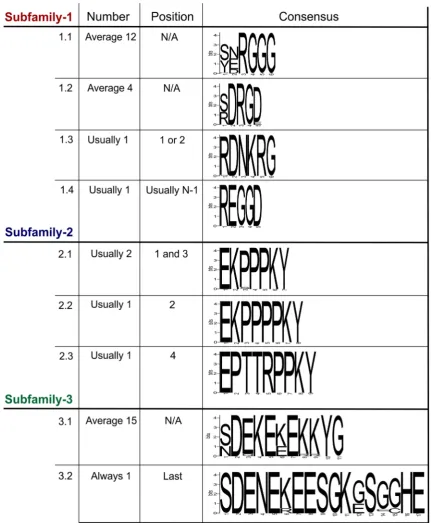
[image:3.612.63.555.524.660.2]Within each subfamily multiple different genomic DNA sequences were amplified, cloned and sequenced. In all cases, the entire tandem repeat region consists of a single open reading frame. ForGp-hyp-1and -3 only, large variation is observed in the number, sequence and order of tandem repeats in the deduced amino acid sequences corresponding to different genomic sequences. Within this region,Gp-hyp-1 genes contain four motifs, two of which are present as tandem repeats with complex organisations (Figure 3). The most common motif (1.1) consists of 6 amino acids, the first two are variable followed by a highly conserved RGGG. This glycine rich motif is present on average 12 Figure 1. Pipeline for identification of genes involved in plant-nematode biotrophy.A) Summarises the process of identifying novel genes, highly up regulated during biotrophy, that encode secreted proteins. B) Example expression data of all candidates with the addition of all other full length or partial HYP effector sequences present in the genome assembly, highlighted in red and green for subfamily-1 and -3 respectively. doi:10.1371/journal.ppat.1004391.g001
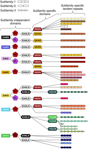
Figure 2. HYP effector gene family schematic and location of primer combinations.All HYP effector genes identified to date encode a secretion signal followed by two highly conserved regions flanking a region of variable number tandem repeats, and a subfamily specific 39UTR. Conserved regions ofGp-hypgenes share.95% identity between subfamilies. Subfamily-2 has a slightly different signal peptide (highlighted). 39UTRs are identical within subfamilies, irrespective of number of tandem repeats. Members of all subfamilies can be amplified using the F-ALL and R-ALL primer pair, subfamilies can be distinguished by PCR using a combination of signal peptide and 39UTR primers (summarised in Table S1). Solid lines represent introns, while dashed lines represent alignments.
times per gene, although this varies greatly. The second motif (1.2) consists of 5 amino acids, with a variable first position followed by conserved DRGD. This motif is present approximately 4 times per gene. The other two motifs (1.3 and 1.4) are usually present no more than once, often at the start or end of the tandem repeat domain (Table S2). Including all variable regions of all motifs in
subfamily-1, 20 unique amino acid sequences are encoded by 43 unique nucleotide sequences.
[image:4.612.89.521.60.584.2]The Gp-hyp-2 tandem repeat region always contains four proline rich tandem repeats, comprised of three different motifs (2.1, 2.2 and 2.3). Motif 2.1 consists of 7 amino acids in the sequence EKPPPKY. Motif 2.2 is identical, except for the Figure 3. Summary of the variation within the tandem repeat domains.Deduced amino acid sequences from the tandem repeat regions of genomic DNA sequences are shown. For each subfamily, the motifs present in the tandem repeat domain are shown along with their average copy number, position and consensus sequence. ForGp-hyp-1 and -3 no pattern has emerged to the positions/organisations of the highest frequency motifs. Motif 1.1 has the greatest variation in tandem repeat number, although motif 3.1 has less variation in the number of tandem repeats it has the greatest variation in the motif consensus sequence.
inclusion of an additional proline in the proline rich repeat (EKPPPPKY) and has no variation in sequence. Motif 2.3 consists of 9 amino acids, also has no sequence variation and is usually present only once (Figure 3). If motif 2.1 is present in positions 1 and 3, the position 3 motifs are more similar at the nucleotide level to other 2.1 motifs in position 3, than they are to 2.1 motifs in position 1, even if they are identical at the amino acid level. The four subfamily-2 tandem repeat variants are encoded by 10 unique nucleotide sequences. Interestingly, both subfamily-1 and -3 also contain the first and last of this type of tandem repeat.
Gp-hyp-3 tandem repeat regions contain two lysine and glutamic acid rich motifs (3.1 and 3.2) the first of which (3.1) occurs in tandem repeats on average 15 times, although this varies greatly. Motif 3.1 consists of 11 amino acids with highly conserved amino acids in position 2, 4, 5, 7, 10 and 11. Motif 3.2 consists of 17 amino acids and is always present as a single copy in the final position of the tandem repeat domain of subfamily-3 (Figure 3 and Table S2). Including all variable regions of all motifs in subfamily-3, 27 unique amino acid sequences are encoded by 42 unique nucleotide sequences.
Organisation of the various motifs within the tandem repeat region of all subfamilies is summarised in Table S2, although no pattern can describe all the variation. The above analysis was carried out using amino acid sequences deduced from genomic DNA sequences. Representatives of all subfamilies are present in cDNA synthesised from mRNA extracted from feeding females and these display similarly variable numbers and arrangements of tandem repeats. However, these are not studied in any detail due to the introduction of non-canonical apparent splice events as a direct result of the reverse transcriptase enzyme activity (described in more detail in Supporting information S2 and Figure S3).
In addition to the variable tandem repeats, within the highly conserved regions several non-synonymous polymorphic sequences have been identified that lead to amino acid changes. Figure 4 shows a schematic representation of these domains and does not represent the order in which they appear in the genes. Each combination of domains can be referred to as a ‘‘type’’. Types are not subfamily specific. Moreover, sequences encoding similar tandem repeat regions can have different amino acid sequence at every domain locus, and similarly identical types at every domain locus can have different tandem repeat regions (Figure 4 and Table S2).
Copy number variation between individuals
The large number of uniqueGp-hypgenomic DNA sequences were cloned from a pool of thousands of individual nematodes. To determine the complement ofGp-hyp sequences present in each nematode, a method was developed to extract high quality DNA, sufficient for multiple PCR reactions, from individual feeding female cyst nematodes. PCR with Gp-hyp subfamily-specific primers (Gp-hyp-1 F-1/3 and R-UTR-1; Gp-hyp-2 F-2 and R-UTR-2;Gp-hyp-3 F-1/3 and R-UTR-3; summarised in Table S1) revealed that individuals within a population differ in the number ofGp-hypsequences, the length ofGp-hypsequences, and in some cases even the presence or absence of Gp-hyp-3 genes. No two nematodes tested had the same complement of HYP effectors (Figure 5). Amplicons from individual PCR reactions were sequenced and this confirmed that a single nematode can contain more than one type, even within subfamilies. These data indicate a profound difference between individuals at the genetic level.
HYP effectors are expressed in large secretory cells associated with the amphids
In situ hybridisation was used to determine the spatial expression patterns ofGp-hypgenes in feedingG. pallidafemales.
A probe designed to target a conserved region of all subfamilies localised transcripts to a paired structure anterior to the metacorpal pump chamber. No such staining pattern was seen using the negative control probe (Figure 6). When compared to the anatomy of a typical tylenchid nematode, the position of the signal is consistent with expression in the amphidial sheath cells. No other paired structures are present in this region of the nematode. Amphid sheath cells are large secretory cells that produce material present in the amphidial canal and at the anterior surface of the nematode [14]. No expression was observed in pre-parasitic J2 nematodes (Figure 6), as expected from the highly significant up-regulation of HYP effectors specifically in all biotrophic stages (p-values range from 1.68*1023 to 5.08*1029 depending on the genomic copy; Figure S1).
Gp-HYP proteins are secreted by the nematode and accumulate at the host-parasite interface
Polyclonal rabbit antibodies were raised against a synthetic peptide derived from Gp-HYP sequences. Anti-Gp-HYP antibody was able to detect several proteins in a pooled nematode extraction and did not detect a signal when using similar quantities of plant root protein tested by western blot (Figure S2 B). Pre-immune serum was unable to detect the same proteins (Figure S3 B). Anti-Gp-HYP antibody specifically detected recombinant Anti-Gp-HYP-2 protein expressed and purified from bacteria, but not an unrelated negative control candidate effector protein similarly produced (Figure S3 A). The anti-Gp-HYP antibody localised a protein present in the apoplasm 14 days post infection at the plant-nematode interface, between the plant-nematode and the syncytial cell wall (Figure 7).
In plantaRNAi targeting HYP effectors causes a reduction in successful plant-parasitism
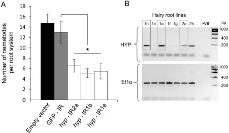
A representativeGp-hyp-1cDNA sequence was used to create an inverted repeat (IR) construct to express double stranded RNA in plantaunder the control of the CaMV35S promoter. As a result of the highly conserved regions, this construct should target all Gp-hyp-1, -2 and -3 transcripts. RT-PCR was used to confirm expression of the inverted repeat constructsin planta(Figure 8B). All potato hairy root lines tested that expressed the Gp-hyp-1 hairpin construct resulted in a 50–60% reduction in the number of nematodes that successfully infected and induced a feeding site compared to GFP IR control (correcting for multiple T-tests p, 0.05), suggesting a role of HYP effectors during parasitism (Figure 8A). Hairy root line 2b was not tested as its growth phenotype was not comparable to the control roots.
HYP effectors are specific to obligate biotrophic plant-parasitic nematodes
Heterodera glycines (patent WO 2007095469). Two partial Gp-hyp-1-like and oneGp-hyp-2-like sequences were also present in the transcriptome of Rotylenchulus reniformis (unpublished and [23]). No HYP effector-like sequences were identifiable from the EST database of the migratory endoparasite Radopholus similis
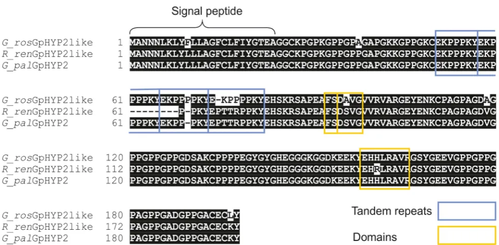
[image:6.612.151.459.61.579.2][24] or the genome sequence of the root-knot nematodes Meloidogyne incognita[25] orMeloidogyne hapla [26]. Interest-ingly theGp-hyp–like sequences from the various nematode species described show a remarkable level of conservation between species and indeed between genera (Figure 10).
Figure 4. Schematic representation of HYP effector domain organisation inG. pallida.Within the conserved regions, non-synonymous SNPs result in various domains. Combinations of domains give rise to ‘‘types’’. Certain types are subfamily specific, whereas others are not. For each column only, the same colour indicates the same sequence. By organising the sequences using types as indicators (not in the order they appear in the genes), HYP effectors do not group by subfamily. The three different subfamily specific tandem repeats are indicated in the final column as different shapes, where the size roughly correlates to number of repeats. Sequences encoding similar tandem repeat regions can have different domains at every locus, and similarly sequences encoding identical domains at every locus can have different tandem repeat regions.
Discussion
For this study we made several assumptions about the characteristics of effectors important throughout the biotrophic interaction of sedentary endoparasitic nematodes. These effectors are expected to be expressed throughout the parasitic stages of the nematode life cycle, and to be absent from all other stages. They are expected to be expressed in a tissue with the capacity to secrete proteins, with no prejudice for the pharyngeal gland cells. The genes are expected to encode proteins that are secreted and which should be detectable inside the host. By following this series of assumptions we have identified the HYP effectors: an incredibly complex and highly conserved gene family present across at least three genera of plant parasitic nematodes infecting mono- and dicotyledons.
The role of amphids during the feeding stages of
Globodera pallida
[image:7.612.124.488.61.263.2]Until recently, nematode effector identification was centred on the dorsal or subventral gland cells. We exploited the highly conserved structure of HYP effectors to design an in situ hybridisation probe that would target all members of all subfamilies, irrespective of the individual genetic complement of hyp genes each nematode had. We were able to demonstrate expression in the amphid sheath cells, large secretory cells associated with the amphids. Previous studies have shown that the cyst nematode feeding plug is continuous with the amphid openings of sedentary females [14,15] and may originate from the amphidial canal [14]. Gp-HYP proteins are secreted from the amphids, however the proteins extend further in the apoplasm Figure 5. HYP effector variation between individuals.Each lane shows PCR products amplified from discrete nematode samples. Each PCR in lane one corresponds to DNA extracted from the same singleG. pallidanematode. PCR on 6 individual nematodes using subfamily specific primers highlights a profound difference between individuals. In particular for subfamily-1 and -3 a reproducible difference can be seen in the complement of HYP effectors between nematodes both within and between subfamilies. The identity of amplification products from various samples was confirmed by sequencing. No two nematodes tested had the same genetic complement of HYP effectors.
doi:10.1371/journal.ppat.1004391.g005
Figure 6. HYP effectors are expressed in the amphid sheath cells of parasitic females.A) Schematic representation ofGlobodera pallida feeding female [64].In situhybridisation probe designed to a conserved region common to all HYP effectors identifies extremely strong and specific dark staining in a paired structure anterior to the metacorpal bulb of 14 days post infection females (C). No such staining pattern is observed with the negative control, or the positive control on second stage juvenile (J2) nematodes (B). Comparison of the staining pattern with a schematic identified this structure as the two lobed amphid sheath cells.
[image:7.612.143.471.532.669.2]than expected when compared to electron micrographs of feeding plugs and so they are probably not structural components of the feeding plug. In Meloidogyne species the importance of the amphids and of the apoplasm as a site of effector action is now being recognised [12,20]. The data presented here also suggest a changing role for the amphids inG. pallida throughout the life cycle. Interestingly, map-1 genes of M. incognita also contain tandem repeats that differ between populations [27], although these have no sequence or structural similarity to HYP effector tandem repeat domains and are relatively simple in comparison.
Recent data suggest these genes may in fact encode CLE-like peptides that originate from the pharyngeal glands [28]. HYP effector tandem repeats have no similarity to CLE peptides.
[image:8.612.100.520.62.214.2]HYP effectors are part of a complex multi-gene family Despite the presence of just three complete Gp-hypsequences across the assembled genome and transcriptome sequences ofG. pallida(one corresponding to each subfamily), the complexity of the HYP effector family was identified by conventional PCR and sequencing of individual clones. In addition, each time more Figure 7. Immunolocalisation of Gp-HYP proteins to the apoplasm.A) 2mm section showing Gp-HYP protein (green) detected between cell walls (blue) in the apoplasm between the feeding site (*) and the anterior end of the nematode (N). Cell walls are stained with calcofluor white (blue). B) Comparison with a schematic highlights the nematode in the context of the syncytium and the root tissue.
doi:10.1371/journal.ppat.1004391.g007
Figure 8. RNA interference ofGp-hypgenes.A) An inverted repeat construct targeting a singleGp-hyp1 gene was transformed into hairy roots of potato. Due to the nature of the conserved regions the construct was expected to target allGp-hypgenes in all subfamilies. For allGp-hyp-IR lines a significant reduction in nematode numbers (p,0.05) was seen when compared to the GFP IR hairy roots (n = 8). Error bars indicate the standard error of the mean. B) Semi-quantitative RT-PCR confirms expression of the inverted repeat construct in the RNAi lines. Line 2b was not tested for resistance as the phenotype varied too greatly from the GFP and empty vector controls.
[image:8.612.80.532.403.666.2]clones were sequenced more unique sequences were identified. This suggests that the 75 unique genomic sequences identified represent a far from exhaustive list, and that the full complexity of HYP effectors is yet to be catalogued. The absence of the full gene family in theG. pallidagenome assembly, and the presence of two fragmentedGp-hypgenes in poly-N regions, highlights a limitation of sequencing and assembly of short reads. AllGp-hypsequences, irrespective of subfamily, share stretches of 410 and 94 nucleotides with.90% identity at the 59and 39ends respectively which may underlie the difficulty in assembly.
[image:9.612.60.291.58.316.2]Comparison of HYP effectors to other pathosystems All unique genomic HYP effector sequences identified to date can be readily assigned to one of three subfamilies primarily, although not only, based on the amino acid sequence of the tandem repeat region. Numbers of subfamily-specific tandem repeats range greatly. It is unclear what the function of the tandem repeats is in the context of plant parasitic nematodes. Variable tandem repeat Transcription Activator-Like (TAL) effectors have been described for Xanthomonas spp [29]. TAL effectors are highly adaptable phytobacterial virulence factors that contain a 34 amino acid tandem repeat present in 17.5 iterations. DNA-binding base pair specificity is conferred by variable di-residues in positions 12 and 13 within each tandem repeat. HYP effectors contain numerous shorter tandem repeats that are considerably more variable in both sequence and organisation. Tandem motif 1.1 does contain highly conserved regions directly preceded by variable di-residues. However, HYP effectors are localised in the apoplasm, and so DNA binding is unlikely, although other ligand binding may be possible. It is unclear what the role of the different subfamilies, variable number/organisation of tandem repeats, or domains is in the context of ligand binding. HYP effector tandem repeats of subfamilies -1, -2, and -3 contain conserved glycine, proline and lysine residues. Glycine residues often create flexible linkers between domains. Taken together, these data may allow Figure 9. Comparison of HYP effector variation between UK
populations ofG. pallidaandG. rostochiensis.ForGp-hypsubfamily -1 and -3 large variation can be seen when amplifying from DNA extracted from a pool of thousands of G. pallida as indicated by multiple bands. Considerably less variation is seen under the same reaction conditions for G. rostochiensis, probably reflecting a more genetically restricted introduction to the UK. Amplicons from each reaction were sequenced to confirm they correspond to HYP effectors. doi:10.1371/journal.ppat.1004391.g009
Figure 10. Conservation ofhypgenes inGloboderaandRotylenchulusspecies.Amino acid alignment of HYP-2 sequences fromG. pallida, G. rostochiensisandR. reniformis. HYP-2 sequences are highly conserved (.93%) across the full length of the protein in the three species. The tandem repeat region, and true locations of the subfamily independent domains are highlighted.
[image:9.612.61.552.443.684.2]different functions. It has been noted however, that subtle amino acid changes that should conserve physiochemical properties can have an impact on effector function [32].
Similarly in oomycete species, the RXLR effectors are a large family of modular effectors with a highly conserved domain involved in membrane translocation [32]. HYP effectors are not characterised by a short conserved amino acid domain: stretches of hundreds of highly conserved amino acids are present at both the C and N-terminal ends of the protein. These highly conserved regions are not just conserved between species, but are conserved across at least three Genera of plant-parasitic nematodes.
Copy number variation of HYP effectors between individuals of the same population
Despite the superficial genetic structural similarity of the HYP effector tandem repeats to TAL effectors, and the similar modularity compared to RXLR and CRN effectors, no effector families, from any pathosystem to date, have been identified with such complex variation between individuals of the same popula-tion. We have demonstrated un-paralleled genomic diversity of HYP effectors between individuals: no two nematodes tested had the same genetic complement of Gp-hyp-1 or -3 sequences. Nematodes differed in the length, number, and even presence/ absence of entire gene subfamilies. It is unclear what the underlying genetic mechanism is that allows such variation between individuals. Due to the nature of the draft genome assembly of G. pallida we are unable to confirm that Gp-hyp sequences are paralogues, although the fact that individual nematodes differ in the size, and particularly number, of sequences within subfamilies suggests this is the case. The number and variation suggests that Gp-hyp genes are under high selection pressure. Gene expansions of cytochrome P450 genes have been described in Anopheles species [33], where estimates are of approximately 30 to 40 genes [34]. This is a good example as CYP450 genes, and in particular their copy number, have been linked to the extremely high selection pressure of resistance to DDT [34,35,36]. It is possible that the expansion of the HYP effector gene family in G. pallida may reflect a similarly high selection pressure from the host. Secreted components are the pathogen factors that are recognised by both pattern recognition receptors and resistance gene products. Diversity in Gp-hyp sequences may reflect the need to evade recognition in order to avoid detection. We have found that a UK population of G. pallidahas considerably more variation in HYP effectors thanG. rostochiensis. It has been suggested that the UK populations of G. pallida are from a broader genetic introduction than G. rostochiensis. If the diversity seen between individuals ofG. pallida for theGp-hypgenes is true on a wider genomic scale, this may explain the difficulty in identifying a broad spectrum resistance against G. pallida compared to G. rostochiensis [37]. Recent
HYP effectors are specific to, and play a role in, plant nematode biotrophic interactions
Transformation of cyst nematodes is not currently possible: In order to carry out gene knockout studies J2s can be soaked in double stranded RNA (dsRNA) or dsRNA can be delivered to feeding nematodes through in planta expression [43,44]. RNA soaking was not attempted in this study as expression of theGp-hyp genes is specific to all feeding stages (Figure S1). Instead, a hairpin construct was expressed in potato hairy roots. It has been reported that dsRNA expression in hairy roots is not ubiquitous and that phenotypes of the root cultures can vary, which may affect nematode infection [45,46]. Phenotypes of empty vector control, GFP IR control, andGp-hyp IR hairy root lines were therefore matched to the best of our ability before infecting with nematodes. Three independent lines expressing dsRNA targeting HYP effectors had reduced infection compared to the controls, suggesting HYP effectors play a role in the nematode infection. Despite the inherent noise of the hairy root system [45], RNAi targeting the Gp-hypgenes resulted in a significant reduction in nematode numbers. Although the dsRNA construct used has the potential to knock out all members of all subfamilies characterised to date, the list of clonedGp-hypsequences we have characterised is unlikely to be complete. Future work will focus on generating transgenic potato plants expressing the inverted repeat constructs of all threeGp-hypgene subfamilies, potentially creating durable resistance across all cyst and reniform nematodes. Due to the patchy nature of hairy root transgene expression [45,46], it is possible that successfully established nematodes had induced feeding sites from cells not expressing the dsRNA. It is therefore challenging to confirm the knockdown ofGp-hypexpression from the nematodes that successfully establish. Similarly due to the amplification step and transitive nature of RNAi in nematodes [47], subfamily-specific knockout studies are not possible.
unknown, although it may be related to regulation of gene expression [50,51].
Using assumptions about the characteristics of genes involved in plant-nematode biotrophic interactions to inform identification strategy, we provide the first description and characterisation of the HYP effectors. HYP effectors may play an important role in plant-nematode interactions and represent a class of effectors with continual expression, and presumably continual function, through-out biotrophy. Future work will focus on elucidating the function/s and specificity of the various gene family members, variable number tandem repeats and various domain arrangements. There are many economically unimportant, and consequently poorly characterised, nematode species that induce syncytial feeding sites in their host roots [52,53,54,55,56]. HYP effectors appear to be specific to plant-nematode interactions involving syncytia; identi-fying HYP effectors in these species may provide an insight into a common underlying feature of a complex endo-parasitic relation-ship.
Methods
Nematode DNA extraction and molecular cloning Genomic DNA was extracted from a pool of thousands of frozen J2G. pallida(‘‘Lindley’’) orG. rostochiensis(Ro1) according to the protocol described by Cottonet al.[22]. PCR was routinely carried out using BioTaq Red DNA polymerase following the manufacturer’s instructions for cycling conditions (Bioline) and the oligonucleotide primers and annealing temperatures listed in Table S1. PCR products were cloned by T-A cloning into the pGEM-T Easy vector (Promega) following the manufacturer’s suggestions. Plasmid DNA was extracted from bacterial culture using a QIAprep Spin Miniprep Kit (Qiagen) following the manufacturer’s instructions. Individual plasmids were sequenced at the service provided by Beckman Coulter Genomics.
Single nematode nucleic acid extraction
IndividualG. pallidafemales, collected 14 days post infection (dpi) of potato plants, were flash frozen in liquid nitrogen in individual 1.5 ml microfuge tubes. Nematodes were suspended in 200ml ‘‘Chaos’’ buffer [57] (4.5 M guanidine thiocyanate, 2% N-lauryl sarcosine, 50 mM EDTA (pH 8.0), 0.1 M 2-mercaptoeth-anol, 0.2% antifoam-A) disrupted with a pipette tip, and lysed by vortexing. One volume of phenol:chloroform:isoamyl alcohol (25:24:1) was added to the sample, vortexed, and centrifuged at 10,000gfor 5 minutes. The upper aqueous phase was transferred to 1 volume of 70% ethanol with the addition of 20 ng carrier RNA (NucleoSpin RNA XS). Total nucleic acid was extracted from the sample using a NucleoSpin RNA XS column, and the NucleoSpin RNA/DNA Buffer Set following manufacturer’s instructions. Two microliters of purified DNA was sufficient per 50ml PCR reaction.
39Rapid Amplification of cDNA Ends (RACE)
RNA was extracted from 14 dpi feeding females ofG. pallida using an RNeasy mini kit (Qiagen) following the manufacturer’s instructions for animal tissues. 39RACE was carried out using the 59/39 RACE Kit (Roche) according to manufacturer’s instruc-tions. The supplied oligo dT primer was used to prime cDNA synthesis. Subfamily-specific forward primers GAGGTTATGAC-GAGCATCATC, GAAAGGGCGGAGACAAAG and TGAG-CATCGTCTCCGTGCTG for subfamily -1, -2 and -3 respec-tively were identified by aligning all cloned sequences and visualising in an alignment viewer (BioEdit). The entire 39RACE PCR reaction was purified with a Qiaquick PCR purification kit
(Qiagen) following the manufacturer’s instructions. Purified amplification products were cloned by T-A cloning into the pGEM-T Easy vector (Promega) following the manufacturer’s suggestions, and positive clones were confirmed by sequencing.
In situhybridisation
In situhybridisation probes were designed to target a region of 134 conserved nucleotides at the 39end of the translated region of Gp-hyp transcripts using oligonucleotide forward (AACACG-GAGGTTATGACGAG), and reverse (GCTTGCGAATG-CAAATAT) primers respectively. Template was amplified from feeding nematode cDNA, and single stranded probes were synthesised from the template to incorporate digoxigenin labelled dUTP (Roche) using PCR by incubating at 94uC for 2 minutes followed by 35 cycles of 94uC for 15 seconds, 55uC annealing for 30 seconds, and 72uC extension for 90 seconds. Incorporation of DIG-labelled dUTP was confirmed by an apparent increase in size on agarose gel electrophoresis compared to template dsDNA.In situ hybridisation was carried out according to the methods described by de Boer et al. [58] with the following alterations. Cleaned potato roots heavily infected with 7–14 dpi feeding female G. pallida were lightly macerated using a bench top blender, and soaked in 10% formaldehyde for 3 days at room temperature. Fixed nematodes were collected by additional blending and subsequent sucrose gradient centrifugation (40% w/v). Feeding females were collected between 200 and 150mm mesh sieves. The protocol was continued as described from the cutting stage.
Root sectioning and immunolabelling
Lengths of potato root 14 days post infection with J2 ofG. pallida were fixed in 4% paraformaldehyde in PEM buffer (50 mM PIPES, 10 mM EGTA, 10 mM MgSO4pH 6.9) for 3 days at 4uC. Samples were dehydrated, resin embedded and sectioned according to Davieset al.[59] with the following alterations. Primary antibodies were raised to a 31 amino acid synthetic peptide VVRVAR-GEYENKCPAGPAGDVGPPGPPGPSG in a conserved region common to all Gp-HYP proteins predicted to have high antigenicity using [60]. The first 20 of which match with 100% identity to a consensus Gp-HYP-1 protein, the first 29 of which match with 100% identity to a consensus Gp-HYP-2 protein and the last 13 of which match with 100% identity to a consensus Gp-HYP-3 protein. Primary antibodies, or pre-immune sera, were hybridised at a dilution of 1 in 5 in 0.5% milk powder in PBS, and detected with a FITC-conjugated anti-rabbit secondary antibody at a dilution of 1 in 100. Plant cell walls were stained using Calcoflour-White at 1 mg/ml. Antibody specificity was tested against protein expressed in, and extracted from, the heterologousE. colisystem (Supporting information S1).
In plantaRNA interference
containing the relevant pART27 IR construct was incubated in 5 ml liquid Luria Bertani medium overnight at 28uC. Potato hairy root transformation was carried out by incubating 1 cm squares of Solanum tuberosum (‘Desire´e’) leaf material in liquid MS20 medium (4.3 g/l Murashige and Skoog (with vitamins), 20 g/l sucrose, pH 5.3–5.6) containing 100ml ofAgrobacteriumculture, for 3 days at room temperature. Leaf squares were dried on filter paper and placed on MS20 agar plates (2.4 g/l agar), containing 50mg/ml kanamycin and 400mg/ml cefotaxime. Roots originat-ing from different locations on each leaf square were considered individual transformation events, and were removed and cultured on MS20 agar plates containing 50mg/ml kanamycin. Inverted repeat construct expression was confirmed by RNA extraction (RNeasy Plant Mini Kit, Qiagen), cDNA synthesis (SuperScript II Reverse Transcriptase, Invitrogen), and PCR using the inverted repeat specific oligonucleotide primers ACGGAGGTTATGAC-GAG and GCTTGCGAATGCAAATATTAA.
Hatched J2 ofG. pallida were sterilised for 20 minutes in an appropriate volume of hexadecyltrimethylammonium bromide (CTAB, 0.5 mg/ml – Sigma) containing 0.1% v/v chlorhexidine digluconate (Sigma) and 0.01% v/v Tween-20, followed by three washes in sterile tap water. J2s were suspended at a concentration of approximately 1 nematode/ml and 30ml of suspension was pipetted onto each infection point. Three infection points were used per hairy root plate and 10 plates were used per line, with three independent lines for the Gp-hyp-IR, one line for GFP control, and one line for empty vector control. Two weeks after infection roots were stained by soaking in 1% sodium hypochlorite for 5 minutes, washing with tap water for 1.5 minutes 3 times, followed by boiling in 16acid fuchsin stain for 2 minutes. Total nematode numbers per root system were counted.
Bioinformatic analyses
Analysis of tandem repeats was carried out on genomic clones of Gp-hyp-1 -2and -3genes. For each subfamily, the middle region of the protein (between the two introns) was translated so that frame would be consistent with a cDNA clone. The software XSTREAM (http://jimcooperlab.mcdb.ucsb.edu/xstream/2013-8-8) was used to analyse the tandem repeats present for each subfamily. Putative signal peptides and transmembrane domains were predicted using SignalP v 4.1 (http://www.cbs.dtu.dk/ services/SignalP/) and TMHMM v 2.0 (http://www.cbs.dtu.dk/ services/TMHMM-2.0/), from the Centre for Biological Se-quence analysis [63].
Accession numbers
HYP effectors present in the G. pallida genome assembly (GPLIN_001208400, GPLIN_001025300, GPLIN_001135100, GPLIN_000907700) available at:
Figure S2 Gp-HYP antibody control. Equal quantities of
whole nematode protein extract and plant root protein extract are electrophoresed alongside a very low concentration of Gp-HYP 2 purified protein. A) HYP antibody specifically detects a Gp-HYP 2 protein expressed and purified from bacteria. B) Gp-Gp-HYP antisera detect a range of proteins from a total nematode extract that are not detected using the same concentrations of pre-immune control. Neither Gp-HYP antisera nor pre-pre-immune control can detect plant proteins at these concentrations. (TIF)
Figure S3 Non-canonical apparent splice events.A single
RNA species was synthesised in vitro. cDNA was reverse transcribed from this RNA followed by PCR analysis. A) For subfamily-1, multiple amplification products can be seen resulting from PCR on cDNA produced with either SuperScript II or Maxima RT. No such banding pattern is observed for PCR on a single gene from plasmid DNA. B) Similar additional banding patterns can be seen for subfamily-3. C) No such band pattern can be seen for a non-tandem repeat containing control GpCys. D) Denaturing RNA gel confirms a single RNA species in each case. Additional amplification products are therefore introduced as a result of the reverse transcriptase enzymes.
(TIF)
Supporting Information S1 HYP effector protein
expres-sion, extraction and antibody specificity. (DOCX)
Supporting Information S2 Non-canonical in vitro
ap-parent splicing of HYP effectors as a result of reverse transcriptase template switching activity.
(DOCX)
Table S1 HYP effector cloning primers and annealing
temperatures. (XLSX)
Table S2 Summary of domains and tandem repeat
motifs in deduced amino acid sequences ofG. pallida
unique genomic HYP effector sequences. (XLSX)
Acknowledgments
We would like to thank Dr Peter J.A. Cock for technical help regarding bioinformatics.
Author Contributions
References
1. Trudgill DL (1997) Parthenogenetic root-knot nematodes (Meloidogynespp); How can these biotrophic endoparasites have such an enormous host range? Plant Pathology 46: 26–32.
2. Nicol JM, Turner SJ, Coyne DL, den Nijs L, Hockland S, et al. (2011) Current Nematode Threats to World Agriculture. In: Jones J, Gheysen G, Fenoll C, editors. Genomics and Molecular Genetics of Plant-Nematode Interactions. pp. 21–43.
3. van Megen H, van den Elsen S, Holterman M, Karssen G, Mooyman P, et al. (2009) A phylogenetic tree of nematodes based on about 1200 full-length small subunit ribosomal DNA sequences. Nematology 11: 927–950.
4. Wyss U (1992) Observations on the feeding behaviour ofHeterodera schachtii
throughout development including events during moulting. Fundamental and Applied Nematology 15: 75–89.
5. Haegeman A, Mantelin S, Jones JT, Gheysen G (2012) Functional roles of effectors of plant-parasitic nematodes. Gene 492: 19–31.
6. Hewezi T, Baum TJ (2013) Manipulation of plant cells by cyst and root-knot nematode effectors. Molecular Plant-Microbe Interactions 26: 9–16. 7. Lilley CJ, Urwin PE, Atkinson HJ, McPherson MJ (1997) Characterization of
cDNAs encoding serine proteinases from the soybean cyst nematodeHeterodera glycines. Molecular and Biochemical Parasitology 89: 195–207.
8. Gheysen G, Fenoll C (2002) Gene expression in nematode feeding sites. Annual Review of Phytopathology 40: 191–219.
9. Wang XH, Mitchum MG, Gao BL, Li CY, Diab H, et al. (2005) A parasitism gene from a plant-parasitic nematode with function similar to CLAVATA3/ ESR (CLE) ofArabidopsis thaliana. Molecular Plant Pathology 6: 187–191. 10. Replogle A, Wang J, Bleckmann A, Hussey RS, Baum TJ, et al. (2011)
Nematode CLE signaling inArabidopsisrequires CLAVATA2 and CORYNE. The Plant Journal 65: 430–440.
11. Semblat J-P, Rosso M-N, Hussey RS, Abad P, Castagnone-Sereno P (2001) Molecular cloning of a cDNA encoding an amphid-secreted putative avirulence protein from the root-knot nematodeMeloidogyne incognita. Molecular Plant-Microbe Interactions 14: 72–79.
12. Vieira P, Danchin EGJ, Neveu C, Crozat C, Jaubert S, et al. (2011) The plant apoplasm is an important recipient compartment for nematode secreted proteins. Journal of Experimental Botany 62: 1241–1253.
13. Rosso M-N, Vieira P, de Almeida-Engler J, Castagnone-Sereno P (2011) Proteins secreted by root-knot nematodes accumulate in the extracellular compartment during root infection. Plant Signal Behav 6: 1232–1234. 14. Endo BY (1978) Feeding plug formation in soybean roots infected with the
soybean cyst nematode. Phytopathology 68: 1022–1031.
15. Sobczak M, Golinowski WA, Grundler FMW (1999) Ultrastructure of feeding plugs and feeding tubes formed byHeterodera schachtii. Nematology 1: 363– 374.
16. Gao BL, Allen R, Maier T, Davis EL, Baum TJ, et al. (2003) The parasitome of the phytonematodeHeterodera glycines. Molecular Plant-Microbe Interactions 16: 720–726.
17. Huang G, Gao B, Maier T, Allen R, Davis EL, et al. (2003) A profile of putative parasitism genes expressed in the esophageal gland cells of the root-knot nematodeMeloidogyne incognita. Molecular Plant-Microbe Interactions 16: 376–381.
18. Perry RN (1996) Chemoreception in plant parasitic nematodes. Annual Review of Phytopathology 34: 181–199.
19. Jones JT, Perry RN, Johnston MRL (1994) Changes in the ultrastructure of the amphids of the potato cyst-nematode,Globodera rostochiensis, during develop-ment and infection. Fundadevelop-mental and Applied Nematology 17: 369–382. 20. Tomalova I, Iachia C, Mulet K, Castagnone-Sereno P (2012) The map-1 gene
family in root-knot nematodes, Meloidogyne spp.: a set of taxonomically restricted genes specific to clonal species. Plos One 7: e38656.
21. Jones J, Reavy B, Smant G, Prior A (2004) Glutathione peroxidases of the potato cyst nematodeGlobodera Rostochiensis. Gene 324: 47–54.
22. Cotton JA, Lilley CJ, Jones LM, Kikuchi T, Reid AJ, et al. (2014) The genome and life-stage specific transcriptomes ofGlobodera pallidaelucidate key aspects of plant parasitism by a cyst nematode. Genome Biology 15: R43.
23. Wubben MJ, Callahan FE, Scheffler BS (2010) Transcript analysis of parasitic females of the sedentary semi-endoparasitic nematodeRotylenchulus reniformis. Molecular and Biochemical Parasitology 172: 31–40.
24. Jacob J, Mitreva M, Vanholme B, Gheysen G (2008) Exploring the transcriptome of the burrowing nematode Radopholus similis. Molecular Genetics and Genomics 280: 1–17.
25. Abad P, Gouzy J, Aury J-M, Castagnone-Sereno P, Danchin EGJ, et al. (2008) Genome sequence of the metazoan plant-parasitic nematode Meloidogyne incognita. Nature Biotechnology 26: 909–915.
26. Opperman CH, Bird DM, Williamson VM, Rokhsar DS, Burke M, et al. (2008) Sequence and genetic map ofMeloidogyne hapla: A compact nematode genome for plant parasitism. Proceedings of the National Academy of Sciences 105: 14802–14807.
27. Castagnone-Sereno P, Semblat J-P, Castagnone C (2009) Modular architecture and evolution of the map-1 gene family in the root-knot nematodeMeloidogyne incognita. Molecular Genetics and Genomics 282: 547–554.
28. Rutter WB, Hewezi T, Maier TR, Mitchum MG, Davis EL, et al. (2014) Members of theMeloidogyneAvirulence protein family contain multiple plant ligand-like motifs. Phytopathology 104: 879–885.
29. Doyle EL, Stoddard BL, Voytas DF, Bogdanove AJ (2013) TAL effectors: highly adaptable phytobacterial virulence factors and readily engineered DNA-targeting proteins. Trends in Cell Biology 23: 390–398.
30. Stam R, Howden AJ, Delgado-Cerezo M, Amaro TM, Motion GB, et al. (2013) Characterization of cell death inducing Phytophthora capsici CRN effectors suggests diverse activities in the host nucleus. Frontiers in plant science 4: 387. 31. Stam R, Jupe J, Howden AJ, Morris JA, Boevink PC, et al. (2013) Identification and characterisation CRN Effectors inPhytophthora capsicishows modularity and functional diversity. PLoS ONE 8: e59517.
32. Whisson SC, Boevink PC, Moleleki L, Avrova AO, Morales JG, et al. (2007) A translocation signal for delivery of oomycete effector proteins into host plant cells. Nature 450: 115–118.
33. Scott JA, Collins FH, Feyereisen R (1994) Diversity of cytochrome-p450 genes in the mosquito, Anopheles albimanus. Biochemical and biophysical research communications 205: 1452–1459.
34. Ranson H, Nikou D, Hutchinson M, Wang X, Roth CW, et al. (2002) Molecular analysis of multiple cytochrome P450 genes from the malaria vector,Anopheles gambiae. Insect Molecular Biology 11: 409–418.
35. Wondji CS, Irving H, Morgan J, Lobo NF, Collins FH, et al. (2009) Two duplicated P450 genes are associated with pyrethroid resistance inAnopheles funestus, a major malaria vector. Genome Research 19: 452–459.
36. Djouaka RF, Bakare AA, Coulibaly ON, Akogbeto MC, Ranson H, et al. (2008) Expression of the cytochrome P450s, CYP6P3 and CYP6M2 are significantly elevated in multiple pyrethroid resistant populations ofAnopheles gambiaes.s. from Southern Benin and Nigeria. Bmc Genomics 9: 538.
37. Barone A, Ritter E, Schachtschabel U, Debener T, Salamini F, et al. (1990) Localization by restriction-fragment-length-polymorphism mapping in potato of a major dominant gene conferring resistance to the potato cyst nematode
Globodera rostochiensis. Molecular & General Genetics 224: 177–182. 38. Sasagawa Y, Nikaido I, Hayashi T, Danno H, Uno KD, et al. (2013)
Quartz-Seq: a highly reproducible and sensitive single-cell RNA sequencing method, reveals non-genetic gene-expression heterogeneity. Genome Biology 14: R31. 39. Alenda C, Gallot-Legrand A, Fouville D, Grenier E (2013) Sequence
polymorphism of nematode effectors highlights molecular differences among the subspecies of the tobacco cyst nematode complex. Physiological and Molecular Plant Pathology 84: 107–114.
40. Plantard O, Picard D, Valette S, Scurrah M, Grenier E, et al. (2008) Origin and genetic diversity of Western European populations of the potato cyst nematode (Globodera pallida) inferred from mitochondrial sequences and microsatellite loci. Molecular Ecology 17: 2208–2218.
41. Thiery M, Fouville D, Mugniery D (1997) Intra- and interspecific variability in Globodera, parasites of Solanaceous plants, revealed by random amplified polymorphic DNA (RAPD) and correlation with biological features. Funda-mental and Applied Nematology 20: 495–504.
42. Blok VC, Malloch G, Harrower B, Phillips MS, Vrain TC (1998) Intraspecific variation in ribosomal DNA in populations of the potato cyst nematode
Globodera pallida. Journal of Nematology 30: 262–274.
43. Lilley CJ, Bakhetia M, Charlton WL, Urwin PE (2007) Recent progress in the development of RNA interference for plant parasitic nematodes. Molecular Plant Pathology 8: 701–711.
44. Lilley C, Davies L, Urwin P (2012) RNA interference in plant parasitic nematodes: a summary of the current status. Parasitology 139: 630–640. 45. Wubben MJ, Callahan FE, Triplett BA, Jenkins JN (2009) Phenotypic and
molecular evaluation of cotton hairy roots as a model system for studying nematode resistance. Plant Cell Reports 28: 1399–1409.
46. Triplett BA, Moss SC, Bland JM, Dowd MK (2008) Induction of hairy root cultures from Gossypium hirsutum and Gossypium barbadense to produce gossypol and related compounds. In Vitro Cellular & Developmental Biology-Plant 44: 508–517.
47. Dalzell JJ, McVeigh P, Warnock ND, Mitreva M, Bird DM, et al. (2011) RNAi effector diversity in nematodes. PLoS neglected tropical diseases 5: e1176. 48. Jones JT, Haegeman A, Danchin EGJ, Gaur HS, Helder J, et al. (2013) Top 10
plant-parasitic nematodes in molecular plant pathology. Molecular Plant Pathology 14: 946–961.
49. Holterman M, Karssen G, van den Elsen S, van Megen H, Bakker J, et al. (2009) Small Subunit rDNA-based phylogeny of the Tylenchida sheds light on relationships among some high-impact plant-parasitic nematodes and the evolution of plant feeding. Phytopathology 99: 227–235.
50. Barrett LW, Fletcher S, Wilton SD (2012) Regulation of eukaryotic gene expression by the untranslated gene regions and other non-coding elements. Cellular and Molecular Life Sciences 69: 3613–3634.
51. Zhao W, Blagev D, Pollack JL, Erle DJ (2011) Toward a systematic understanding of mRNA 39untranslated regions. Proceedings of the American Thoracic Society 8: 163–166.
52. Suarez Z, Moss CS, Inserra RN (1985) Anatomical changes induced by
Punctodera chalcoensisin corn roots. Journal of Nematology 17: 242–244. 53. Cohn E, Mordechai M (1982) Biology and host parasite relations of a species of